
NIPT. Indication. Advantages and limitation. Ten marks question. PG of Obs and gynae.
Reading File
Now let me search the medical library and PubMed simultaneously for comprehensive NIPT coverage.
Finding Sources
Finding Sources
Searching PubMed
"non-invasive prenatal testing" NIPT indications
Good hits from Harrison's 22E and Creasy & Resnik. Let me now read those sections in parallel.
Reading File
Reading File
Finding Sources
Excellent content from Harrison's 22E and Tietz. Let me now read more of the Harrison's block and the Thompson & Thompson section for complete clinical details.
Reading File
Reading File
Finding Sources
Excellent - I now have comprehensive material from Harrison's 22E, Tietz, and Thompson & Thompson. Let me read a bit more of the Harrison's block for the limitations, advantages, and recent expansions section.
Reading File
Reading File
I now have all the material needed for a comprehensive 10-mark answer. Let me also fetch the NIPT diagram from Thompson & Thompson for visual illustration.
Loading Image
I now have all the content needed for a thorough 10-mark PG-level answer. Here is the complete structured response:
Non-Invasive Prenatal Testing (NIPT)
PG Obstetrics & Gynaecology | 10 Marks
Introduction
Non-invasive prenatal testing (NIPT), also called cell-free DNA (cfDNA) screening or NIPS (non-invasive prenatal screening), is a method of screening for fetal chromosomal abnormalities using circulating cell-free DNA (cfDNA) extracted from a maternal peripheral blood sample.
The biological basis was established in 1997 when Lo and colleagues demonstrated that fetal cfDNA - originating from apoptotic trophoblast cells of the placenta - is present in maternal plasma and can be distinguished and analyzed. This placental cfDNA shares the same genome as the fetus and constitutes approximately 5-20% of total circulating cfDNA (the "fetal fraction") after 10 weeks' gestation.
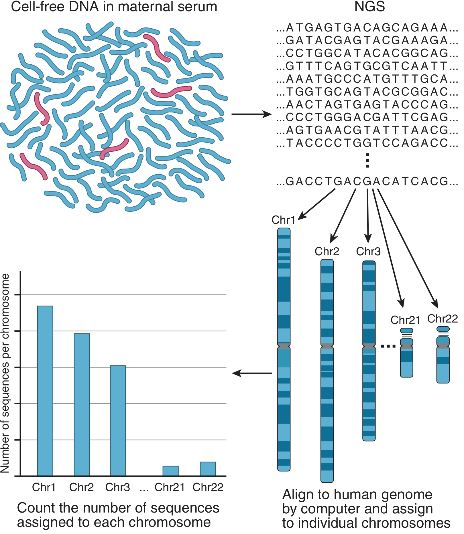
Principle / How It Works
Two main methodologies are used:
-
Quantitative counting (massively parallel sequencing / MPS): Total cfDNA (maternal + fetal) is sequenced using next-generation sequencing. Millions of reads are mapped back to each chromosome. If the fetus has trisomy 21, the proportion of sequences mapping to chromosome 21 exceeds the expected ~1.5%, signaling aneuploidy. This method works with low fetal fraction and is applicable in twin pregnancies.
-
SNP genotyping method: Targets single nucleotide polymorphisms (SNPs) on chromosomes of interest. By comparing maternal plasma DNA (maternal + fetal) against pure maternal leukocyte DNA, fetal genotype is inferred. This is the only method that can determine parent of origin of aneuploidy and detect triploidy. It cannot be used in egg-donor or twin pregnancies.
Indications
Traditional (High-Risk) Indications
| Category | Detail |
|---|---|
| Advanced maternal age (AMA) | ≥35 years at delivery; higher a priori risk for trisomy |
| Abnormal first-trimester serum screen | Raised or low PAPP-A, β-hCG, or abnormal NT measurement |
| Abnormal second-trimester quad screen | Abnormal AFP, estriol, inhibin A, β-hCG |
| Prior pregnancy with chromosomal aneuploidy | History of Down syndrome, Edwards, or Patau pregnancy |
| Parent carrying balanced chromosomal translocation | Increased risk of unbalanced offspring |
| Fetal structural anomaly on ultrasound | Cardiac defects, choroid plexus cysts, echogenic bowel (soft markers) |
| Parental anxiety | After counselling, as per patient choice |
Expanded (Population-Level / Low-Risk) Indications
Contemporary evidence confirms equivalent utility in low-risk as well as high-risk populations. Major society guidelines (ACOG, SMFM, ISUOG) now endorse offering NIPT to all pregnant women regardless of age or risk category.
Extended Indications (Beyond Common Aneuploidies)
- Sex chromosome aneuploidies: 45,X (Turner), 47,XXY (Klinefelter), 47,XXX, 47,XYY
- Microdeletion/microduplication syndromes: 22q11.2 deletion (DiGeorge), Prader-Willi/Angelman, 1p36 deletion, 5p- (cri-du-chat)
- Rare autosomal trisomies (RAT) - chromosomes other than 13, 18, 21
- Single-gene disorders (emerging: autosomal dominant de novo conditions, e.g., achondroplasia)
- Fetal sex determination (in X-linked disorders)
- Rhesus D genotyping in RhD-negative mothers
Timing
- Can be performed as early as 9-10 weeks' gestation (most programs recommend ≥10 weeks)
- cfDNA is detectable from 4-5 weeks but fetal fraction is too low before 10 weeks for reliable results
- This early window allows ample time for confirmatory invasive testing (CVS at 10-13 weeks or amniocentesis at 15-20 weeks) if NIPT is positive
Advantages
Clinical Advantages
| Advantage | Detail |
|---|---|
| No fetal risk | Non-invasive; maternal venepuncture only, no procedure-related miscarriage |
| High sensitivity for trisomy 21 | >99% detection rate (sensitivity and specificity approaching 99%) - far superior to traditional serum screening (~80-85%) |
| Low false-positive rate | ~0.1-0.5% for common aneuploidies; traditional quad screen has ~5% FPR |
| Early gestation | Reportable from 10 weeks, earlier than amniocentesis |
| Reduces unnecessary invasive procedures | Negative NIPT markedly lowers probability of aneuploidy and avoids amniocentesis / CVS in most patients |
| Fetal sex determination | Useful in X-linked conditions |
| Expanded detection capability | Sex chromosome aneuploidies, select microdeletions, rare trisomies |
| Single-gene disorder detection | Emerging platforms for de novo dominant disorders (achondroplasia, FGFR-related conditions) |
| Multiple gestations | More recent large cohorts show detection rates in twins mirror singleton performance |
| Psychological benefit | Early reassurance or earlier preparation for affected pregnancies |
Technical Advantages
- Rapid turnaround (7-14 days)
- No cell culture required (unlike karyotype)
- Genome-wide analysis is performed even when only select results are reported
Limitations
Technical Limitations
| Limitation | Explanation |
|---|---|
| It is a screening test, NOT a diagnostic test | Positive result must always be confirmed by CVS or amniocentesis before any irreversible decision |
| Low fetal fraction | If fetal fraction <4%, the result is unreportable ("no-call"). This is more common at <10 weeks, in maternal obesity, with autoimmune disease, maternal malignancy, or vanishing twin. A no-call result itself carries an increased risk of aneuploidy |
| Confined placental mosaicism (CPM) | cfDNA is placental in origin, not directly fetal. CPM can cause false-positive or false-negative NIPT results, particularly for smaller genomic variants |
| Maternal genomic abnormalities | Undetected maternal sex chromosome mosaicism, maternal chromosomal deletion, or maternal malignancy can distort results |
| Cannot detect all chromosomal anomalies | Open neural tube defects, structural anomalies, and most single-gene disorders are not detectable |
| Positive Predictive Value (PPV) varies | PPV is highly dependent on a priori risk (maternal age). In a 20-year-old, PPV for trisomy 21 may be only ~40-50% even with a "high-risk" NIPT result |
| Lower accuracy for sex chromosome aneuploidies | Sensitivity and PPV for 45,X, 47,XXY etc. are lower than for trisomies 21/18/13 due to mosaicism |
| Microdeletion detection limitations | High false-positive rates for microdeletion syndromes; CPM is a frequent confound; not yet recommended as standard for all populations |
| Rare autosomal trisomies | Variable significance; some are associated with adverse outcomes, others are benign mosaics - management remains controversial |
| Cannot replace anatomy scan | Fetal structural abnormalities require ultrasound evaluation |
| Single-gene disorders | Technically challenging; limited clinical availability; mostly available only for a small set of conditions |
| Egg donor / gestational carrier pregnancies | SNP-based methods cannot be used; results reflect donor's genetic contribution |
| Cost | Remains high in many healthcare systems; not universally covered |
| Incidental findings | Whole-genome NIPT may reveal copy number variants of uncertain significance, creating parental anxiety and ethical dilemmas |
Performance Characteristics
| Condition | Sensitivity | Specificity | PPV (varies with maternal age) |
|---|---|---|---|
| Trisomy 21 (Down) | >99% | >99% | ~90-99% (high risk); ~40-80% (low risk) |
| Trisomy 18 (Edwards) | ~96-98% | >99% | Lower than T21 |
| Trisomy 13 (Patau) | ~90-99% | >99% | Lower than T18 |
| Sex chromosome aneuploidies | 88-95% | ~99% | Lowest PPV |
| Microdeletions | 75-90% | ~99% | Very low (high FPR) |
Important Counseling Points (Examiner Favourites)
- NIPT is a screening test - a positive result is not a diagnosis.
- A negative NIPT does not exclude all chromosomal or structural abnormalities - anatomy scan and other screening modalities are still required.
- Positive NIPT must be confirmed by CVS or amniocentesis before pregnancy termination or major clinical decisions.
- Low fetal fraction ("no-call") result requires either repeat testing or invasive diagnostic testing.
- The test does not screen for all causes of intellectual disability or congenital abnormality.
- Pre-test and post-test genetic counselling is strongly recommended.
Comparison with Traditional Screening
| Parameter | Serum Screening (Combined / Quad) | NIPT |
|---|---|---|
| Timing | 10-14 wk (combined); 15-20 wk (quad) | From 10 weeks |
| Sensitivity (T21) | 80-90% | >99% |
| False-positive rate | ~5% | <0.1% |
| Invasive procedure risk | Indirect (for follow-up) | None |
| Diagnostic | No | No (screening only) |
| Cost | Lower | Higher |
| Structural anomaly detection | Via NT/USS component only | No |
Conclusion
NIPT represents a major advance in prenatal screening. Its high sensitivity and specificity for the common trisomies, zero procedural risk to the fetus, and early gestational window of application make it the preferred first-tier or second-tier screening option in contemporary obstetric practice. However, it remains a screening tool: results must be interpreted in the context of a priori risk, fetal fraction adequacy, and confirmed with diagnostic testing when positive. Awareness of its limitations - particularly for microdeletions and sex chromosome aneuploidies - is essential for appropriate clinical application and patient counselling.
Sources:
- Harrison's Principles of Internal Medicine, 22nd ed. (2025), Chapter 503 - Cell-free fetal DNA testing sections
- Tietz Textbook of Laboratory Medicine, 7th ed. - Chapter 68, Noninvasive Prenatal Testing
- Thompson & Thompson Genetics and Genomics in Medicine, 9th ed. - Chapter 18, Noninvasive Prenatal Screening by cfDNA Analysis
- Eltabbakh et al., Front Med 2024 - Advancements of NIPT: the role of obstetricians
This is a shared conversation. Sign in to Orris to start your own chat.