
Cream for daiper rash in babies
diaper rash cream babies treatment recommendations 2025
diaper rash baby skin treatment
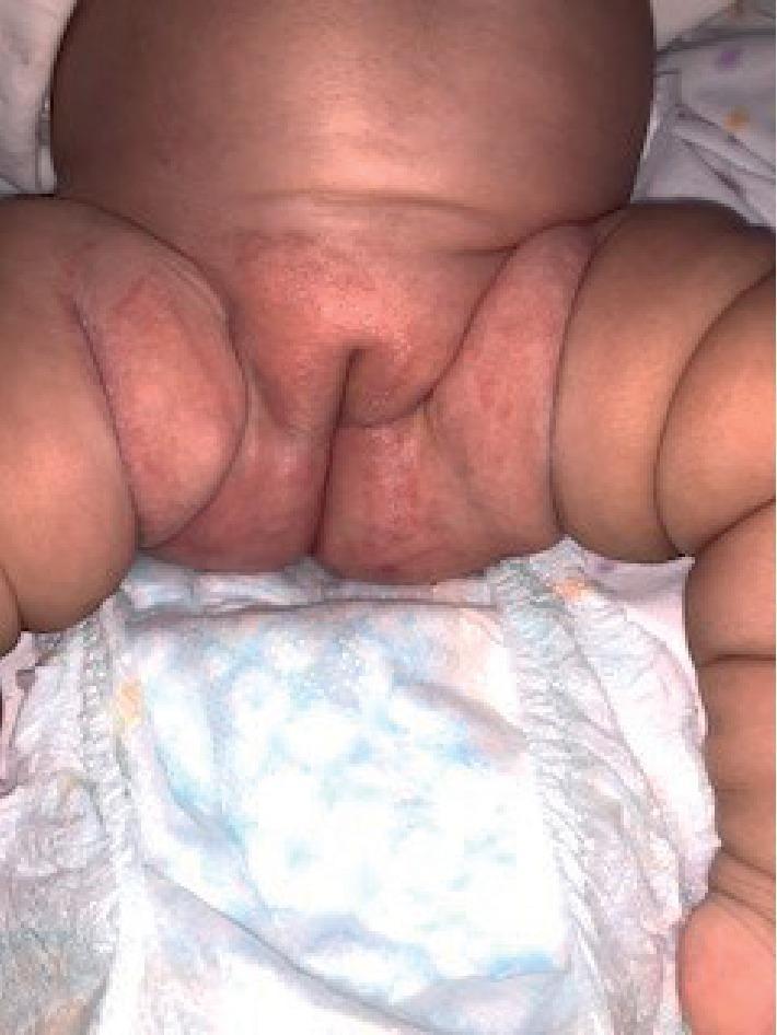
This clinical photograph shows the perineal and inguinal regions of an infant, demonstrating an extensive napkin (diaper) rash. The image reveals diffuse erythema and inflammation across the groin, suprapubic area, and inner thighs. Notable features include the involvement of the skin folds (intertriginous areas), which is characteristic of certain types of primary or secondary diaper dermatitis, such as candidiasis. The affected skin appears slightly macerated and damaged, indicating a breakdown of the epidermal barrier. A disposable diaper is visible at the bottom of the frame, providing clinical context for the distribution of the lesions. This visual represents a common pediatric dermatological condition where the moist, occlusive environment of a diaper leads to skin irritation or secondary infection. The image serves as an educational tool for identifying inflammatory skin changes in the diaper area and assessing the severity of the dermatitis.
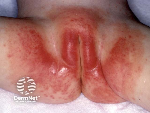
Imaging modality: Clinical photography. This is an external frontal view of the diaper area, encompassing the perineum, buttocks, and surrounding inguinal skin. The primary subject is napkin dermatitis (diaper dermatitis) in an infant, a common irritant contact dermatitis related to prolonged moisture and friction. The skin shows diffuse bright red erythema with marked maceration in diaper-covered regions; surface appears moist, shiny, and sensitive, with mild desquamation along the creases. The rash is bilateral and predominantly within the diaper distribution, extending to the perianal area and inner thighs while sparing some higher folds. No vesicles, pustules, or crusts are evident in this image. The observed pattern is most consistent with irritant dermatitis from urine and feces exposure, though candidal infection can yield satellite lesions and more confluent involvement. Pathophysiology involves inflammation due to irritants, impaired barrier function, and frictional injury in occluded skin. Diagnostic significance: visually consistent with diaper dermatitis; supports barrier-focused management over antibiotics alone. Differential diagnoses include candida diaper dermatitis, bacterial dermatitis, intertrigo, and less likely eczema. Clinical correlation: prioritize frequent diaper changes, thorough cleaning, air exposure, gentle drying, and barrier creams (zinc oxide, petrolatum); consider antifungal therapy if satellite lesions or persistent symptoms. Useful for education, triage, and research on pediatric irritant dermatoses.
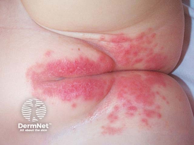
Clinical photography of the diaper area in a young infant showing napkin dermatitis (diaper dermatitis) with diffuse erythematous, moist plaques confined to the perineal, buttock, and inguinal regions within the diaper border. The rash is bilateral and symmetrical, with well-demarcated margins where diaper coverage ends, and a shiny, glazed appearance consistent with irritant contact dermatitis. Involvement extends to the inner thighs and intertriginous folds, without deep ulceration or necrosis. No discrete vesicles are evident, but papules and patchy confluent erythema are present. The surrounding skin may appear slightly desquamative in places, reflecting repetitive friction and moisture. The clinical picture favors irritant diaper dermatitis from prolonged exposure to urine and feces, minimized barrier protection, and occlusion by diapers. Secondary infection with Candida albicans is a consideration if satellite lesions or intensification of erythema occurs at flexural creases, though this image does not clearly show candidal features such as satellite pustules. Appropriate management includes frequent diaper changes, gentle cleansing, air exposure, and barrier creams containing zinc oxide or petrolatum; consider antifungal therapy if candidal involvement is suspected. This image is relevant for education on differential diagnosis with fungal diaper dermatitis, contact dermatitis to wipes, and pediatric dermatology case discussions.
Diaper Rash (Diaper Dermatitis) in Babies
What It Looks Like
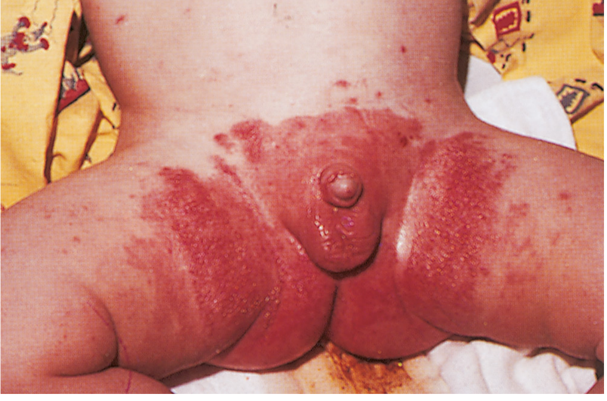
- Caused by prolonged contact with urine/stool, or during diarrheal illness
- Erythematous (red), macular or papular rash with well-demarcated borders
- Spares the deep skin folds
- Differential: perianal streptococcal infection (warm, macular, with desquamation)
- Caused by Candida species (ubiquitous in the infant GI tract)
- Intensely red with satellite lesions - small pustules beyond the margins of the main rash
- Scalloped border, sharply demarcated edge, scaling around margins
- Can coexist with oral thrush - always check the mouth
Creams and Treatment
For Irritant (Contact) Diaper Rash
| Product | Key Ingredient | Notes |
|---|---|---|
| Desitin Maximum Strength | 40% Zinc Oxide | Best for active rash; thick barrier; hypoallergenic |
| Triple Paste | Zinc Oxide | Highly rated; free of harmful chemicals |
| A+D Original Ointment | Petrolatum + Lanolin | Good barrier ointment; no zinc oxide |
| Aquaphor Baby | Petrolatum | Multi-purpose barrier; gentle; good for everyday use |
| Balmex / Boudreaux's Butt Paste | Zinc Oxide | Strong barrier formulas |
- Apply thickly - as if "frosting a cake" (per the American Academy of Dermatology)
- Zinc oxide is the gold standard for active rash - it provides both barrier protection and treats irritation. The Consumer Reports guide recommends having a zinc oxide cream for active rash AND a plain petrolatum/barrier balm for everyday prevention
- Choose fragrance-free products to avoid allergens and irritants
- Apply after every diaper change when a rash is present
For Candidal Diaper Rash (the type with satellite lesions)
- Nystatin cream 100,000 units/gram - applied 3 times daily for 10-14 days - this is first-line treatment
- Apply in this order: nystatin first → then zinc oxide barrier cream on top
- For severely inflamed lesions: add 1-2% hydrocortisone cream - apply after nystatin but before zinc oxide
- If oral thrush is also present: treat with oral nystatin 100,000 units/mL (2 mL four times daily in infants)
From Tintinalli's Emergency Medicine: "Treatment is with topical antifungal agents, most commonly nystatin cream applied in combination with barrier ointments (zinc oxide applied after nystatin) and topical steroids for severely inflamed lesions."
General Prevention and Care
- Change diapers frequently - do not wait until fully soaked
- Cleanse gently with baby wipes or warm water after each change
- Pat dry (do not rub) or air-dry before applying cream
- Apply barrier cream at every change as a preventive layer
- Make sure the diaper fits correctly - too tight = chafing
When to See a Doctor
- Rash worsens or does not improve after 2-3 days of home treatment
- Satellite lesions (pustules around the rash) - suggests Candida, needs antifungal
- Rash accompanied by fever
- Blisters, ulcers, or bleeding skin
- Spreading beyond the diaper area