
Renal cell carcinoma for 10 marks
I'll look up renal cell carcinoma from the medical library for a comprehensive 10-mark answer.
Reading File
Now let me search the medical library for renal cell carcinoma content in parallel:
Finding Sources
Finding Sources
Excellent sources found. Let me read the most comprehensive ones in parallel - Robbins Pathology and Campbell Walsh Urology:
Reading File
Reading File
Reading File
Good content so far. Let me now read more from Robbins (morphology, clinical features, staging) and Goldman-Cecil (clinical features and management):
Reading File
Reading File
Let me get the clinical features, morphology, and management sections from the Robbins and Goldman-Cecil blocks:
Reading File
Reading File
Excellent - I have rich content. Let me get the treatment/management section and the clinical features/morphology more completely:
Reading File
Reading File
Let me get the treatment continuation and clinical features from Goldman-Cecil, plus read the Robbins morphology section:
Reading File
Reading File
I now have comprehensive content from multiple authoritative sources. Let me also fetch the key pathology images and get the metastatic treatment content:
Reading File
Reading File
Loading Image
I now have everything needed for a comprehensive 10-mark answer. Let me also get the histology image:
Loading Image
Reading File
I now have all the content needed. Let me compile the comprehensive 10-mark answer:
Loading Image
I have everything needed. Here is the comprehensive 10-mark answer on Renal Cell Carcinoma:
Renal Cell Carcinoma (RCC)
Definition
Renal cell carcinoma (RCC) is a group of malignant epithelial neoplasms arising from the renal tubular epithelium, located predominantly in the cortex. It is not a single entity but a collection of distinguishable tumors, each with a characteristic morphology and genetic profile. RCC accounts for 80-85% of all primary malignant renal neoplasms and 2-3% of all adult cancers. Classically known as "hypernephroma" or "clear cell carcinoma," it is now understood to encompass several subtypes.
Epidemiology
- Sixth most common cancer in men, ninth most common in women
- Male: Female ratio = 2:1 to 3:1
- Peak incidence in the 6th-7th decade of life
- Over 75,000 new cases diagnosed annually in the US; ~14,000 deaths/year
- Highest incidence in Black Americans; lowest in Asians/Pacific Islanders
Risk Factors:
- Cigarette smoking (most modifiable risk factor; risk decreases ~20 years after cessation)
- Obesity (risk rises with increasing BMI)
- Hypertension and diuretic use
- Acquired polycystic kidney disease (complication of chronic dialysis) - 30-fold increased risk
- Pre-existing renal disease: polycystic kidney disease, horseshoe kidney, chronic renal failure on hemodialysis
- Occupational exposure to cadmium
- Genetic predisposition (VHL disease, hereditary papillary RCC)
Classification and Pathogenesis
RCC is classified into three major subtypes based on morphology and molecular genetics:
1. Clear Cell Carcinoma (65% of RCC)
- Most common subtype; derived from the proximal convoluted tubule
- Accounts for ~90% of all metastases from RCC
- Molecular hallmark: Loss/inactivation of both copies of the VHL (von Hippel-Lindau) gene on chromosome 3p25
- In VHL disease (autosomal dominant), germline mutation of VHL predisposes to bilateral, multifocal clear cell RCC in 40-60% of patients
- Without VHL protein, hypoxia-inducible factors (HIFs) are not degraded, remaining high even under normoxic conditions
- HIFs stimulate VEGF expression, driving angiogenesis and tumor vascularization
- HIF also collaborates with MYC to alter cellular metabolism and promote growth
- Recent genomic studies also show frequent loss-of-function mutations in genes regulating histone methylation (epigenomic changes)
2. Papillary Renal Cell Carcinoma (10-15% of RCC)
- Frequently multifocal and bilateral; tends to present at an early stage
- Unifying feature: Genetic abnormalities activating MET (tyrosine kinase receptor on chromosome 7q)
- Sporadic form: increased MET copy number or somatic activating mutations in MET
- Familial form: germline activating MET mutations
- Accounts for ~25% of total tumors but only 10% of metastases - more indolent than clear cell
3. Chromophobe Renal Cell Carcinoma (5% of RCC)
- Arises from intercalated cells of collecting ducts
- Characterized by multiple losses of entire chromosomes, leading to extreme hypoploidy
- Generally has a favorable prognosis
- Cells have eosinophilic (pale, chromophobe) cytoplasm with distinct cell membranes and perinuclear haloes
Morphology
Gross:
- Clear cell carcinoma: Large, solitary, spherical mass (3-15 cm), arising in the cortex; cut surface is yellow to orange due to abundant cytoplasmic lipid and glycogen, with areas of hemorrhage and cystic change
- Tumor may fungate through the calyceal system into the ureter
- Classic feature: Invasion of the renal vein, with tumor growing as a solid column - may extend all the way into the inferior vena cava and even into the right heart
- Papillary RCC: Less vividly yellow (lower lipid content); shows necrosis, hemorrhage, cystic degeneration
- Chromophobe RCC: Tan-brown gross appearance
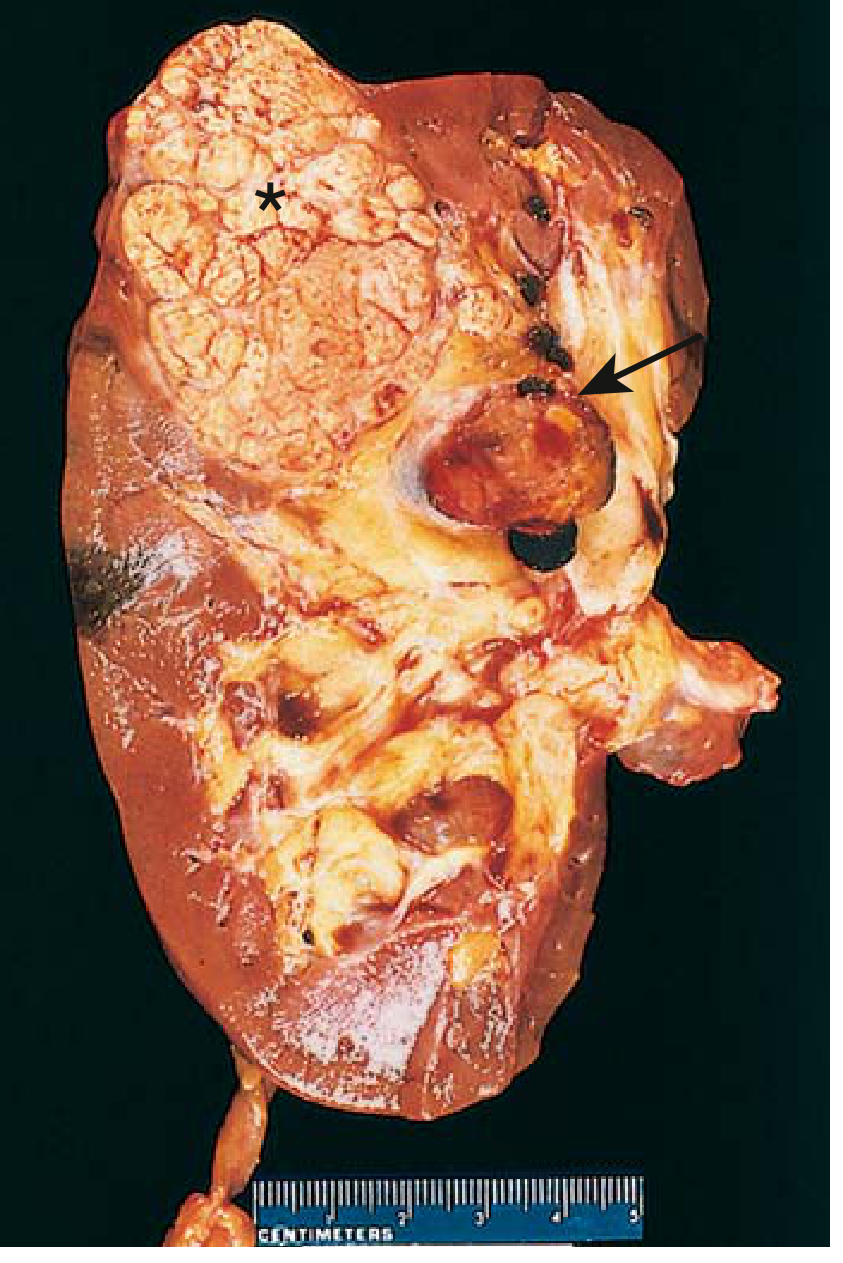
FIG: Cross-section of kidney showing clear cell RCC (asterisk) - yellowish-orange spherical neoplasm in upper pole, with tumor thrombus in dilated renal vein (arrow). (Robbins & Kumar Basic Pathology)
Microscopic:
- Clear cell type (A): Cells with vacuolated, clear cytoplasm (lipid and glycogen dissolved out) and distinct cell membranes; arranged in nests separated by delicate fibrovascular stroma; small, round nuclei
- Papillary type (B): Cuboidal to low columnar cells lining papillae with fibrovascular cores; cuboidal cells with eosinophilic to clear cytoplasm
- Chromophobe type (C): Eosinophilic cytoplasm mixed with pale granular cells; very prominent distinct cell membranes, perinuclear haloes
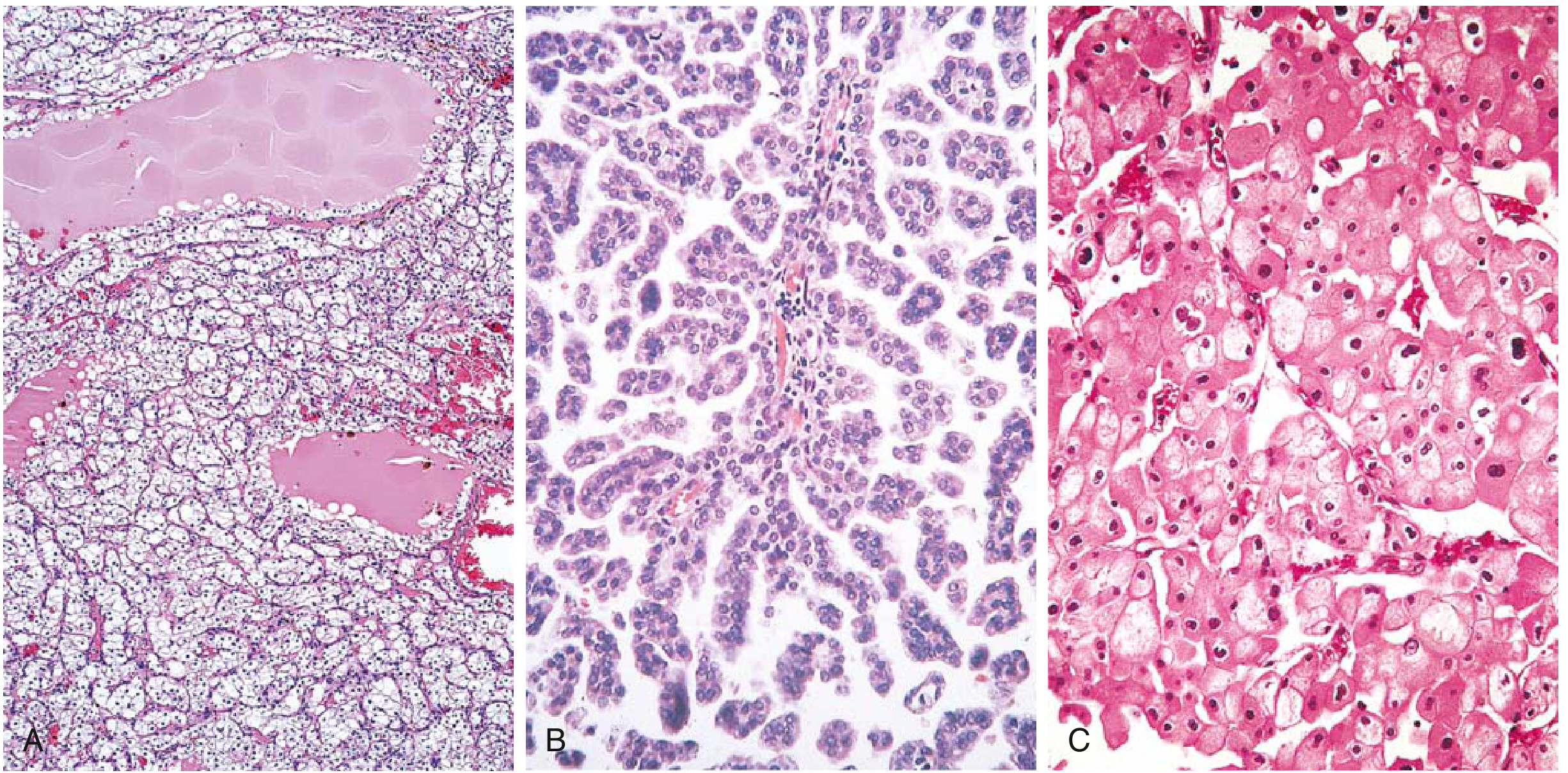
FIG: RCC histological subtypes - (A) Clear cell, (B) Papillary, (C) Chromophobe. (Robbins & Kumar Basic Pathology)
Clinical Features
The classic triad is:
- Painless hematuria (gross or microscopic)
- Palpable flank/abdominal mass
- Dull flank pain
However, all three are present simultaneously in only 10% of cases. Hematuria is the single most frequent presenting symptom (>50% of cases); it is typically intermittent and macroscopic over a background of persistent microscopic hematuria.
Many tumors today are detected incidentally on imaging (CT/MRI done for other reasons).
Paraneoplastic Manifestations (important for exam!)
RCC is notorious for producing systemic effects distant from the tumor:
| Paraneoplastic Feature | Mechanism | Frequency |
|---|---|---|
| Anemia | Chronic disease / blood loss | 52% |
| Erythrocytosis/Polycythemia | Ectopic erythropoietin production | 4-10% |
| Hypercalcemia | PTHrP production | 13% |
| Hypertension | Renin production | 3% |
| Fever / night sweats | Cytokine (IL-6) secretion | 8% |
| Hepatic dysfunction (Stauffer syndrome) | Non-metastatic hepatopathy | 32% |
| Cushing syndrome | ACTH production | Rare |
| Weight loss / malaise | Systemic | 19-23% |
Metastatic patterns: Lungs (most common), bone, liver, brain, contralateral kidney. Occasionally presents as a pathological fracture or lung nodule before the primary is discovered.
Diagnosis
Imaging:
- Ultrasound: Initial screening; can differentiate solid from cystic, but low sensitivity for small lesions
- Multiphasic contrast-enhanced CT (gold standard): Best for evaluating solid lesions; done in pre-contrast, arterial (~25 sec), nephrographic (~90 sec), and excretory phases. Key: enhancement on nephrographic phase differentiates malignant from benign
- Clear cell RCC: Avid enhancement in corticomedullary phase; becomes hypoenhancing in nephrographic phase
- Papillary RCC: Hypointense on T2-weighted MRI; hypoenhancing on all contrast phases
- Chromophobe RCC: Avid corticomedullary enhancement, less than clear cell
- MRI: Preferred when contrast is contraindicated (poor renal function); excellent for delineating venous thrombus extension into renal vein/IVC; helpful for complex cysts
Workup:
- CBC, chemistry profile (liver function, calcium, creatinine)
- CT chest, abdomen, pelvis
- Bone scan if: advanced disease, hypercalcemia, bone pain, elevated alkaline phosphatase
- Staging by TNM system (AJCC 8th edition)
TNM Staging
| Stage | T | N | M |
|---|---|---|---|
| I | T1 (≤7 cm, confined to kidney) | N0 | M0 |
| II | T2 (>7 cm, confined to kidney) | N0 | M0 |
| III | T1-T3, N1 or T3 (extends into major veins/perinephric tissue) | N0 or N1 | M0 |
| IV | T4 (beyond Gerota's fascia) or any T/N | Any | M1 |
T3 sub-stages (important):
- T3a: Extends into renal vein/segmental branches, or perinephric fat
- T3b: Extends into vena cava below diaphragm
- T3c: Extends into vena cava above diaphragm or invades IVC wall
Treatment
Localized Disease (Stages I-III)
- Partial nephrectomy (nephron-sparing surgery): Preferred for tumors ≤7 cm; provides equivalent local control and survival to radical nephrectomy while preserving renal function; open or laparoscopic technique
- Radical nephrectomy: Preferred for large, centrally located tumors; tumors replacing most of the parenchyma; tumors with regional adenopathy; tumors extending into the IVC; can be open or laparoscopic
- Ipsilateral adrenal gland removed; regional lymph node dissection is optional/controversial
- Adjuvant therapy: RCC is resistant to both radiation and cytotoxic chemotherapy
- Sunitinib (VEGF tyrosine kinase inhibitor) or pembrolizumab (anti-PD-1) recommended for high-risk cases after nephrectomy
Metastatic Disease (Stage IV)
- ~30% of patients present with metastatic disease; another 20-30% relapse after surgery
- First-line systemic therapy: Anti-PD-1 checkpoint inhibitors + tyrosine kinase inhibitors (TKIs):
- Nivolumab + cabozantinib or nivolumab + ipilimumab (anti-CTLA-4) - significantly superior to sunitinib monotherapy
- Pembrolizumab + axitinib or pembrolizumab + lenvatinib - recommended in all risk groups
- mTOR inhibitors: Everolimus, temsirolimus - used for previously treated patients
- Belzutifan (HIF-2α inhibitor) - useful for VHL-mutation associated tumors
- Radiation therapy: Palliative role for bone and brain metastases
- Metastasectomy: May extend survival in selected patients (disease-free interval >1 year, solitary lung metastasis)
Prognosis
| Stage | 5-Year Survival |
|---|---|
| Stage I (localized, ≤7 cm) | 90-95% |
| Stage II-III | 40-70% |
| Stage IV (metastatic) | <10% |
Prognostic factors: tumor size, histologic grade (Fuhrman/WHO-ISUP grading), pathologic stage, venous involvement, and histologic subtype (clear cell > papillary/chromophobe in terms of aggressiveness).
Sources:
- Robbins & Kumar Basic Pathology, Chapter 12 (Kidney)
- Goldman-Cecil Medicine, Chapter 182 (Renal Cell Carcinoma)
- National Kidney Foundation Primer on Kidney Diseases, 8th ed., Chapter 6
- Campbell-Walsh-Wein Urology, Part XI
This is a shared conversation. Sign in to Orris to start your own chat.