

1000237629.jpg
Pt was brought to er with c/o b/l ankle nd foot swelling since 2days No comorbs H/o insect bite -3days back Pitting + tenderness+ multiple lesion wound swab taken 34yrs
analyse_media
Reading File
Now let me search the medical library for relevant content on this presentation:
Finding Sources
Finding Sources
Finding Sources
Finding Sources
Finding Sources
Finding Sources
Excellent - the image from Andrews shows a relevant comparison. Let me fetch it and read the key sections:
Loading Image
Reading File
Reading File
The textbook image from Andrews' is a near-perfect match to the patient's presentation. Now I have enough to give a thorough, well-referenced answer.
Clinical Assessment: 34F - Bilateral Ankle/Foot Swelling Post Insect Bite
Image Findings (correlating with clinical history)
The bilateral feet and ankles show:
- Multiple discrete to grouped erythematous papules and nodules, 5-15 mm, on the dorsum of both feet and lower legs
- Several lesions with central crusting/erosion (secondary impetiginization)
- Diffuse pitting edema of both feet/ankles
- Post-inflammatory hyperpigmentation on older resolving lesions
- No frank spreading erythema of cellulitis, no bullae
The textbook image from Dermatology 2-Volume Set 5e (Andrews') shows an almost identical pattern in a similar skin tone - this represents a classic exaggerated insect bite hypersensitivity reaction with secondary infection.
Most Likely Diagnosis
Exaggerated/Hypersensitivity Insect Bite Reaction (Papular Urticaria) with Secondary Bacterial Superinfection
The bilateral pitting edema is the result of the intense local inflammatory response and lymphatic compromise from multiple bite sites, NOT a primary cardiac/renal cause. This is a recognized presentation - per Dermatology 2-Volume Set 5e:
"Characteristic insect bite reactions are grouped or disseminated, 2-8 mm, erythematous urticarial papules that are markedly pruritic and often excoriated... Chronic or resolving lesions are often hyperpigmented... prurigo nodularis-like lesions may develop, especially on the extremities. Secondary infection is common; although staphylococcal infections are seen most frequently, streptococcal infections also occur."
Differential Diagnoses (to consider/exclude)
| Dx | Points For | Points Against |
|---|---|---|
| Exaggerated insect bite reaction (top Dx) | H/o bite 3d ago, bilateral papular nodular pattern, edema, pruriginous | - |
| Eosinophilic Cellulitis (Wells Syndrome) | Triggered by insect bite, edematous plaques, bilateral limb involvement, often misdiagnosed as cellulitis | Less pruritic, more plaque-like, requires biopsy (flame figures) |
| Bacterial Cellulitis with secondary superinfection | Pitting edema, tenderness, wound crusting, bite portal of entry | No spreading erythema/warmth, lesions appear multiple discrete papules not a single spreading area |
| Scabies | Bilateral foot/ankle, papules, pruritus | No burrows, not in web spaces, acute onset 2-3 days |
| Papular urticaria (chronic) | Pattern identical | Usually children; in adults, often underdiagnosed |
Workup Recommended
Immediate / Bedside:
- ✅ Wound swab (already sent - good) - C&S for Staph aureus, Streptococcus
- CBC with differential - look for eosinophilia (supports Wells syndrome or hypersensitivity)
- CRP/ESR (to gauge systemic inflammation)
- RFT, LFT, urine protein - to rule out systemic cause of edema
- Blood glucose (exclude undiagnosed DM)
- KOH mount/fungal culture if any scaling
If no improvement or diagnosis unclear:
- Skin biopsy (punch biopsy from a fresh lesion) - essential to differentiate:
- Wells syndrome (eosinophilic infiltrate + flame figures)
- Prurigo nodularis
- Lymphomatoid papulosis (rare but in differential)
- Serum IgE level
- APTT/PT if blistering occurs
Management
1. Antibiotics (cover secondary infection - the wound swab guides definitive therapy):
- Empirical: Amoxicillin-Clavulanate 625 mg TDS OR Cephalexin 500 mg QID x 5-7 days
- If MRSA suspected: Co-trimoxazole DS BD
2. Anti-inflammatory / anti-pruritic (address the hypersensitivity response):
- Oral antihistamine: Cetirizine 10 mg OD or Fexofenadine 180 mg OD (non-sedating, daytime)
- Moderate-potency topical steroid: Mometasone furoate 0.1% cream BD to the lesions
- Short course oral prednisolone (0.5 mg/kg/day x 5 days) - if edema and itch are severe and no contraindication - this significantly reduces the immune-driven inflammatory response
3. Supportive:
- Limb elevation (reduces pitting edema)
- Avoid scratching (prevents further superinfection)
- Antiseptic wash (chlorhexidine or povidone-iodine) to crusted lesions daily
4. Insect protection:
- Identify the biting arthropod (mosquito, flea, mite, bug)
- Insect repellent (DEET-based), protective clothing
- Treat home/environment if infestation suspected (fleas, bed bugs)
Red Flags to Watch For (escalate if present)
- Spreading erythema/warmth suggestive of true cellulitis -> IV antibiotics
- Fever > 38.5°C, rigors -> blood cultures, IV therapy
- Rising creatinine / new proteinuria -> nephrology consult
- Bullae formation -> consider Wells syndrome, bullous pemphigoid (biopsy)
- No improvement in 48-72 hrs on oral antibiotics -> re-evaluate
Summary
This 34-year-old female most likely has an exaggerated insect bite hypersensitivity reaction with secondary bacterial superinfection as the cause of bilateral ankle/foot swelling. The edema is inflammatory in origin, not systemic. Treat with antibiotics (guided by wound swab), antihistamines, topical/short-course oral steroids, and limb elevation. CBC differential and biopsy will help exclude Wells syndrome if the clinical response is inadequate.
Source: Dermatology 2-Volume Set 5e (Andrews'), Ch. 85 - Arthropod bites; Ch. 26 - Eosinophilic Cellulitis (Wells Syndrome)
Wells syndrome images
Searching sources...
Finding Sources
Finding Sources
Searching the Web
Wells syndrome eosinophilic cellulitis clinical images dermatology
Finding Sources
Loading Image
Loading Image
Loading Image
Loading Image
Now let me get the histopathology image and the textbook images:
Loading Image
Searching the Web
Wells syndrome eosinophilic cellulitis histopathology flame figures microscopy image
Loading Image
Loading Image
Loading Image
Loading Image
Here is a comprehensive visual gallery of Wells Syndrome (Eosinophilic Cellulitis) with clinical and histopathological images:
Wells Syndrome (Eosinophilic Cellulitis) - Image Gallery
Clinical Images
1. Early/acute phase - erythematous papules and plaques (thigh)
(Dermatology Advisor - JCAD case)

Multiple raised, erythematous-to-urticarial papules/plaques - resembling insect bite reactions. This early phase can look identical to your patient's presentation.
2. Resolving phase - erythematous nodular lesions (popliteal fossa)
(DermNet NZ)
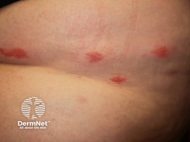
Discrete erythematous nodules in a resolving phase. Post-inflammatory hyperpigmentation develops as lesions heal.
3. Eroded/crusted nodule - close-up
(DermNet NZ)
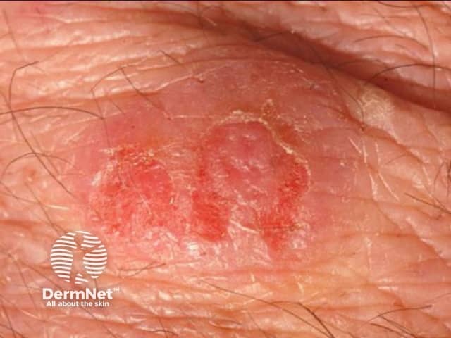
A solitary eroded nodule with serous crusting - exactly the type of lesion seen on your patient's lower leg.
4. Classic cellulitis-mimicking phase - lower leg
(PCDS - Primary Care Dermatology Society)

Diffuse erythematous, edematous, tender plaque on the lower leg - the classic presentation that mimics bacterial cellulitis. This is why it is frequently misdiagnosed and given antibiotics that fail.
5. Indurated erythematous plaque - lower leg
(PCDS)

Indurated, darkening erythematous plaque - the intermediate phase. Lesions progress from bright red to pink-brown to slate-gray as they resolve over 4-8 weeks.
Histopathology Images (H&E stain)
6. High power - Classic "Flame Figure" (pathognomonic)
(PathologyOutlines - Temiz)
The large bright-pink eosinophilic aggregate in the center is the flame figure - eosinophil granule proteins coating degenerating collagen bundles, surrounded by histiocytes and lymphocytes. This is the hallmark finding on biopsy.

7. Medium power - Dermal eosinophilic infiltrate with flame figures
(PathologyOutlines - Temiz)
Dense interstitial eosinophilic infiltrate throughout the dermis. Multiple early flame figures visible as bright-pink collagen deposits surrounded by inflammatory cells.

8. Medium power - Flame figures with eosinophil-rich infiltrate (PMC)
(J Dermatol Case Reports - PMC open access)
Clusters of degranulating eosinophils forming flame figures within a background of edematous dermis and mixed inflammatory infiltrate.

Key Diagnostic Summary
| Feature | Wells Syndrome |
|---|---|
| Trigger | Insect bite/sting (most common), drugs, vaccines, infections |
| Clinical | Edematous erythematous plaques/papules/nodules, bilateral extremities, markedly pruritic |
| Labs | Peripheral eosinophilia (common), elevated IgE |
| Biopsy | Dense dermal eosinophilic infiltrate + flame figures (eosinophil granule proteins coating collagen) |
| Course | Resolves in 4-8 weeks, but recurs |
| Treatment | Oral prednisolone (1st line), antihistamines, dapsone (refractory) |
The DermNet NZ Wells syndrome page and PathologyOutlines have additional case images and biopsy criteria if you need further reference.
This is a shared conversation. Sign in to Orris to start your own chat.