
Every treatment protocal and medicine for snake bite
snakebite treatment protocol antivenom WHO guidelines 2024
"snakebite" AND "antivenom" AND "treatment"
https://moh.gov.bt/wp-content/uploads/2025/05/National-Guide…
snakebite envenomation treatment antivenom clinical presentation
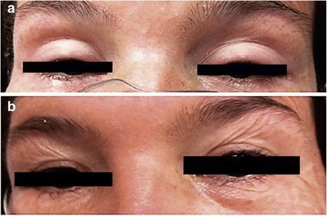
This clinical comparison photograph displays the periocular region of a patient before and after treatment for elapid envenomation. Panel (a) illustrates bilateral palpebral ptosis, a significant drooping of the upper eyelids that obscures much of the globe, which is a classic neurotoxic sign of Micrurus (coral snake) envenomation. The surrounding facial musculature appears paretic. Panel (b) shows the same patient after the administration of monovalent coral antivenom, demonstrating a total neurological recovery. Key visible changes in panel (b) include the restoration of the normal palpebral fissure width and the presence of dynamic skin wrinkling around the eyebrows, indicating active frontalis muscle contraction and the resolution of neurotoxic paralysis. This comparison serves as an educational tool for identifying the clinical presentation and therapeutic response of ophthalmic neurological deficits in snakebite victims.
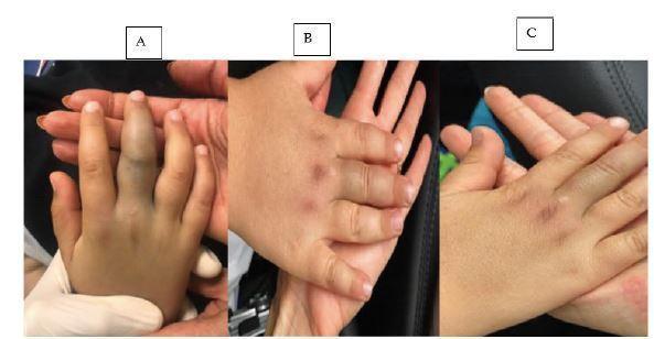
This sequence of three clinical photographs (A, B, and C) illustrates the clinical progression and resolution of localized soft tissue envenomation on a pediatric patient's right hand following a Montivipera bornmuelleri snakebite. Image A shows the acute phase: the right middle finger is severely edematous, primarily over the middle phalanx, with deep bluish-purple ecchymosis and tense swelling compared to adjacent unaffected digits. Image B demonstrates the follow-up 24 hours after antivenom administration, showing a marked reduction in digital girth and lightening of the ecchymotic discoloration. Image C shows the status four days post-injury, where the swelling has almost entirely resolved, and the ecchymosis has faded to faint, yellowish-brown residual bruising. The series serves as an educational example of the local cytotoxic and hemotoxic effects of certain viperid venoms and the efficacy of timely antivenom treatment in managing pediatric envenomation cases. Key clinical signs include edema, ecchymosis, and eventual resolution of localized tissue trauma.
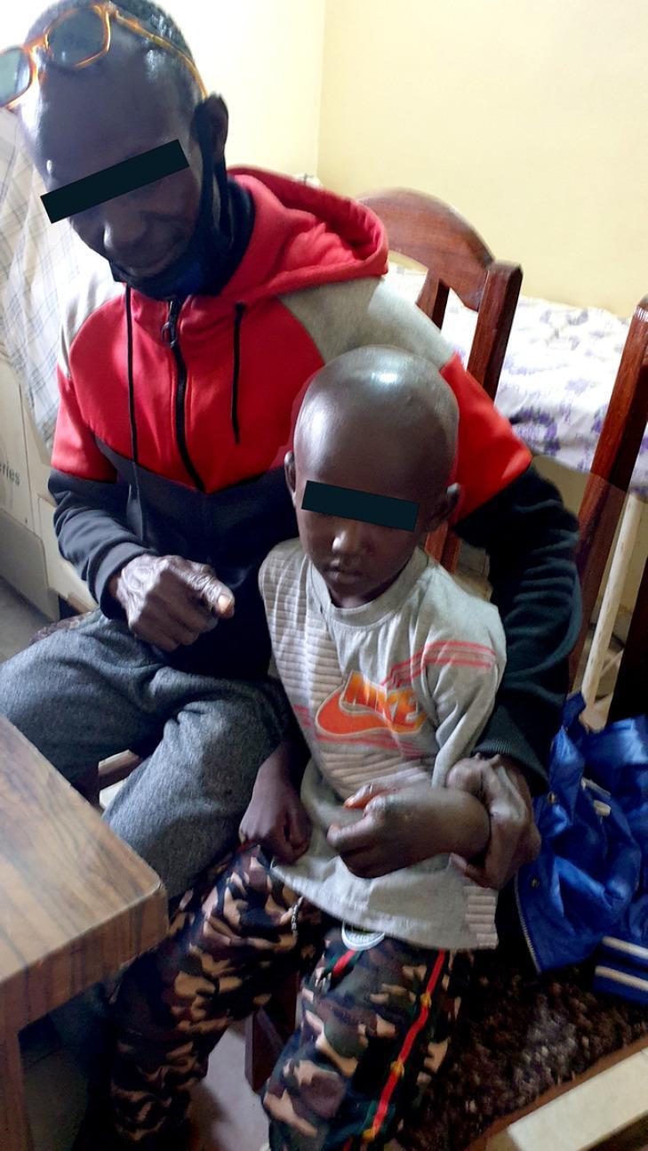
Clinical photograph documenting the 'Guérison' (recovery) phase of a pediatric patient following severe snakebite envenomation. The image shows a young child of African descent seated with an adult, illustrating the long-term clinical outcome after intensive medical intervention. The child's left hand, which previously underwent a surgical amputation of the index finger due to necrotic complications (gas gangrene and grade 3 edema), is visible. The hand appears healed with significant functional and aesthetic sequelae resulting from the venom-induced local tissue necrosis and subsequent surgical management. The photograph serves as a medical case study of successful recovery from a life-threatening ophidian accident involving coagulopathy and severe local infection, highlighting the result of combined treatment with polyvalent antivenom, surgical debridement/amputation, and broad-spectrum antibiotic therapy.
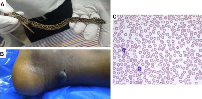
A multi-panel figure documenting a snakebite case. Panel A displays a clinical photograph of a juvenile Russell’s viper (Daboia russelii) held by gloved hands, identified by its characteristic dorsal brown blotches. Panel B is a clinical photograph of a patient's right lower leg and heel showing local envenomation effects, including a prominent dark, hemorrhagic, fluid-filled blister and two adjacent small puncture wounds consistent with fang marks. Panel C shows a diagnostic light microscopy image of a peripheral blood smear (Wright-Giemsa stain). The smear demonstrates normocytic, normochromic erythrocytes with regular morphology and no evidence of fragmentation, schistocytes, or dysmorphic red cells. Scattered multi-lobed leukocytes (neutrophils) are visible against the background of mature red blood cells. The educational focus is on the clinical presentation of viperine envenomation (local tissue destruction) and the exclusion of microangiopathic hemolytic anemia via blood film examination.
Complete Snakebite Treatment Protocol and Medicines
1. Epidemiology & Overview
| Family | Examples | Dominant Toxicity |
|---|---|---|
| Viperidae (vipers, pit vipers) | Russell's viper, saw-scaled viper, rattlesnake, copperhead, cottonmouth | Hemotoxic, local necrosis |
| Elapidae | Cobra, krait, mamba, coral snake, Australian brown/tiger snake | Neurotoxic (+ cytotoxic for cobras) |
| Hydrophiidae | Sea snakes | Neurotoxic + myotoxic |
| Colubridae (rear-fanged) | Boomslang, twig snake | Hemotoxic (rare) |
2. First Aid (Pre-Hospital)
What TO DO:
- Immobilize the bitten limb below heart level (reduces lymphatic venom spread)
- Pressure immobilization bandaging (PIB) - indicated ONLY for neurotoxic bites (elapids, Australian snakes, sea snakes): apply firm crepe bandage from distal to proximal over entire limb, then splint; do NOT remove until antivenom is ready
- Keep the patient calm and still (activity speeds venom absorption)
- Remove rings, watches, tight clothing from bitten limb before swelling begins
- Transport to hospital immediately
- Note the time of bite and, if safe, describe or photograph the snake
What NOT TO DO (Harmful Practices - absolutely avoid):
| Harmful Action | Why it's harmful |
|---|---|
| Incision and suction at bite site | Increases infection risk, causes tissue damage, does not remove venom |
| Tourniquets | Causes ischemia, compartment syndrome, limb loss |
| Electric shock treatment | No evidence, causes burns |
| Applying ice / immersing in cold water | Worsens local tissue injury |
| Applying herbs, chemicals, or traditional remedies | Ineffective, delays care |
| Giving alcohol | Vasodilation increases absorption |
| Pressure bandaging for cytotoxic bites (vipers) | Concentrates venom, worsens necrosis |
3. Hospital Assessment
History & Physical
- Time since bite, snake description, symptoms onset
- Fang marks (1 or 2 puncture wounds), local swelling, bruising, blistering, necrosis
- Systemic: bleeding, neurological signs, cardiovascular signs
Severity Grading (Dart/Sabiston system)
| Grade | Features |
|---|---|
| 0 - Dry bite | No envenomation signs, fang marks only |
| 1 - Minimal | Local reaction only, no systemic signs |
| 2 - Moderate | Extends beyond bite site, non-life-threatening systemic signs (nausea, mild hypotension, tachycardia) |
| 3 - Severe | Extensive local + severe systemic: severe hypotension, DIC, respiratory failure, coma |
Essential Investigations
- 20-Minute Whole Blood Clotting Test (20WBCT): Place 2 mL fresh blood in clean glass tube, leave undisturbed for 20 min - if blood does NOT clot = coagulopathy (positive test = hemotoxic envenomation)
- CBC, coagulation profile (PT, aPTT, fibrinogen, D-dimer)
- BMP / RFT: creatinine, BUN, electrolytes (AKI risk)
- Urinalysis: hematuria, hemoglobinuria, myoglobinuria
- LFTs, CK (rhabdomyolysis)
- ECG and cardiac monitoring in severe cases
- Chest X-ray if pulmonary edema suspected
4. Definitive Treatment: Anti-Snake Venom (Antivenom / ASV)
Indications for Antivenom (any one of the following):
- Haemotoxicity: bleeding from any site, clotting failure (20WBCT positive), thrombocytopenia
- Neurotoxicity: ptosis, diplopia, dysarthria, dysphagia, respiratory muscle weakness
- Cardiovascular: hypotension, shock, ECG changes
- Renal: oliguria, hematuria, rising creatinine
- Myotoxicity: myalgia, myoglobinuria (sea snake / some elapid bites)
- Rapidly progressive swelling beyond the bite site
- Enlarged tender draining lymph node
- Local necrosis
Note: Pregnancy is NOT a contraindication to antivenom. Dry bites (~30% of cases) do NOT require antivenom.
Types of Antivenom
| Type | Coverage | Examples |
|---|---|---|
| Monovalent | One species | King cobra ASV, Russell's viper ASV |
| Polyvalent | Multiple species | Indian polyvalent ASV (covers Big 4 Indian snakes), African polyvalent |
| Fab fragment (CroFab) | North American pit vipers | Crotalidae polyvalent immune Fab (ovine) - FDA approved |
| F(ab')2 fragment (Anavip) | North American pit vipers | Crotalidae immune F(ab')2 (equine) - FDA approved |
Antivenom Dosing Protocol (WHO / National Guidelines 2024)
- Subcutaneous adrenaline (epinephrine) 0.25 mg (adults) / 0.01 mg/kg (children) - given 15-20 min before to reduce reactions
- Antihistamine (promethazine 25 mg IM or chlorpheniramine 10 mg IM)
- Have anaphylaxis-dose epinephrine drawn up and at bedside
Skin sensitivity test is NOT recommended (unreliable and delays treatment)
| Envenomation Type | Initial Dose | Repeat Dose | Maximum |
|---|---|---|---|
| Neurotoxic | 10 vials over 60 min | If neuroparalysis worsens at 2 hours: repeat 10 vials | 20 vials |
| Hemotoxic | 10 vials over 60 min | Repeat 20WBCT at 6 hrs; if still positive: 5 vials q6h until blood clots; if continued bleeding: repeat initial dose at 1 hour | 30 vials |
| Local envenomation | Follow hemotoxic protocol | - | - |
Children receive the same dose as adults (venom dose is not weight-dependent)
- Hypotension: improves within 30-60 minutes
- Coagulopathy: resolves over 6-24 hours (clotting factors are synthesized anew)
- Neurotoxicity: may take hours to days to reverse (antivenom less effective once toxin is bound)
Antivenom Reactions & Management
- Stop infusion temporarily
- Epinephrine (adrenaline) 0.5 mg IM (adult) into thigh - repeat every 5-15 min as needed
- High-flow oxygen
- IV fluids for hypotension
- Antihistamine (diphenhydramine / chlorpheniramine)
- Restart ASV at slower rate after stabilization
- Stop infusion temporarily
- Paracetamol 500 mg-1 g (adults); 10-15 mg/kg (children) orally or rectally
- Cool the patient
- IV fluids
- Restart ASV slowly if still indicated
- Prednisolone 5 mg orally every 6 hours for 5-7 days
- Chlorpheniramine 2 mg orally every 6 hours
5. Specific Treatment by Toxidrome
A. Neurotoxic Envenomation (Elapids - Cobra, Krait, Mamba, Coral Snake)
- Give atropine 0.6 mg IV, then edrophonium 10 mg IV (or neostigmine 1.5 mg IM)
- Improvement in ptosis/weakness within 5 min = positive (cobra-type postsynaptic neurotoxin)
- No improvement = presynaptic toxin (krait, mamba) - anticholinesterases unhelpful
- Neostigmine 0.5-2.5 mg IM/IV/SC with Atropine every 1-3 hours (adults); neostigmine 0.01-0.04 mg/kg + atropine (children)
- Maximum neostigmine 10 mg/24 hours (adults)
- OR oral neostigmine 15 mg QID + oral atropine 0.6 mg BD
- Stop if: complete recovery, fasciculations, bradycardia, or no response after 3 doses
- Early intubation and mechanical ventilation for respiratory muscle weakness (inspiratory force < -20 cmH2O or VC < 10-15 mL/kg)
- Prolonged ventilation may be needed (days to weeks for severe krait poisoning)
- Baseline and serial vital capacity measurements
- 3-5 vials Anti-venin (M. fulvius) IV even in asymptomatic confirmed bites (effects cannot be reversed once established)
- Arizona (Sonoran) coral snake - antivenom usually not needed
B. Hemotoxic Envenomation (Vipers - Russell's Viper, Saw-Scaled Viper, Rattlesnake)
- Antivenom as above (hemotoxic protocol)
- DO NOT give heparin (ineffective against venom-induced thrombin, may worsen bleeding)
- DO NOT give fresh frozen plasma (FFP) or platelets before/instead of antivenom (will be consumed by ongoing venom effect - give antivenom first)
- Blood products (FFP, cryoprecipitate, platelets) only AFTER antivenom has been given AND venom neutralized (i.e., after >6 hours of antivenom)
- IV fluids for hypovolemia
- Monitor urine output; catheterize if oliguric
C. Cytotoxic Envenomation (Cobras, some vipers - local tissue necrosis)
- Antivenom is most effective if given early (does not reverse established necrosis)
- Do NOT apply pressure bandage (concentrates venom locally, worsens ischemia)
- Wound care: clean with antiseptic, debridement of necrotic tissue
- Tetanus prophylaxis: tetanus toxoid 0.5 mL IM if not up to date; tetanus immunoglobulin 250-500 IU IM if unimmunized
- Antibiotics: only if signs of secondary infection (not prophylactically routinely); amoxicillin-clavulanate or ciprofloxacin ± metronidazole
- Watch for compartment syndrome: measure compartment pressures if suspected; fasciotomy is a LAST resort (rarely needed if antivenom is given promptly)
- Reconstructive surgery / skin grafting for large necrotic wounds
D. Spitting Cobra Ophthalmia (Venom in Eyes)
- Irrigate eye immediately and copiously with large volumes of water or normal saline
- Local anesthetic eye drops (tetracaine) for pain relief
- Examine for corneal ulceration (fluorescein staining)
- Topical antibiotics to prevent secondary infection
- NO systemic antivenom needed (no systemic absorption through cornea)
6. Supportive Care Medicines
| Problem | Treatment |
|---|---|
| Shock / hypotension | IV crystalloids (normal saline, Ringer's lactate); vasopressors (norepinephrine/dopamine) if fluid-refractory |
| Acute Kidney Injury (AKI) | IV fluids to maintain urine output >0.5 mL/kg/hr; dialysis (hemodialysis or peritoneal dialysis) if oliguria persists or severe uremia; avoid nephrotoxic drugs (NSAIDs, aminoglycosides) |
| Respiratory failure | Oxygen, intubation, mechanical ventilation |
| DIC / coagulopathy | Antivenom first; blood products (FFP, cryoprecipitate, platelets) only after antivenom; no heparin |
| Rhabdomyolysis / myoglobinuria | IV fluids to maintain urine output; alkalinize urine with IV sodium bicarbonate (target urine pH > 6.5) |
| Pain | Paracetamol (acetaminophen); opioids (morphine, tramadol) for severe pain; avoid NSAIDs (bleeding risk) |
| Tetanus prophylaxis | Tetanus toxoid + TIG if unimmunized |
| Secondary infection | Amoxicillin-clavulanate 625 mg TID, OR ciprofloxacin 500 mg BD ± metronidazole 400 mg TID |
| Serum sickness | Prednisolone 5 mg q6h x 5-7 days + antihistamine |
| Bradycardia / muscarinic excess from neostigmine | Atropine 0.6 mg IV bolus |
7. Disposition and Monitoring
| Scenario | Duration |
|---|---|
| Suspected dry bite (no symptoms) | Observe minimum 8 hours; recheck labs before discharge |
| Minimal envenomation, no antivenom needed | Observe 12-24 hours for progression |
| Any confirmed envenomation | Admit to monitored bed, minimum 24 hours |
| Severe envenomation (Grade 3) | ICU admission |
| Post-antivenom | Monitor for minimum 2 hours after starting ASV |
| Discharge instructions | Warn patient about serum sickness (days 5-24); return precautions for recurrent coagulopathy (can recur up to 2 weeks) |
8. Quick Drug Reference Card
| Drug | Dose (Adult) | Dose (Child) | Indication |
|---|---|---|---|
| Anti-snake venom (ASV) | 10 vials IV in 100-400 mL NS/D5W over 60 min | Same dose as adult | Envenomation |
| Adrenaline (epinephrine) | 0.25 mg SC (premedication); 0.5 mg IM (anaphylaxis) | 0.01 mg/kg SC/IM | Pre-ASV / anaphylaxis |
| Neostigmine | 0.5-2.5 mg IM/IV q1-3h (max 10 mg/24h) | 0.01-0.04 mg/kg q2-4h | Cobra postsynaptic neurotoxin |
| Atropine | 0.6 mg IV with each neostigmine dose | 0.02 mg/kg | Prevent neostigmine side effects |
| Paracetamol | 500 mg-1 g q4-6h (max 4 g/day) | 10-15 mg/kg (max 100 mg/kg/day) | Pyrogenic reaction, pain |
| Chlorpheniramine | 10 mg IM or 2 mg oral q6h | 0.1 mg/kg | Antivenom reaction |
| Prednisolone | 5 mg q6h x 5-7 days | 1 mg/kg/day | Serum sickness |
| Tetanus toxoid | 0.5 mL IM | Same | Wound prophylaxis |
| Sodium bicarbonate | 1-2 mEq/kg IV in D5W | Same | Myoglobinuria (rhabdomyolysis) |
| Morphine / Tramadol | As per pain protocol | Age-appropriate dose | Severe pain (avoid NSAIDs) |
9. Clinical Images
10. Key Points to Remember
- Antivenom is the only specific treatment - give it promptly when indicated; it is safe in pregnancy.
- PIB is only for neurotoxic (elapid) bites - never for cytotoxic (viper) bites.
- Dose does NOT change for children - venom dose is independent of the patient's weight.
- Do not give heparin or FFP/platelets before antivenom in coagulopathy.
- Do not perform wound incision, suction, or tourniquet - all are harmful.
- Dry bites (30%) require no antivenom but need 8+ hours observation.
- Krait bites may present with minimal local signs but severe delayed neurotoxicity (12-18 hours) - extended observation is mandatory.
- Serum sickness can occur 5-24 days after antivenom (treat with prednisolone).
- AKI is common with Russell's viper - monitor urine output and renal function closely.
- Always contact a clinical toxicologist or poisons information centre for complex cases.