
Absence of bile salts in small intestine causea steatorrhea
bile salts micelle formation fat absorption small intestine diagram
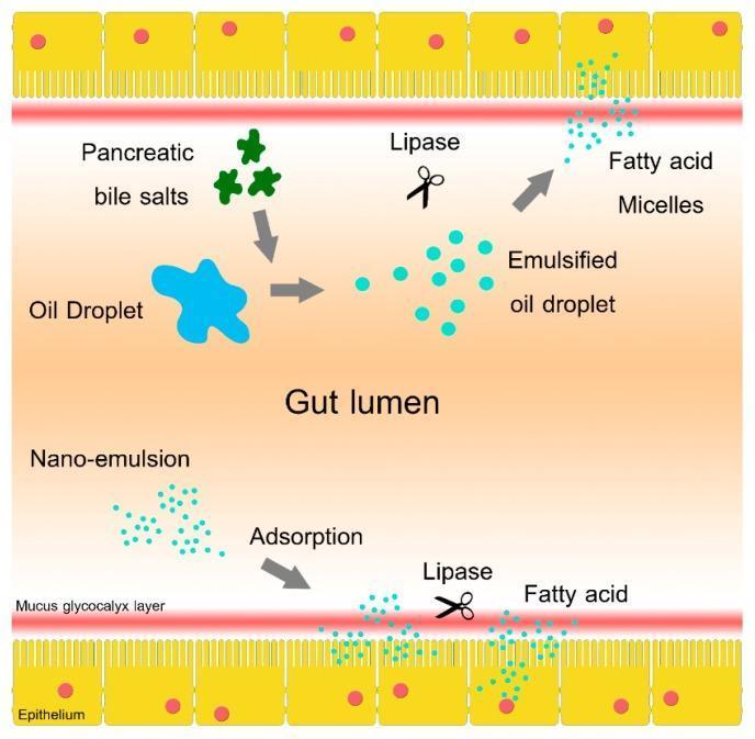
A pathophysiology diagram illustrating two comparative mechanisms of lipid digestion and absorption within the gut lumen. The diagram is bounded by an epithelial layer (yellow columnar cells with red nuclei and microvilli) and a red mucus glycocalyx layer. The upper pathway depicts conventional dietary oil absorption: a large, irregular blue oil droplet is emulsified by green pancreatic bile salts into smaller, uniform turquoise droplets. Lipase (represented by a scissor icon) then acts on these droplets in the central lumen to form fatty acid micelles, which migrate toward the epithelium. The lower pathway depicts a self-emulsified dietary oil mechanism: a nano-emulsion of small turquoise droplets undergoes direct adsorption onto the mucus glycocalyx layer. Lipase activity occurs locally at the glycocalyx-epithelium interface, resulting in the direct release and absorption of free fatty acids. This visual compares standard luminal digestion versus localized adsorption-enhanced bioavailability, focusing on the roles of bile salts, lipase location, and the unstirred mucus layer in nutrient translocation.
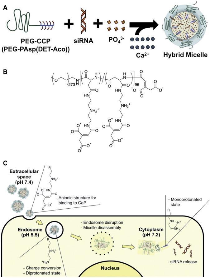
Educational diagram illustrating the synthesis and intracellular delivery mechanism of a hybrid micelle nanocarrier for siRNA. Panel A (Pathophysiology Diagram) depicts the formation of a hybrid micelle through the self-assembly of polyethylene glycol-block-charge-conversional polymer (PEG-CCP), small interfering RNA (siRNA), phosphate ions (PO4 3-), and calcium ions (Ca2+). Panel B (Chemical Structure) shows the molecular formula for PEG-PAsp(DET-Aco), highlighting the pH-responsive side chains. Panel C (Flowchart/Algorithm) illustrates the cellular uptake and trafficking mechanism: 1) Extracellular space (pH 7.4), where the complex maintains an anionic structure for calcium phosphate (CaP) binding; 2) Endocytosis into an endosome (pH 5.5), where acidic conditions trigger charge conversion to a diprotonated state, causing endosome disruption and micelle disassembly; 3) Release into the cytoplasm (pH 7.2), where the polymer shifts to a monoprotonated state, facilitating siRNA release for gene silencing. This visual serves as a pharmacological resource for drug delivery systems and nanomedicine.
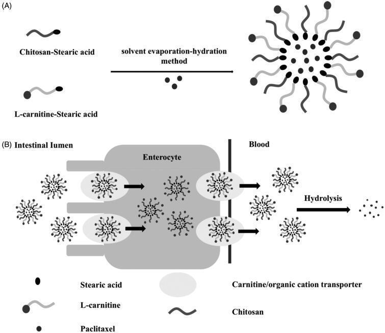
A two-part schematic diagram detailing the preparation and absorption mechanism of paclitaxel-loaded polymeric micelles for oral delivery. (A) Preparation phase: Illustrates the synthesis of PTX-loaded LC-SA/CS-SA micelles using the solvent evaporation-hydration method. Individual components including Chitosan-Stearic acid (CS-SA), L-carnitine-Stearic acid (LC-SA), and paclitaxel molecules are shown self-assembling into a spherical micelle structure with a hydrophobic core and a functionalized outer shell. (B) Absorption mechanism: Depicts the intestinal transport of intact micelles from the intestinal lumen across the enterocyte membrane. The process is facilitated by carnitine/organic cation transporters (OCTN2), represented by oval docking sites on the brush border. Following transepithelial transport, the micelles enter the bloodstream where they undergo hydrolysis, releasing the encapsulated paclitaxel along with stearic acid, L-carnitine, and chitosan components. This illustration demonstrates a targeted drug delivery strategy to improve the bioavailability of lipophilic drugs like paclitaxel via the carnitine transporter pathway.
Absence of Bile Salts in the Small Intestine Causes Steatorrhea
Normal Role of Bile Salts in Fat Absorption
"The products of lipolysis interact with bile salts and phospholipids to form mixed micelles, which also incorporate cholesterol and fat-soluble vitamins (D, A, K, and E) in their hydrophobic centers." - [Goldman-Cecil Medicine, 26e]
What Happens Without Bile Salts
- Steatorrhea: Up to 40% of ingested fat is lost in the feces. As stated in Guyton & Hall: "Without the presence of bile salts in the intestinal tract, excessive amounts of fat are excreted in the feces, a condition called steatorrhea; up to 40% of the ingested fats may be lost."
- Fat-soluble vitamin deficiency: Vitamins A, D, E, and K malabsorption is particularly severe because micelle formation is essential for their absorption (unlike fatty acids, which can form lamellar structures and be partially absorbed without micelles).
- Metabolic deficit: From the caloric and nutrient loss.
Causes of Bile Salt Deficiency in the Lumen
| Mechanism | Example |
|---|---|
| Decreased synthesis | Severe liver disease (cirrhosis) |
| Decreased delivery | Cholestasis, bile duct obstruction |
| Bacterial deconjugation | Small intestinal bacterial overgrowth (anaerobes deconjugate bile salts, raising pKa so they are passively absorbed before reaching micellar concentration) |
| Terminal ileal disease/resection | Loss of the active reabsorption site (enterohepatic circulation disrupted) |
| Drug binding | Cholestyramine (bile acid sequestrant) |
| Acid hypersecretion | Low pH precipitates bile salts (Zollinger-Ellison syndrome) |
The Enterohepatic Circulation Link
- Guyton and Hall Medical Physiology, 14e, p. 810 - Function of Bile Salts in Fat Digestion and Absorption
- Goldman-Cecil Medicine, 26e - Conditions that Impair Intraluminal Digestion; Impaired Micelle Formation