
Imaging techniques and treatment of CP angle tumor in detail for 20 marker long question answers in simplified manner from standard textbooks
MRI cerebellopontine angle vestibular schwannoma acoustic neuroma gadolinium T1
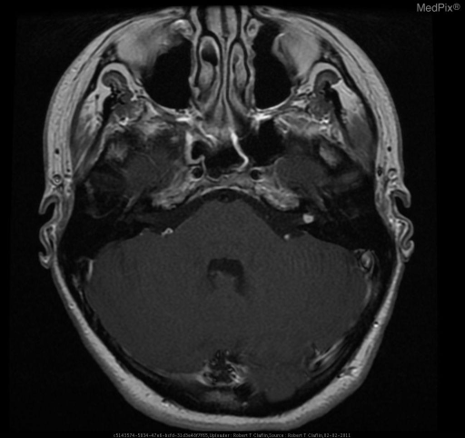
**Imaging Modality:** Gadolinium-enhanced, T1-weighted Magnetic Resonance Imaging (MRI), axial section.
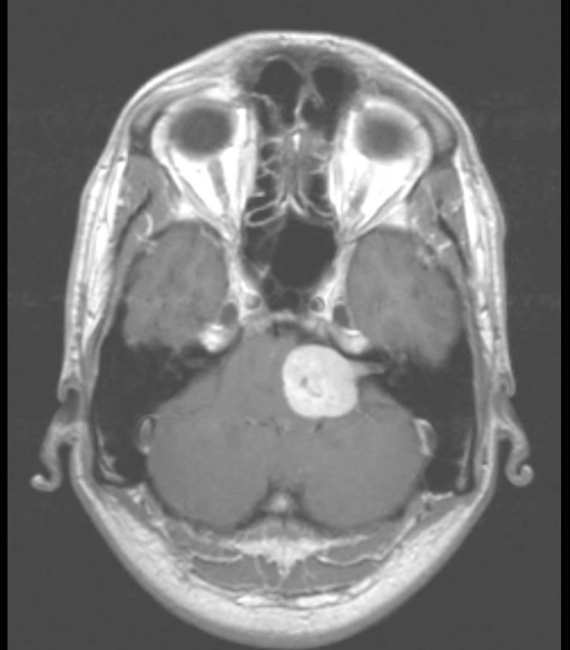
**Imaging Modality:** Contrast-enhanced T1-weighted Magnetic Resonance Imaging (MRI), axial section.
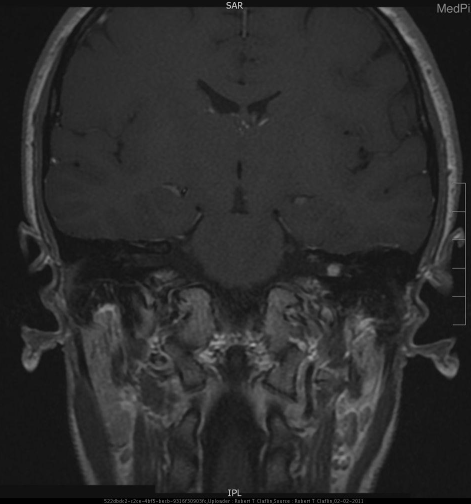
**Imaging Modality:** Coronal T1-weighted post-contrast Magnetic Resonance Imaging (MRI).
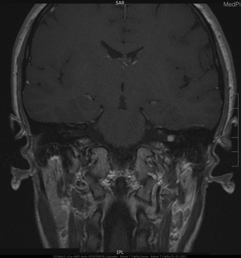
**Imaging Modality:** Contrast-enhanced Magnetic Resonance Imaging (MRI), Coronal T1-weighted sequence.
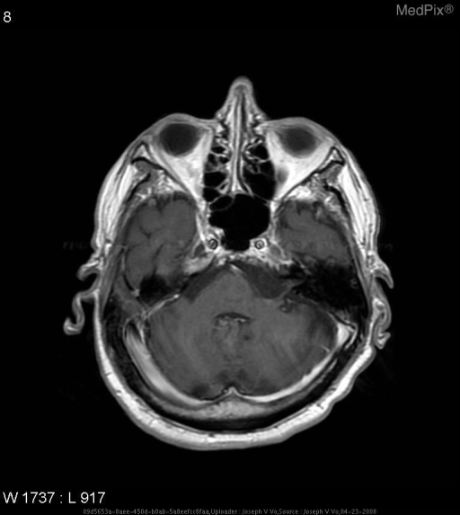
**Imaging Modality:** Magnetic Resonance Imaging (MRI), Axial T1-weighted post-contrast sequence.
"vestibular schwannoma" AND management
Cerebellopontine (CP) Angle Tumor: Imaging and Treatment
Introduction
Clinical Background (brief)
- Progressive unilateral sensorineural hearing loss with disproportionate loss of speech discrimination (retrocochlear pattern)
- Tinnitus and disequilibrium (rather than true vertigo, since compensation is gradual)
- Facial hypesthesia (5th nerve) as the tumor enlarges beyond ~3 cm
- Later: ataxia, headache, and hydrocephalus from brainstem/4th ventricle compression
IMAGING TECHNIQUES
1. MRI with gadolinium contrast - investigation of choice
- T1-weighted pre- and post-gadolinium: Acoustic neuroma is isointense/mildly hypointense to brain before contrast, and enhances markedly (often inhomogeneously) after gadolinium.
- T2-weighted / CISS (constructed interference in steady state) sequences: The tumor appears moderately hyperintense; CISS clearly delineates the cochlea, nerve, and CSF cleft around the tumor without needing contrast - useful as a screening protocol and in patients where gadolinium should be avoided.
- Classic "ice-cream cone" sign: A globular cisternal component continuous with a narrow intracanalicular extension into a widened IAC - highly characteristic of vestibular schwannoma.
| Feature | Acoustic Neuroma | Meningioma | Epidermoid |
|---|---|---|---|
| Location | Centered on IAC | Eccentric to IAC | Antero/postero-lateral to brainstem |
| Bone change | Widens IAC | Occasional hyperostosis | Occasional erosion |
| Shape/angle | Spherical/ovoid, acute bone-tumor angle | Hemispherical, obtuse angle, dural "tail" | Irregular, dumbbells around structures |
| CT density | Isodense | Slightly hypodense, may calcify | Hypodense |
| Enhancement | Moderate-marked, inhomogeneous | Marked, homogeneous | Non-enhancing |
| T2 MRI | Hypointense | Hypointense | Hyperintense, restricted diffusion on DWI |
2. CT scan
- Shows bony anatomy - IAC widening, erosion, hyperostosis (helps distinguish meningioma from schwannoma), and honeycomb/spiculated bone in facial nerve tumors.
- Useful when MRI is contraindicated (cardiac pacemaker, cochlear implant, severe claustrophobia).
- Contrast CT can miss tumors smaller than 1.5 cm, especially purely intracanalicular ones - a major limitation that led to MRI becoming the standard.
3. Audiometric and electrophysiological tests
- Pure tone audiometry (PTA): unilateral, asymmetric sensorineural hearing loss - the commonest audiometric abnormality.
- Speech discrimination score: disproportionately poor relative to the pure-tone loss - a classic retrocochlear sign.
- Auditory brainstem response (ABR): prolonged interpeak latency (wave I-V) or absent response on the affected side; used historically as a screening tool for small intracanalicular tumors, though MRI has superseded it in accuracy. Stacked ABR protocols improve sensitivity for small tumors.
- Tone decay and reflex decay tests, electronystagmography/caloric testing: show retrocochlear/vestibular nerve involvement (reduced or absent caloric response on the tumor side) but are non-specific and largely of historical value now.
4. Other investigations
- Oxygen cisternography - historically used for small intracanalicular lesions before MRI; now obsolete.
- Genetic testing - for suspected NF2 in bilateral or young-onset cases (chromosome 22, merlin/schwannomin gene).
TREATMENT
1. Observation ("Wait and Scan")
- Suitable for small (<1.5-2 cm), asymptomatic or minimally symptomatic tumors, especially in elderly or high-risk patients, since average growth rate is only about 0.2 cm/year and many tumors do not grow at all.
- Protocol: serial gadolinium MRI (e.g., at 6-12 months, then annually) for at least 5 years; intervention is offered if there is significant growth or new symptoms.
- Advantage: avoids surgical risk in tumors that may never progress; disadvantage: delayed treatment may reduce chances of hearing preservation later.
2. Stereotactic Radiosurgery (Gamma Knife / CyberKnife)
- Best suited to tumors less than 3 cm in maximum diameter, predominantly solid, especially in elderly patients or those unfit for surgery, and for residual/recurrent tumor after surgery.
- Delivers a single high dose of focused radiation to arrest tumor growth (not necessarily to shrink it) while sparing surrounding structures.
- Excellent tumor control rates with a lower incidence of cranial neuropathy (facial nerve palsy) than open surgery, though hearing preservation still declines over time.
- Not ideal for large tumors causing significant brainstem compression, as it does not provide immediate decompression.
3. Microsurgical Excision
- Via transmastoid labyrinthectomy, sacrificing hearing on that side.
- Indicated for medium/large tumors, or any tumor in a patient who already has no serviceable hearing, since hearing preservation is unlikely for tumors >2 cm regardless of approach.
- Advantages: direct, early identification of the facial nerve at the fundus of the IAC, giving the highest rate of facial nerve preservation; avoids cerebellar retraction.
- Also used for meningiomas, non-acoustic schwannomas, gliomas, chordomas needing CPA exposure.
- Transmastoid decompression of the sigmoid sinus with a retrosigmoid craniotomy, without disturbing the labyrinth.
- Indicated when hearing preservation is desired, in patients with serviceable hearing and limited IAC involvement (any size tumor medial to IAC can be approached).
- Drawback: cerebellum lies between surgeon and tumor, requiring cerebellar retraction after CSF drainage; risk of postoperative headache is higher than with other approaches.
- Temporal craniotomy exposing the IAC from above, via the floor of the middle fossa.
- Ideal for small, purely intracanalicular tumors in patients with good hearing; CPA extension beyond 1 cm is a relative contraindication because exposure is limited.
- Preserves hearing while permitting decompression of the internal auditory canal.
- Extension of the translabyrinthine approach with posterior transposition of the facial nerve and removal of the cochlea, for petroclival meningiomas, petrous apex epidermoids, or extensive non-acoustic tumors extending to the clivus.
| Approach | Hearing preserved? | Best for |
|---|---|---|
| Middle fossa | Yes | Small, intracanalicular tumors |
| Retrosigmoid | Yes | Tumors with limited IAC extension, serviceable hearing |
| Translabyrinthine | No | Medium/large tumors, or no useful hearing, best facial nerve outcome |
| Transcochlear | No | Extensive petroclival/clival extension |