
Mouth cancer surgery
What aspect of mouth cancer surgery are you most interested in? · Who is this for?
oral cavity cancer surgery resection reconstruction
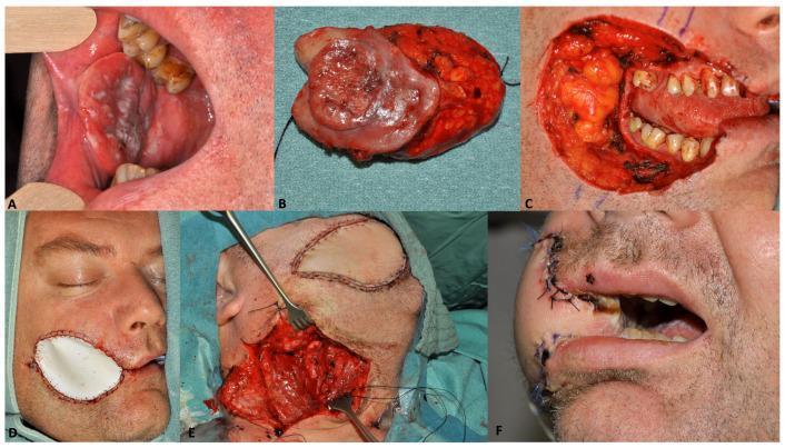
This composite clinical and intraoperative photograph series documents a two-staged, delayed reconstruction for oral cavity cancer (pT2N2bM0) in a 44-year-old male. Panel A shows the preoperative clinical photograph of a large, ulcerated, and heterogeneous malignant mass involving the buccal mucosa. Panel B displays the resected specimen, illustrating the tumor's margins and deep tissue involvement. Panel C captures the significant full-thickness surgical defect immediately after resection, exposing the tongue and dentition. Panel D illustrates temporary wound management using a double-layered artificial skin substitute (EpiGARD®) as a temporary lining to protect the oral mucosa and external facial defect. Panel E depicts the second-stage definitive reconstruction involving the harvest and microsurgical placement of a radial forearm free flap to provide vascularized soft tissue coverage. Panel F shows the patient two weeks post-operatively, demonstrating initial healing with visible sutures and the successful restoration of facial contour. The series serves as an educational resource for surgical oncology, plastic surgery, and maxillofacial reconstruction, highlighting strategies for managing complex head and neck defects when immediate reconstruction is deferred.
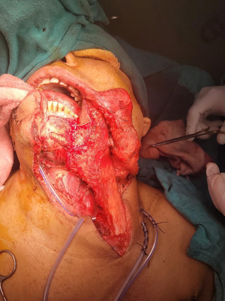
This intraoperative clinical photograph displays a complex head and neck reconstruction using a pectoralis major myocutaneous (PMMC) flap in a patient following oncological resection for oral cavity cancer. The surgical site spans the lower face, oral cavity, and neck. A bulky, reddish-pink, vascularized muscle flap is visible being tunneled and transposed from the chest to reconstruct a significant defect in the buccal mucosa and oral floor. Key anatomical landmarks include the exposed mandibular teeth, oral vestibule, and the submental region. The neck is opened as part of a modified radical neck dissection, with surgical sutures clearly visible at the inferior incision margin. Two clear suction drainage tubes are positioned within the neck wound bed to prevent fluid accumulation. Surgical instruments, including forceps and scissors, are visible in the periphery, manipulated by gloved surgical staff. The image illustrates the clinical application of a pedicled myocutaneous flap for primary reconstruction of large-volume soft tissue defects in head and neck surgery.
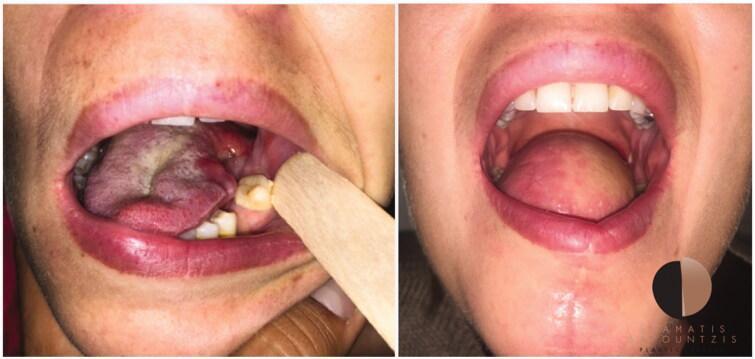
This clinical comparison photograph illustrates a case of tongue reconstruction following oncological resection. The left panel shows the intra-oral state after a hemiglossectomy for tongue cancer. The residual tongue exhibits significant asymmetry, tissue loss, and surgical trauma, with observable erythema, edema, and a whitish fibrinous exudate or slough on the surgical bed. A wooden tongue depressor is used to retract the buccal mucosa for better visualization of the defect. The right panel demonstrates the post-operative result of free-flap reconstruction, specifically a radial forearm free flap (RFFF). The reconstructed tongue shows restored volume and bulk, filling the oral cavity defect. The flap tissue appears viable with healthy pink coloration and a smooth contour, though it lacks the characteristic papillary texture of the native lingual mucosa. The image serves as an educational tool for head and neck surgery, demonstrating surgical oncology outcomes and the efficacy of microvascular free tissue transfer in restoring oral anatomy and function.
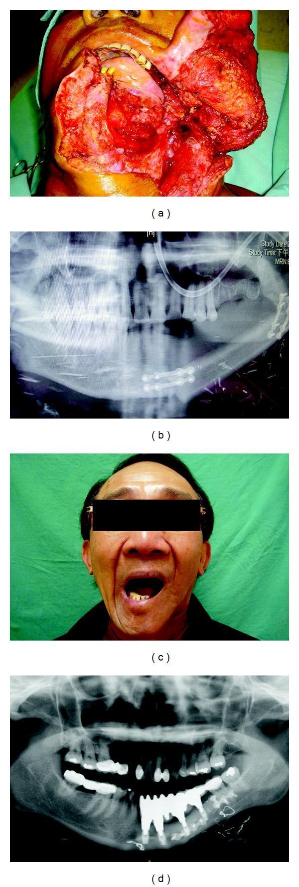
A multi-panel series of clinical and diagnostic images illustrating oromandibular reconstruction following lower gum cancer ablation. (a) Intraoperative clinical photograph showing a massive anterolateral oromandibular defect after tumor resection, exposing oral cavity structures. (b) Panoramic radiograph (orthopantomogram) demonstrating immediate post-operative results of a fibular osteocutaneous free flap reconstruction. The fibular bone segment is integrated into the mandibular arc and secured using radiopaque surgical miniplates and screws for bony fixation. (c) Post-operative clinical photograph of the patient showing successful aesthetic restoration and functional recovery, evidenced by an adequate range of mouth opening. (d) Follow-up panoramic radiograph showing long-term structural outcomes, including the successful osseointegration of multiple dental implants within the reconstructed fibula flap bone. The image highlights the suitability of fibular bone architecture for supporting dental prosthetics and maintaining mandibular continuity. This series serves as an educational resource for plastic and reconstructive surgery, specifically focusing on microvascular free flap techniques and oral rehabilitation.
"oral cavity cancer"[MeSH Terms] AND "surgery"[MeSH Terms]
Mouth Cancer (Oral Cavity Carcinoma) Surgery - Comprehensive Review
1. Overview and Histology
- >90% are squamous cell carcinoma (SCC); the remainder are minor salivary gland tumors (adenoid cystic, mucoepidermoid) or basal cell carcinoma (rare, upper lip)
- Oral cavity = lip, oral tongue (anterior 2/3), floor of mouth, hard palate, buccal mucosa, upper/lower alveolar ridges, retromolar trigone (RMT)
- Key risk factors: tobacco, alcohol, betel nut, chronic trauma, sunlight (lower lip), immunosuppression, poor oral hygiene
2. Staging (AJCC 8th Edition) - Exam High-Yield
| Stage | Definition |
|---|---|
| T1 | Tumor ≤2 cm, DOI ≤5 mm |
| T2 | Tumor ≤2 cm with DOI >5 mm; OR tumor >2 cm and ≤4 cm with DOI ≤10 mm |
| T3 | Tumor >2-4 cm with DOI >10 mm; OR tumor >4 cm with DOI ≤10 mm |
| T4a | Tumor >4 cm with DOI >10 mm; OR invades cortical bone or facial skin |
| T4b | Invades masticator space, pterygoid plates, skull base; encases internal carotid artery |
- Nodal staging (N) uses extranodal extension (ENE): N3b = any node with clinically overt ENE(+)
- Positive lymph nodes decrease chance of cure by ~50% in non-HPV cancers
3. Surgical Principles
Surgical Margins
- Clear (free) margins are paramount; positive or close margins mandate re-excision or adjuvant radiotherapy
- Perineural invasion and lymphovascular invasion at margins are high-risk features
Depth of Invasion and Elective Neck Dissection
- Tumors with DOI >4 mm: recommend elective neck dissection (levels I-III)
- Thin tumors (<2 mm): low regional metastatic potential
- Sentinel lymph node biopsy (SLNB): considered in T1 N0 tumors with DOI 2-4 mm in experienced centres
4. Surgical Procedures by Subsite
A. Lip Cancer (Most common oral cavity site - 95% lower lip)
- T1/T2: Surgery (preferred) OR radiation alone
- T3/T4: Multimodal (surgery + RT)
- Reconstruction - critical rule: align vermilion border precisely in 4 layers:
- <1/4 lip involved: V-shaped wedge excision + primary closure
- 1/4-1/2: Abbe flap (near commissure) or Estlander flap (at commissure)
- 1/2-2/3: Karapandzic flap (neurovascular rotation flap, preserves orbicularis)
-
2/3 to total: Bernard-Burrow or Gillies fan flap; free tissue transfer
- Outcomes: Stage I/II >90% 5-year survival; lip cancer overall 91% 5-year survival
B. Oral Tongue Cancer (Second most common - 30%)
- Arises most often on lateral borders
- DOI >2-4 mm + perineural invasion = higher regional metastasis, recurrence, mortality
- Glossectomy types:
- Partial glossectomy: T1-T2 tumors
- Hemiglossectomy: lateral tumors involving up to half the tongue
- Total/near-total glossectomy: large T3-T4 tumors (requires reconstruction to restore swallowing/speech)
- Primary nodal drainage: levels I-III
- T1-T2: may close primarily or with skin/acellular dermis graft; larger defects need free flap (radial forearm free flap - RFFF)
- 5-year survival: Stage I 64-80%, Stage II 61-84%, Stage III 28-68%, Stage IV 6-36%
C. Floor of Mouth
- Often involves mandible early due to proximity
- DOI critical - thin tumors have better prognosis
- Resection may require partial mandibulectomy (marginal or segmental)
D. Buccal Mucosa & Hard Palate
- High tendency for bone involvement
- Surgical access may require lip-splitting incision (translabial approach) + mandibulotomy
- Full-thickness through-and-through defects of cheek require layered reconstruction
E. Retromolar Trigone (RMT)
- Thin mucosa overlying mandible - bone invasion occurs early
- Inferior alveolar nerve (V3) involvement early due to proximity to mandibular foramen
- Regional mets at presentation: 10-20%, drains to upper jugular nodes (level II)
- If periosteum or minimal cortical erosion only: marginal mandibulectomy
- Advanced bone invasion: composite resection (mandibulectomy) via transcervical or translabial + lateral mandibulotomy approach
- Reconstruction: fibula osteocutaneous free flap for segmental mandibular defects
- Outcomes: 5-year locoregional control overall 71%; disease-specific survival overall 69%
5. Neck Dissection
Types:
| Type | Nodes removed | Structures sacrificed |
|---|---|---|
| Radical (Crile, 1906) | Levels I-V | SCM + IJV + CN XI (spinal accessory) |
| Modified Radical (MRND) | Levels I-V | Spares 1-3 of above structures |
| Selective Neck Dissection | Specific levels only | All 3 structures preserved |
Oral Cavity Drainage and Levels:
- Oral cavity cancers: dissection of levels I-III (± level IV depending on nodal burden)
- Oropharynx/larynx/hypopharynx: levels II-IV
- Level V: rarely involved except in nasopharyngeal carcinoma (NPC) and advanced skin cancers
Elective Neck Dissection Rule:
- Performed for N0 neck when risk of occult nodal metastasis >15%
- Sentinel lymph node biopsy (SLNB) may replace elective neck dissection in early-stage N0 oral cavity cancer
Key Structures to Protect in Neck Dissection:
- Level IB: marginal mandibular branch of CN VII (must identify before submandibular gland resection)
- Level IV left: thoracic duct (ligate lymphatics here)
- Level V: spinal accessory nerve (CN XI) above Erb's point
- Deep cervical fascia: protects phrenic nerve and brachial plexus
6. Reconstruction After Resection
| Defect size / Type | Reconstruction |
|---|---|
| Small soft tissue (<3 cm) | Primary closure or skin graft |
| Floor of mouth / tongue (small-medium) | Acellular dermis, split-thickness skin graft |
| Tongue (partial to hemi) | Radial Forearm Free Flap (RFFF) - thin, pliable, sensate option |
| Large tongue/floor of mouth | Anterolateral thigh (ALT) free flap |
| Segmental mandibular defect | Fibula osteocutaneous free flap - bone length, supports dental implants |
| Large soft tissue / chest | Pectoralis major myocutaneous (PMMC) flap - pedicled, reliable workhorse |
| Full-thickness cheek | Layered reconstruction (mucosa + soft tissue + skin) |
- Microvascular free tissue transfer is the mainstay for oral cavity oncology surgery
- Fibula free flap allows dental implant osseointegration for oral rehabilitation
- PMMC flap is tunneled from chest and used when free flap is not feasible
7. Adjuvant Therapy Indications (Post-Surgery RT/CRT)
- T3-T4 (locally advanced) disease
- Positive surgical margins
- Perineural invasion at primary site
- Lymph node metastases (especially with ENE)
- Multiple positive nodes
- Lymphovascular invasion
8. Complications of Surgery and Adjuvant Treatment
| Complication | Context |
|---|---|
| Fistula (orocutaneous/pharyngocutaneous) | Post-resection, especially after prior radiation |
| Flap failure/necrosis | Free flap (1-5% failure rate in experienced centres) |
| Wound infection, haematoma | Universal post-op risks |
| Trismus | Post-radiation fibrosis of masticatory muscles |
| Osteoradionecrosis of mandible | Primary reason surgery is preferred over radiation for oral cavity |
| Marginal mandibular nerve (CN VII) injury | Neck dissection |
| Spinal accessory nerve (CN XI) injury | Neck dissection - shoulder droop |
| Chyle leak | Level IV dissection (thoracic duct) |
| Dysphagia / aspiration | After tongue resection |
| Altered speech | After tongue resection (especially total glossectomy) |
9. Follow-Up and Surveillance
- Pain is a sensitive indicator of tumor recurrence - should serve as a warning sign (K.J. Lee's, p. 846)
- In the absence of symptoms or abnormal exam, NCCN guidelines recommend against routine imaging screening
- Regular interval visits for clinical examination
- TSH monitoring if thyroid irradiated
Key Exam Mnemonics / High-Yield Points
- DOI not tumor thickness - AJCC 8th edition oral cavity staging uses Depth of Invasion
- Surgery > radiation for oral cavity because of osteoradionecrosis risk with radiation
- Oral cavity nodes: levels I-III; oropharynx: II-IV
- Elective neck dissection if occult nodal risk >15%
- Fibula free flap = best for mandibular reconstruction (supports dental implants)
- RFFF = best for tongue/floor of mouth (thin, pliable)
- PMMC flap = pedicled workhorse when free flap not possible
- Marginal mandibulectomy if only periosteum/minimal cortex involved; segmental if full bone invasion
- Karapandzic flap for 1/2-2/3 lip defects (neurovascular-preserving)
- RMT = retromolar trigone; early bone invasion; needs fibula if segmental resection needed