
Clavicle ossification
Reading File
The clavicle is unique among long bones - it ossifies by intramembranous ossification (not endochondral), and it is the first bone to ossify in the fetus (around week 5-6). A secondary ossification center appears at the sternal end, which is the last epiphysis to fuse in the body (around age 22-25).
Searching sources...
Finding Sources
Finding Sources
Good results. Let me read the most detailed sources on clavicle ossification - the imaging anatomy atlas and the embryology textbook.
Reading File
Reading File
Reading File
Excellent - rich detail gathered. Let me fetch the clavicle anatomy diagram image and also get the forensic/age estimation angle from another source.
Loading Image
Finding Sources
Reading File
Finding Sources
I have comprehensive information from multiple authoritative textbooks. Here is the full answer:
Clavicle Ossification
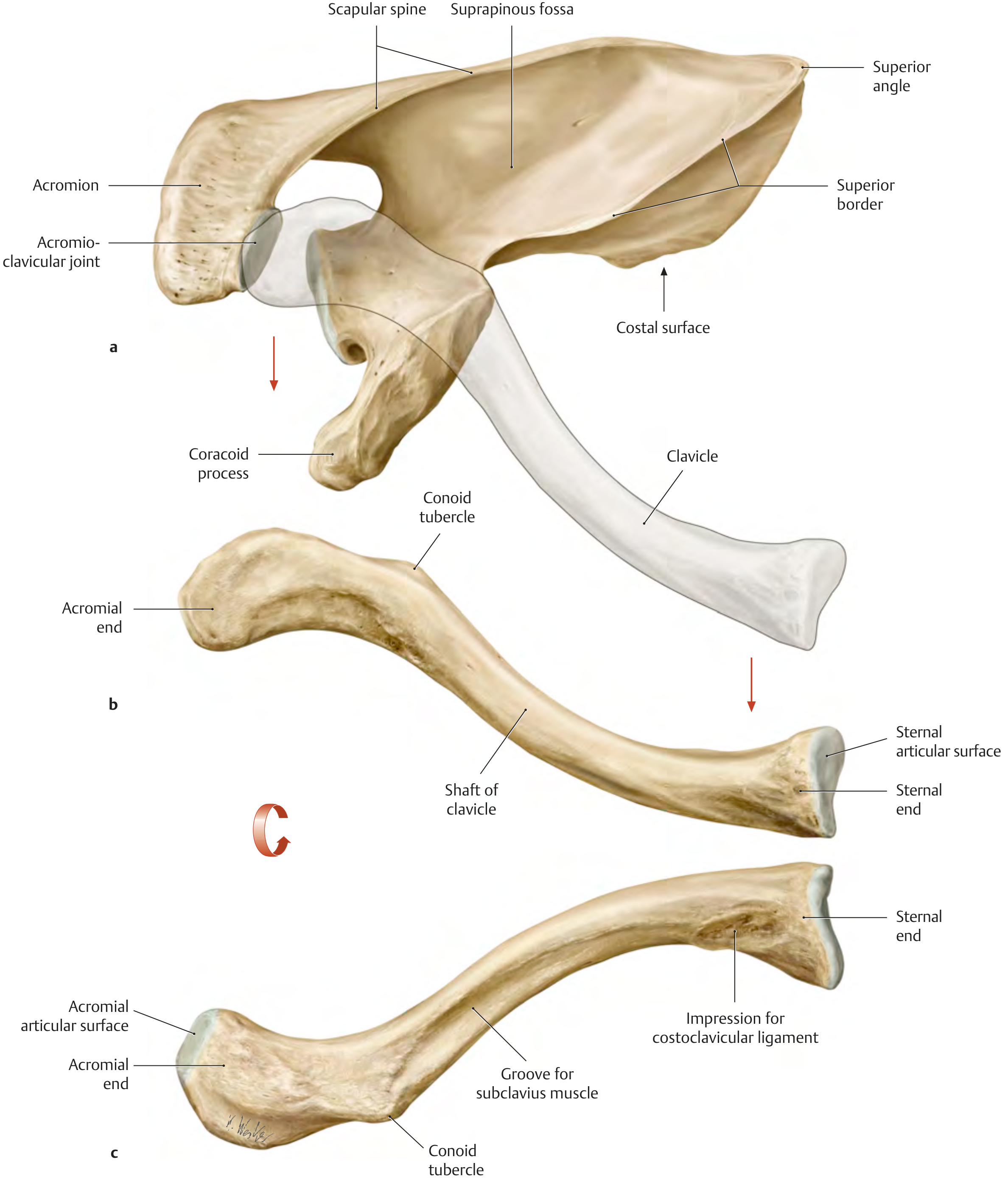
Key Facts at a Glance
| Feature | Detail |
|---|---|
| First bone to ossify in the body | Yes - 5th gestational week |
| Method | Primarily intramembranous (desmal) |
| Number of epiphyses | One (sternal/medial end only) |
| Last epiphysis to fuse | Yes - last in the entire body |
| Age of fusion | 22-25 years |
Type of Ossification
The clavicle is unique because it is the only bone in the postcranial skeleton (and in the limbs) that is not preformed in cartilage. Instead it ossifies directly from connective tissue:
- Acromial (lateral) end - intramembranous ossification
- Medial (sternal) end - endochondral ossification
- The clavicle initially develops by intramembranous ossification and later forms growth cartilages at both ends
This mixed pattern explains why it has only one true epiphysis - at the sternal end - where endochondral ossification occurs.
All other postcranial long bones develop endochondrally (via a cartilage template), so the clavicle stands apart. Histologically, flat bones of the skull, jaws, and the clavicle all share this intramembranous pattern.
- Histology: A Text and Atlas (Ross & Pawlina): "Flat bones of the skull, mandible, and clavicle develop by intramembranous ossification; all other bones develop by endochondral ossification."
- Junqueira's Basic Histology, 17e: "Most bones of the skull and jaws, as well as the scapula and clavicle, develop by intramembranous ossification."
Timeline of Ossification
| Stage | Timing |
|---|---|
| First ossification center appears | ~5th gestational week (earliest of any bone) |
| Primary ossification (diaphysis) | 5th-6th gestational week |
| Growth cartilages form at both ends | Later in fetal life |
| Secondary ossification center (sternal end) appears | ~18-20 years |
| Sternal epiphysis fully fuses | 22-25 years (last epiphysis in the entire body) |
The femora are the next bones after the clavicle to show traces of ossification.
- The Developing Human, 11e (Moore & Persaud): "The clavicle initially develops by intramembranous ossification, and it later forms growth cartilages at both ends... The clavicles begin to ossify before other bones in the body."
- Imaging Anatomy, Vol. 3: "The clavicle is the first bone in the human body to ossify, usually in the 5th gestational week... it has only one epiphysis located at its sternal end with complete ossification at 22 to 25 years of age, the last one in the body to fuse."
Clinical and Forensic Relevance
1. Cleidocranial Dysostosis (CCD)
- Failure of the connective tissue from which the clavicle forms results in aplasia or hypoplasia of the clavicles
- Associated ossification defects in the cranial vault occur because both structures form by membranous ossification
- Autosomal dominant inheritance; affects all bones formed by intramembranous ossification (clavicles, cranium, pelvis)
- General Anatomy and Musculoskeletal System (THIEME Atlas): "A congenital failure or abnormality in the development of this connective tissue results in an anomaly called cleidocranial dysostosis. There may be associated ossification defects in the cranial vault, which are also formed by membranous ossification."
2. Congenital Pseudarthrosis of the Clavicle
- Caused by failure of ossification of a precartilaginous bridge that normally connects the two ossification centers, or by direct pressure from the subclavian artery on the immature clavicle. (Campbell's Operative Orthopaedics, 15th Ed)
3. Forensic Age Estimation
- Because the sternal epiphysis is the last to fuse in the entire skeleton (mid-1920s), the clavicle is a valuable forensic tool for estimating age in young adults up to the late 20s
- Age-related changes in the medial epiphysis surface are used in skeletal age estimation methods
- Forensic Anthropology: A Comprehensive Introduction, 2nd Ed: "The clavicle is the first bone to ossify in utero and the last bone to fuse... it provides accurate age estimates into the mid-1920s to late 20s."
4. Fractures
- Middle-third clavicle fractures are among the most common fractures in children and adults
- Obstetric trauma causes clavicle fractures in 1-2% of all newborns
- The clavicle is frequently a site of healed fractures seen in child abuse cases
- In children, ~50% of all clavicular fractures occur before age 6
Summary Mnemonic
"First in, Last out" - The clavicle is the first bone to ossify in fetal life (~week 5) and has the last epiphysis to fuse in the body (~22-25 years). It ossifies mainly by intramembranous (desmal) ossification - no cartilage template needed.
What is intramembranous
Intramembranous ossification is bone formation that occurs directly within a sheet of fibrous connective tissue (mesenchyme), without a cartilage intermediate. Mesenchymal cells differentiate straight into osteoblasts, which lay down bone matrix right in place.
Searching sources...
Finding Sources
Finding Sources
Reading File
Reading File
Reading File
Loading Image
Loading Image
Intramembranous Ossification
Intramembranous ossification is bone formation that occurs directly within a condensed sheet ("membrane") of embryonic connective tissue (mesenchyme), without any cartilage intermediate. The word "intramembranous" literally means "within a membrane" - referring to this fibrous mesenchymal sheet.
It contrasts with endochondral ossification, where a cartilage template is laid down first and then replaced by bone.
Step-by-Step Process
Here is the process illustrated:
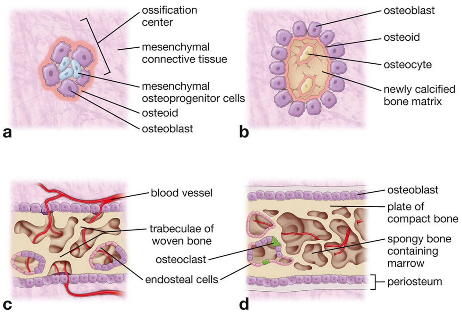
Step 1 - Condensation (panel a)
Spindle-shaped mesenchymal cells migrate and aggregate at specific sites, forming an ossification center. These cells express the CBFA1 (RUNX2) transcription factor, which drives them to become osteoprogenitor cells, then osteoblasts. The cytoplasm shifts from eosinophilic to basophilic as the cell differentiates.
Step 2 - Osteoid secretion (panel a & b)
Osteoblasts secrete osteoid - unmineralized bone matrix rich in type I collagen, bone sialoproteins, and osteocalcin. The mesenchyme also becomes highly vascular at this stage (Wnt signaling drives osteoblast differentiation). Osteoblasts accumulate at the periphery of the center and keep secreting osteoid inward.
Step 3 - Mineralization and osteocyte trapping (panel b)
Osteoid undergoes calcification as calcium phosphate deposits in the matrix. Osteoblasts get trapped within the matrix and become osteocytes, connected to each other by thin cytoplasmic processes running through canaliculi.
Step 4 - Woven bone formation (panel c)
The newly formed tissue appears as irregular spicules and trabeculae of woven (immature) bone - a spongy scaffold with osteoblasts lining the trabeculae and blood vessels running through the spaces. This is structurally weak.
Step 5 - Remodeling into mature bone (panel d)
Osteoclasts resorb woven bone; osteoblasts simultaneously lay down compact bone plates on the outer surfaces (inner and outer tables). The intervening spongy bone persists, and mesenchyme in its interstices differentiates into bone marrow. The remaining condensed mesenchyme that does not ossify becomes the periosteum and endosteum.
Histological appearance
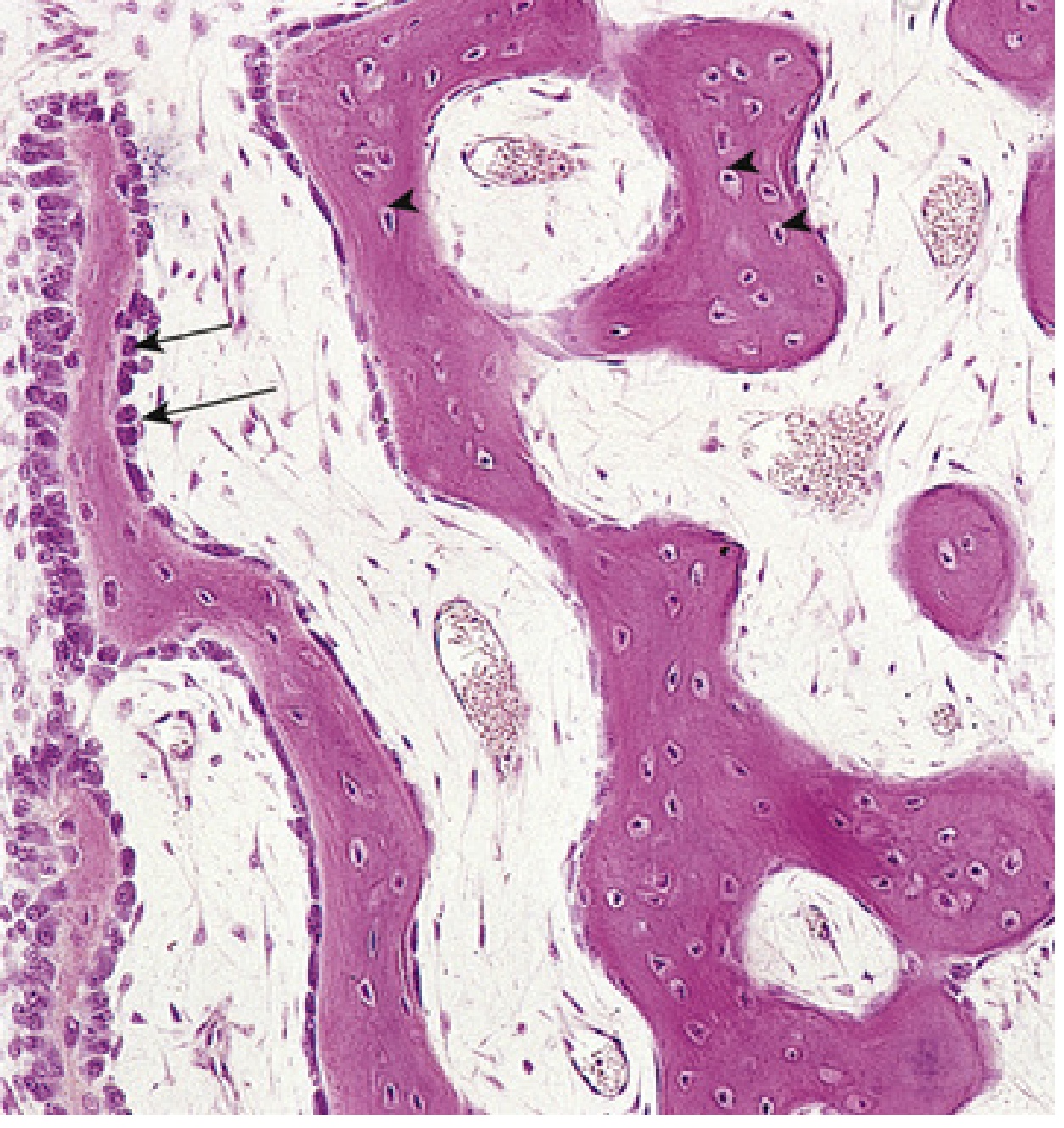
In this H&E section you can see:
- Bone trabeculae being formed (dark pink)
- Osteoblasts lining the trabeculae surface (arrows)
- Osteocytes trapped in lacunae (arrowheads)
- Primordial osteons with central blood capillaries forming
Which Bones Form This Way?
| Bones | Route |
|---|---|
| Flat bones of the skull (frontal, parietal, etc.) | Intramembranous |
| Mandible and maxilla | Intramembranous |
| Clavicle (mainly) | Intramembranous |
| Scapula (partly) | Intramembranous |
| All other long bones | Endochondral |
Key Molecular Signal
CBFA1/RUNX2 is the master transcription factor. It is essential for osteoblast differentiation and drives expression of genes needed for both intramembranous and endochondral ossification. Loss of CBFA1 is the molecular basis of cleidocranial dysostosis - where the clavicle and skull bones (both intramembranous) fail to develop properly.
Summary Comparison
| Feature | Intramembranous | Endochondral |
|---|---|---|
| Starting tissue | Mesenchyme (fibrous membrane) | Hyaline cartilage model |
| Cartilage intermediate | No | Yes |
| Bones formed | Skull, jaw, clavicle | All long bones, vertebrae, ribs |
| First cells | Mesenchymal → osteoblast | Chondrocytes → replaced by osteoblasts |
| Speed | Faster | Slower, more complex |
- Histology: A Text and Atlas (Ross & Pawlina, 8e): "In intramembranous ossification, bone formation is initiated by condensation of mesenchymal cells that differentiate into osteoblasts."
- The Developing Human, 11e (Moore & Persaud): "Intramembranous ossification occurs in mesenchyme... and produces osseous tissue without prior cartilage formation."
- Junqueira's Basic Histology, 17e: "Intramembranous ossification... takes place within condensed sheets ('membranes') of embryonic mesenchymal tissue."
This is a shared conversation. Sign in to Orris to start your own chat.