
Various anatomical sites involved in OSA in detail in simplified manner according to standard ENT textbook also give detailed answer on medial maxillectomy too. Though both of them are totally different and do not correlate with each other, you give different separate answers for each
Reading File
Now let me search for both topics in parallel.
Finding Sources
Finding Sources
Finding Sources
Finding Sources
Reading File
Reading File
Finding Sources
Reading File
Finding Sources
Finding Sources
Finding Sources
Reading File
Finding Sources
Finding Sources
Now I have enough from the textbook. Let me read the Cummings OSA section for anatomical site details, and also look at the Fishman's reference on DISE and obstruction sites.
Reading File
Reading File
Reading File
Loading Image
I now have excellent textbook content from Cummings Otolaryngology for both topics. Let me compile the comprehensive answer.
PART 1: Anatomical Sites Involved in OSA
(Source: Cummings Otolaryngology Head and Neck Surgery; Fishman's Pulmonary Diseases and Disorders)
Understanding Why the Airway Collapses in OSA
The key concept is this: the pharynx is the only segment of the upper airway that has no rigid bony or cartilaginous support. It is entirely a soft-tissue tube held open by muscle activity. During sleep, muscle tone falls, and if the tube is already narrowed or the muscles respond poorly, it collapses - producing apnea.
There are four core traits that drive this collapse (Cummings, p.306):
| Trait | What it means simply |
|---|---|
| 1. Impaired upper airway anatomy | Narrow or floppy pharynx from obesity, soft-tissue bulk, or jaw position |
| 2. Low arousal threshold | Patient wakes too easily from airway narrowing, never reaching deep sleep where muscles stabilize |
| 3. Poor upper airway dilator muscle response | Genioglossus & tensor palatini don't "kick in" enough to hold the airway open |
| 4. High loop gain | Over-sensitive breathing control - small CO2 rise triggers a big ventilatory effort, causing large negative luminal pressures that suck the airway shut |
The Three Major Anatomical Sites of Obstruction
Cummings states: "The three major areas of obstruction are the nose, palate, and hypopharynx." These map to the Fujita classification (1984), which is the standard ENT classification:
Fujita Classification of OSA Obstruction
| Type | Site of Collapse | Region |
|---|---|---|
| Type I | Retropalatal only | Soft palate / nasopharynx region |
| Type II | Retropalatal + Retrolingual | Both palate and tongue base |
| Type III | Retrolingual only | Tongue base / hypopharynx |
Site 1: THE NOSE
What contributes:
- Deviated nasal septum
- Inferior turbinate hypertrophy
- Nasal polyps
- Adenoid hypertrophy (in children and some adults)
- Allergic/non-allergic rhinitis
How it causes OSA:
Nasal obstruction increases upstream airway resistance. It forces mouth breathing during sleep. Open-mouth breathing drops the mandible, which posteriorly displaces the tongue, narrowing the oropharynx. Nasal obstruction alone is rarely the sole cause of OSA - it is a contributing/aggravating factor that worsens pharyngeal collapse (Cummings, p.307).
Site 2: THE RETROPALATAL REGION (Soft Palate / Oropharynx)
This is the most common site of collapse in OSA.
What contributes:
- Elongated/redundant soft palate
- Large uvula
- Lateral pharyngeal wall narrowing
- Tonsillar hypertrophy (especially in children - most common cause in pediatric OSA)
- Lateral pharyngeal fat deposition (with obesity)
How it collapses:
The velopharynx (region behind the soft palate) is the narrowest segment of the pharynx at rest. During sleep, the tensor palatini relaxes, the soft palate sags posteriorly, and lateral walls close in - producing retropalatal obstruction. Studies confirm retropalatal obstruction occurs in 100% of OSA patients vs. 70% of simple snorers (Cummings, p.307).
Key muscles involved:
- Tensor palatini - the main palatal dilator; its reduced activity during sleep is central to retropalatal collapse
- Levator veli palatini
- Superior pharyngeal constrictor (lateral wall narrowing)
Site 3: THE RETROLINGUAL REGION (Tongue Base / Hypopharynx)
What contributes:
- Macroglossia (enlarged tongue)
- Posterior tongue fat deposition (obesity)
- Short mandible / retrognathia - pushes the tongue backwards even without macroglossia
- Low hyoid position
- Micrognathia, retrognathia (craniofacial abnormalities)
How it collapses:
The genioglossus is the single most important pharyngeal dilator. It pulls the tongue forward and opens the retrolingual space. During sleep, genioglossus activity falls. Patients with OSA actually have higher awake genioglossus activity (compensating for a narrowed airway), but lose this compensation during sleep. Studies show retrolingual obstruction in 77% of OSA patients vs. 40% of simple snorers (Cummings, p.307).
Site 4: THE HYPOPHARYNX AND LARYNX
Less commonly discussed but recognized:
- Epiglottis - omega-shaped, floppy, or posterior-displaced epiglottis (epiglottis is included in the VOTE classification used in DISE)
- Bilateral vocal cord paralysis - can produce laryngeal-level OSA
- Laryngomalacia in adults/children
- Obstructing laryngeal lesions
The VOTE Classification (Modern Standard for DISE)
Drug-Induced Sleep Endoscopy (DISE) - where a flexible scope is passed during pharmacologic sedation mimicking sleep - uses the VOTE classification to map all collapsing sites (Fishman's, p.1735):
| Letter | Structure |
|---|---|
| V | Velum (soft palate) |
| O | Oropharyngeal lateral walls (tonsillar pillars) |
| T | Tongue base |
| E | Epiglottis |
Each structure is graded for degree of collapse (none, partial, complete) and pattern (anteroposterior vs. concentric vs. lateral). DISE is now essential before surgical planning, especially before hypoglossal nerve stimulator implantation (Inspire therapy) - a complete concentric palatal collapse on DISE is a contraindication.
Summary: Anatomical Site → Clinical Implication → ENT Surgery
| Anatomical Site | Key Pathology | Relevant Surgery |
|---|---|---|
| Nose | Septal deviation, turbinate hypertrophy | Septoplasty, turbinoplasty |
| Retropalatal | Redundant palate, uvula, tonsils | UPPP (uvulopalatopharyngoplasty), tonsillectomy |
| Oropharyngeal walls | Lateral wall collapse | Expansion sphincter pharyngoplasty |
| Tongue base / retrolingual | Macroglossia, retrognathia | Genioglossus advancement, hyoid suspension, TORS tongue base reduction |
| Mandible/skeletal | Retrognathia, micrognathia | Maxillomandibular advancement (MMA) - most effective surgical option |
| Hypopharynx/epiglottis | Epiglottic collapse | Epiglottoplasty |
PART 2: Medial Maxillectomy
(Source: Cummings Otolaryngology Head and Neck Surgery; Scott-Brown's Otorhinolaryngology Head & Neck Surgery)
Definition
Medial maxillectomy refers to the surgical removal of the medial wall of the maxillary sinus - which is also the lateral wall of the nasal cavity - along with the associated structures (inferior turbinate, uncinate process, and orbital lamina if involved). It essentially opens the maxillary sinus widely into the nasal cavity by removing the partition between them.
Indications
Medial maxillectomy is performed primarily for:
- Sinonasal tumors - the most common indication
- Inverted papilloma (most frequent benign indication)
- Low-grade malignancies confined to the medial maxillary wall
- Adenocarcinoma, squamous cell carcinoma of the lateral nasal wall
- Malignant melanoma
- Angiofibroma extension into the maxillary sinus
- Fungal disease with bony erosion (e.g., invasive aspergillosis)
- Rarely: refractory chronic sinusitis with lateral wall pathology
Anatomical Structures Removed
According to Cummings (p.1757), the anatomic segments removed in a medial maxillectomy include:
- Medial maxillary wall (down to the hard palate)
- Inferior turbinate (with its attachment)
- Uncinate process
- Orbital lamina (lamina papyracea) - if involved by tumor
- Posterior margin can extend to the nasopharynx
- Middle turbinate with its skull base and orbital attachments can also be resected
The lateral margin of resection does NOT cross the plane of the maxillary division of the trigeminal nerve (V2) in the orbital floor - this is the key anatomical boundary.
Two Main Approaches
A. Endoscopic (Endonasal) Medial Maxillectomy
The preferred modern approach for most cases.
Steps (Cummings, p.1757):
- Begin with standard functional endoscopic sinus surgery steps - maxillary antrostomy, clearance of uncinate process
- Remove the medial maxillary wall from the hard palate inferiorly up to the orbital floor superiorly
- The inferior turbinate is removed at its attachment
- The ethmoid cavities are cleared to the bony skull base
- The sphenoid sinus and frontal sinus outflow tract are opened (for clearance of superior margins and to allow post-op surveillance)
- If the anterior maxillary sinus needs exposure: Denker's extension is added
Denker's Extension:
- An incision is made at the internal nasal vestibular aperture
- Periosteum of the anterior facial soft tissues is elevated to the infraorbital nerve
- The lateral nasal piriform aperture is fully exposed and removed with drills/bone-cutting instruments
- The nasolacrimal duct is usually cut at the orbital floor level (routine stenting not required)
- This gives full access to the entire anterior wall of the maxillary sinus
Advantages of endoscopic approach:
- No external incision
- Better cosmesis
- Faster recovery
- Equivalent oncological outcomes for select tumors
- Allows post-operative endoscopic surveillance
B. Medial Maxillectomy via Lateral Rhinotomy (Open Approach)
Described in Scott-Brown's (p.7299) and used when wider access is needed or for larger tumors.
Incision:
- Runs along the lateral border of the nose to the upper edge of the alar margin
- Upper end starts just above the medial canthus level
- Cosmetically acceptable as it follows natural facial contours
- Can be extended into the nasal cavity for larger resections without compromising cosmetic outcome
Steps:
- Incision as above
- Orbital periosteum elevated (as for external ethmoidectomy) and extended laterally over the maxilla to the infraorbital nerve
- Lacrimal sac and duct exposed by removing overlying bone
- Orbital contents freed medially by:
- Dividing the lacrimal sac low down
- Clipping and dividing the anterior ethmoidal artery
- Freeing the insertion of the trochlea by sharp dissection
- Bony window extended as required
- Mucosa of nose, antrum, and ethmoids resected along with the entire lateral nasal wall
- Resection can be extended into sphenoid and frontal sinuses or into the pterygopalatine fossa
- Packing is rarely required after resection
Advantages:
- Excellent visualization of all compartments
- Allows en bloc resection
- Can be combined with anterior craniofacial approach for very extensive tumors
Boundaries of Resection (Endoscopic Medial Maxillectomy)
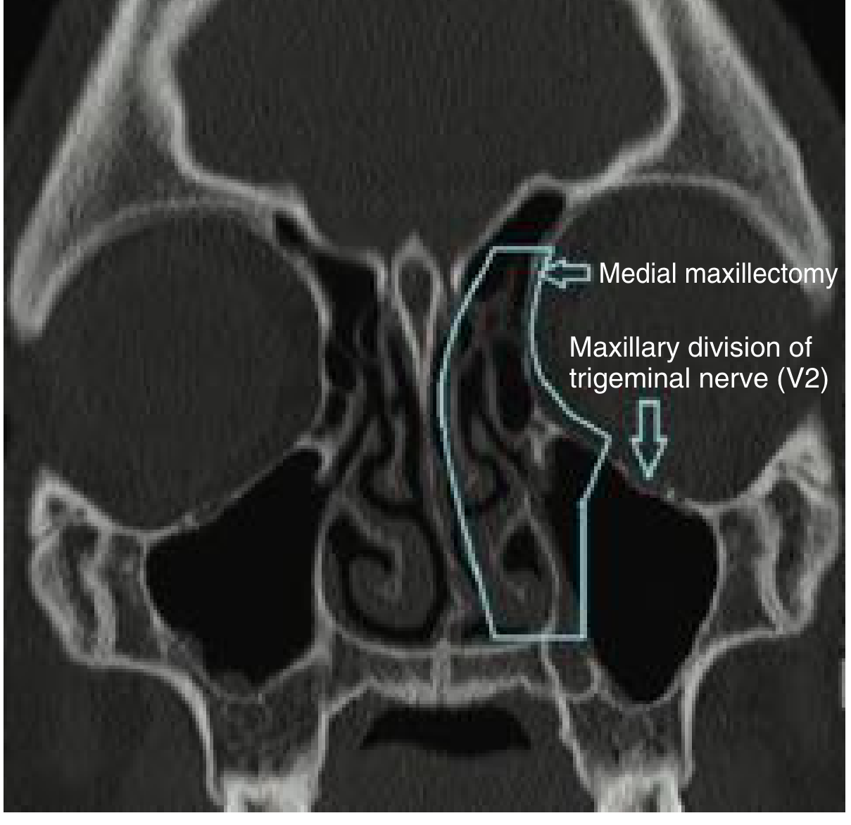
Fig: Boundaries of the endoscopic medial maxillectomy. Resection involves the medial maxillary wall to the palate, uncinate process, inferior turbinate, and orbital lamina. The lateral margin does not cross V2 in the orbital floor. (Cummings Otolaryngology)
Complications
From Cummings (p.1757-1758):
| Complication | Mechanism |
|---|---|
| Epiphora | Damage to nasolacrimal duct |
| Diplopia | Orbital fat herniation or medial rectus damage |
| Orbital hematoma | Bleeding from anterior/posterior ethmoidal vessels |
| Vision loss | Optic nerve injury (rare, more in posterior dissection) |
| Bleeding | Anterior ethmoidal artery, pterygopalatine plexus |
| V2 nerve damage | Hyperaesthesia/numbness of cheek, upper teeth, lip |
| CSF leak | If dissection approaches skull base |
| Infection/meningitis | If skull base breached |
| Olfactory loss | Disruption of olfactory epithelium |
| Sinonasal/vestibular scarring | Contracture of resection cavity |
Post-operative Prosthesis (Open Approach)
After open resection, the cavity is immediately fitted with a temporary prosthesis to:
- Cover the palate
- Restore normal facial contours
This is secured with circumzygomatic wires or fixation screws into remaining hard palate. The prosthesis is changed at 14 days and repeatedly adjusted until the cavity heals, at which point a final prosthesis is constructed (Scott-Brown's, p.7296).
Quick Comparison: Endoscopic vs. Open Medial Maxillectomy
| Feature | Endoscopic | Lateral Rhinotomy (Open) |
|---|---|---|
| External scar | None | Yes (lateral nasal border - cosmetically acceptable) |
| Access | Limited to endonasal | Full face and orbit |
| Best for | Inverted papilloma, early/limited tumors | Large tumors, orbital involvement, extensive disease |
| En bloc resection | Piecemeal (acceptable for benign) | True en bloc possible |
| Surveillance | Excellent (endoscopic) | Requires external access |
| Combination with craniofacial approach | Yes (with skull base surgeons) | Yes (preferred for very extensive cases) |
References:
- Cummings Otolaryngology Head and Neck Surgery, Chapter 15 (OSA Pathophysiology) and Chapter 94 (Endoscopic Sinonasal Tumor Surgery), pp. 305-307, 1757-1758
- Scott-Brown's Otorhinolaryngology Head & Neck Surgery, Chapter 7, pp. 7296-7317
- Fishman's Pulmonary Diseases and Disorders, Chapter 98, pp. 1735-1736
This is a shared conversation. Sign in to Orris to start your own chat.