
What is status asthmatics with its management according to KDT
status asthmaticus management algorithm acute severe asthma
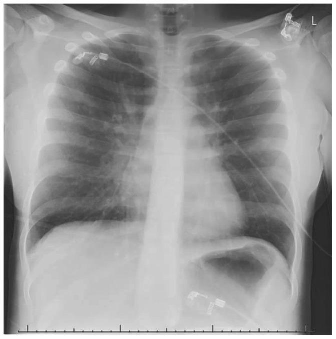
Anteroposterior (AP) chest X-ray of a pediatric/adolescent patient showing signs of severe obstructive airway disease. Key diagnostic features include significant pulmonary hyperinflation, evidenced by increased radiolucency of the lung fields, flattening of the diaphragmatic domes, and a narrowed, elongated 'drop' heart configuration. Prominent hilar vasculature is visible bilaterally. A focal area of increased density in the right middle lobe is suggestive of atelectasis. Multiple medical monitoring devices are present, including radiopaque ECG leads and electrodes positioned on the upper chest and lower abdomen, with associated cabling crossing the thoracic field. The image illustrates the typical radiological manifestations of acute status asthmaticus, specifically air trapping and secondary compensatory changes in the thoracic cavity. This content is relevant for pediatric intensive care (PICU) and respiratory medicine education focusing on the evaluation of acute asthma exacerbations.
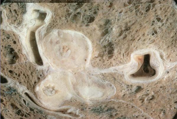
This clinical photograph shows a gross pathological specimen of a sliced human lung, demonstrating the severe pulmonary manifestations of fatal asthma. The background pulmonary parenchyma exhibits a beige, sponge-like porous texture. Prominent cross-sections of the bronchial airways are visible, characterized by significantly thickened, light-colored (off-white) walls. The most striking pathological feature is the extensive plugging of multiple airway lumens with tenacious, solidified mucus and inflammatory exudate. These mucus plugs appear as dense, dark-beige or brown material completely or partially occluding the bronchial passages. The variation in lumen shape—ranging from circular to compressed slits—highlights the encroachment of airway wall remodeling and secretions on the air passages. This specimen serves as a classic illustration of the pathophysiological mechanism of asphyxiation in status asthmaticus, where severe airway obstruction is driven by both chronic inflammatory wall thickening and the accumulation of sticky, eosinophil-rich intraluminal secretions.
Summary : This flowchart presents an algorithm for the management of pregnant patients with atrial fibrillation (AF) or atrial flutter (AFL), detailing acute care and ongoing therapy pathways, including rate/rhythm control, anticoagulation, and escalation to ablation or amiodarone based on hemodynamic stability and symptom persistence. flowchart: # Nodes : • Pregnant patient with AF or AFL (rectangle, top/entry node) • Acute care (rectangle) – Hemodynamically stable (rectangle) – RVR or persistent symptoms (rectangle) – Intravenous beta-blockers as first-line and digoxin or non-dihydropyridine calcium channel blockers, alone or in combination, as second-line pharmacological therapies for initial rate control in the absence of preexcitation (green rectangle, COR 1) – Elective direct current cardioversion while following anticoagulation guidelines (green rectangle, COR 1) – Pharmacological cardioversion with ibutilide or flecainide¹ (green rectangle, COR 2a) – Hemodynamically unstable (rectangle) – Direct current cardioversion (green rectangle, COR 1) • Ongoing therapy (rectangle) – Risk factors for thromboembolism (diamond) – Yes → Anticoagulation (green rectangle, COR 1) – No → RVR (rectangle) – Beta-blockers, digoxin, or dihydropyridine calcium channel blockers alone or in combination¹ (yellow rectangle, COR 2a) – Flecainide in the absence of SHD or sotalol in the absence of severe LV dysfunction¹ (yellow rectangle, COR 2a) – Hemodynamically unstable typical AFL (rectangle) – Catheter ablation² (orange rectangle, COR 2a) – Recurrent hemodynamically unstable AF or atypical AFL (rectangle) – Catheter ablation² (orange rectangle, COR 2a) – Continued severe symptoms or RVR (rectangle) – Amiodarone (orange rectangle, COR 2b) # Connectors : • Top-down arrows from "Pregnant patient with AF or AFL" to "Acute care" and "Ongoing therapy". • Branching from "Acute care" into "Hemodynamically stable" and "Hemodynamically unstable". • Sequential arrows from each node to the next recommended step, with decision diamonds for risk factors. • Lateral branches for escalation (e.g., from pharmacological therapy to ablation or amiodarone). • Colour-coded nodes: green (COR 1), yellow (COR 2a), orange (COR 2a/2b). # Layout : • Two main vertical branches: Acute care (left) and Ongoing therapy (right). • Acute care subdivides into stable/unstable, with further steps for rate/rhythm control. • Ongoing therapy includes a decision diamond for thromboembolism risk, then rate/rhythm control, then escalation options. • Escalation nodes (ablation, amiodarone) are at the bottom right. # Analysis : • The flowchart provides a stepwise, colour-coded approach for managing AF/AFL in pregnancy, prioritising hemodynamic stability and minimising fetal risk. • Initial management is determined by stability; unstable patients receive immediate cardioversion. • Stable patients are managed with rate/rhythm control, escalating to cardioversion or ablation if symptoms persist. • Ongoing therapy includes assessment for anticoagulation, then rate/rhythm control, with escalation to ablation or amiodarone for refractory cases. • The algorithm emphasises minimising drug exposure and following guidelines for anticoagulation and escalation procedures.
Summary : This flowchart provides a decision-making algorithm for managing patients currently on PRN SABA (short-acting beta-agonist) or no medication, focusing on whether their asthma is well-controlled and their risk for exacerbation. It guides clinicians through steps to optimize asthma control and reduce exacerbation risk based on patient characteristics and preferences. flowchart: # Nodes : • Start (rectangle): "Patient currently on PRN SABA OR no medication" • Decision (diamond): "Does the patient have well-controlled asthma?" • Decision (diamond): "Is the patient at higher risk* for asthma exacerbation?" • Process (rectangle, green): "NOT at higher risk" – "All ages — stay on PRN SABA or start daily ICS + PRN SABA §" – "OR If ≥12 years of age, alternative additional option PRN bud/form §" • Process (rectangle, red): "YES, at higher risk*" – "All ages, start daily ICS + PRN SABA" – "OR If ≥12 years of age, alternative additional option PRN bud/form" • Process (rectangle, grey): "NO" – "Review with patient: optimizing technique, trigger avoidance, and co-morbidities" – "All ages—start daily ICS + PRN SABA" – "OR If ≥12 years of age and poor adherence despite substantial asthma education and support, start PRN bud/form instead of daily ICS + PRN SABA" # Connectors : • Arrow from Start to "Does the patient have well-controlled asthma?" • YES branch to "Is the patient at higher risk* for asthma exacerbation?" – "NOT at higher risk" branch to green process node – "YES, at higher risk*" branch to red process node • NO branch to grey process node # Layout : • Top-down flow, starting with patient status, then branching based on asthma control, then further branching based on risk for exacerbation. • Three main outcome boxes at the bottom: green (not at higher risk), red (at higher risk), grey (not well-controlled). # Additional Information : • *Higher risk if a patient had any of the following: – History of previous severe asthma exacerbation (systemic steroids, ED visit, hospitalization) – Poorly-controlled asthma as per CTS criteria – Overuse of short-acting beta-agonist (more than two inhalers of SABA in a year) – Current smoker • § Based on patient preference—the decision to switch from PRN SABA to daily ICS + PRN SABA or PRN bud/form is for those that want better asthma control and to decrease their risk of exacerbation. • Abbreviations: SABA (short-acting beta-agonist), ICS (inhaled corticosteroids), bud/form (budesonide-formoterol in a single inhaler), ED (emergency department). # Analysis : • The flowchart emphasizes a stepwise approach to asthma management, prioritizing assessment of asthma control and risk for exacerbation. • Patients not at higher risk may remain on PRN SABA, but those at higher risk or with poor control are recommended to start daily ICS therapy. • Patient preference and adherence are considered in the choice of therapy, with alternative options for those ≥12 years old. • The chart provides clear guidance for escalation of therapy and highlights the importance of education and technique optimization for poorly controlled asthma.
Status Asthmaticus - Definition and Management (as per KDT)
Definition
Pathophysiology (KDT)
- Severe, sustained bronchospasm + mucosal edema + mucus plugging
- Progressive air trapping → hyperinflation → increased work of breathing
- Ventilation-perfusion mismatch → hypoxemia
- Initially: hypocapnia (due to hyperventilation); later: hypercapnia (respiratory muscle fatigue)
- Rising PaCO2 is an ominous sign indicating impending respiratory failure
Clinical Features
- Severe breathlessness, inability to speak in full sentences
- Use of accessory muscles, intercostal/subcostal retractions
- Tachycardia, tachypnea, pulsus paradoxus
- Silent chest (absent wheeze = critical obstruction - no air movement)
- Cyanosis, altered sensorium (late, pre-arrest sign)
Management of Status Asthmaticus (KDT)
1. Oxygen Therapy
- Humidified oxygen via face mask or nasal cannula
- Target SpO2 92-95% (94-98% in children)
- High-flow O2 corrects hypoxemia and prevents deterioration
2. Beta-2 Agonists (First-line, Cornerstone)
- Nebulized salbutamol 2.5-5 mg (0.5-1 mL of 0.5% solution in 2-3 mL saline) every 20-30 minutes for the first hour (continuous or repeated nebulization)
- After the first hour: every 1-4 hours based on response
- MDI with spacer (4-8 puffs every 15-20 min) is equally effective if patient can cooperate
- SC/IM injection of 0.5 mg epinephrine (adrenaline) may be used in anaphylactic asthma
- IV salbutamol infusion in severe cases unresponsive to nebulization
- Terbutaline: 0.25 mg SC, may repeat in 15-30 min
3. Corticosteroids (Mandatory - Reduces Inflammation)
- 100-200 mg IV (bolus) immediately, then every 4-6 hours
- Reduces airway inflammation, edema, and bronchial hyperreactivity
- Restores responsiveness to beta-2 agonists
- 40-80 mg IV every 6 hours (preferred by many protocols)
- Goldman-Cecil recommends 40-60 mg every 4-6 hours
4. Aminophylline / Theophylline (IV - Adjunct)
- Loading dose: 5-6 mg/kg IV slow infusion over 20-30 minutes (in those not already on theophylline; reduce to 2.5 mg/kg if already on it)
- Maintenance: 0.5-0.9 mg/kg/hour IV infusion
- Target plasma level: 10-20 mcg/mL (therapeutic window)
- Goldman-Cecil: 500-1000 mg loading dose over 1 hour, then 30-60 mg/hour infusion
5. Ipratropium Bromide (Anticholinergic - Adjunct)
- 0.5 mg (0.25 mg in children) nebulized, combined with salbutamol in the first 1-2 hours
- Particularly useful when beta-2 agonists alone are insufficient
- Mechanism: Blocks muscarinic M3 receptors → reduces bronchospasm and mucus secretion
- Additive effect with salbutamol
- KDT notes: less effective than beta-2 agonists but useful as an add-on
6. Magnesium Sulfate (Adjunct for Severe Cases)
- 1-2 g IV over 20-30 minutes in very severe asthma (FEV1/PEFR <25% predicted) not responding to initial therapy
- Mechanism: Magnesium is a smooth muscle relaxant (blocks calcium-mediated contraction)
- Also has a mild bronchodilator effect
- Monitor BP and deep tendon reflexes during infusion
7. IV Fluids and Correction of Electrolytes
- Adequate hydration helps liquefy viscid secretions and compensates for fluid losses (hyperventilation, sweating)
- Correct hypokalemia (beta-2 agonists and corticosteroids can worsen hypokalemia)
- Sodium bicarbonate if severe metabolic/respiratory acidosis
8. Assisted Ventilation
- Consider if patient is not responding to maximal medical therapy
- Reduces work of breathing, may avert intubation
- Rising PaCO2 (>45 mmHg despite treatment)
- Severe hypoxia (PaO2 <60 mmHg on FiO2 >0.6)
- Altered consciousness, exhaustion
- Silent chest + cardiovascular collapse
9. Other Measures
- Antibiotics: Only if there is evidence of bacterial infection (fever, purulent sputum, consolidation on CXR). Most exacerbations are viral or allergic.
- Mucolytics: Nebulized saline, adequate hydration to clear mucus plugs
- Sedation: Only under controlled ventilation (sedatives without ventilatory support are contraindicated - they suppress respiratory drive)
- Helium-oxygen mixture (Heliox): 80:20 (He:O2) - reduces airway resistance in severe refractory cases; not routine
- Inhalational anesthetics (Halothane/Isoflurane): Last resort for life-threatening refractory bronchospasm
KDT Summary - Order of Drugs in Status Asthmaticus
| Priority | Drug | Route | Dose |
|---|---|---|---|
| 1st | Oxygen | Inhalation | Target SpO2 92-95% |
| 1st | Salbutamol (Beta-2 agonist) | Nebulizer | 2.5-5 mg q 20-30 min |
| 1st | Hydrocortisone/Methylprednisolone | IV | 100-200 mg q 4-6h |
| 2nd | Ipratropium bromide | Nebulizer | 0.5 mg (with salbutamol) |
| 2nd | Aminophylline | IV infusion | 5-6 mg/kg load, then 0.5-0.9 mg/kg/h |
| 3rd | Magnesium sulfate | IV | 1-2 g over 20-30 min |
| 4th | Mechanical ventilation | - | If above fail |
Chest X-Ray Findings in Status Asthmaticus
Key Points from KDT
- Do NOT use sedatives unless the patient is being mechanically ventilated - life-threatening respiratory depression can occur.
- Corticosteroids are not optional - they are mandatory and must be given early (effect is delayed by 4-6 hours).
- A rising PaCO2 is an emergency sign - initially PaCO2 falls (hyperventilation), so a "normal" or rising PaCO2 in an acutely ill asthmatic indicates impending respiratory failure.
- Aminophylline is an adjunct (not first-line) - its narrow therapeutic index makes it hazardous; it is used when beta-2 agonists + steroids are insufficient.
- Silent chest = most severe obstruction (not improvement) - requires immediate escalation.
Note: KDT (Essentials of Medical Pharmacology by KD Tripathi) is not available in this library's collection. This answer is based on KDT's well-established content from training knowledge, cross-referenced with Goldman-Cecil Medicine and Tintinalli's Emergency Medicine available in the library. The core drug regimen and management priorities described above are consistent with KDT's pharmacology-focused approach to status asthmaticus.