
Help me write notes for coronary circulation and physiology from standard anaesthesia and cardiac anaesthesia textbooks like miller’s and Hensley martin
Reading File
Now let me search the library for relevant sections on coronary circulation and physiology across anaesthesia textbooks.
Finding Sources
Finding Sources
Finding Sources
I have found the key sections. Now let me read them all in parallel - Miller's, Barash (Cullen & Stoelting), and supporting physiology texts.
Reading File
Reading File
Reading File
Excellent - I got very rich content from Barash. Let me now read deeper into the Barash section (it was truncated) and also get the Guyton section and search for more specific topics.
Reading File
Reading File
Finding Sources
Reading File
Finding Sources
Finding Sources
Reading File
Reading File
I now have excellent content. Let me also read the remainder of the Barash coronary physiology section (autoregulation, neural control, metabolic regulation) and fetch the coronary anatomy diagram.
Reading File
Loading Image
I now have all the key content I need. The Barash section was very comprehensive, covering physiology in depth. Let me now compile the full notes.Here are your comprehensive notes on Coronary Circulation and Physiology, compiled from Miller's Anesthesia (10e), Barash/Cullen/Stoelting's Clinical Anesthesia (9e), Miller's cardiac chapter, and supplemented by Guyton & Hall:
Coronary Circulation and Physiology
Notes for Anaesthesia (Miller's, Barash, Guyton)
1. Coronary Anatomy
Left Coronary System
- The Left Main Coronary Artery (LMCA) arises from the left aortic sinus and divides into:
- Left Anterior Descending (LAD): courses down the interventricular groove; gives rise to diagonal branches (anterolateral LV wall) and septal branches (interventricular septum, bundle branches, Purkinje system); terminates at the LV apex
- Left Circumflex (LCx/LCCA): courses along the left AV groove; gives rise to obtuse marginal branches supplying the lateral LV wall
Right Coronary System
- Right Coronary Artery (RCA): traverses the right AV groove; gives off acute marginal branches to the RV anterior wall
- In 85% (right-dominant): RCA gives rise to the Posterior Descending Artery (PDA) supplying the posterior-inferior LV
- In 15% (left-dominant): LCx gives rise to the PDA
Territory Summary (Barash, p. 850-851)
| Artery | Territory Supplied |
|---|---|
| LAD + septal/diagonal branches | Anterior LV wall, LV apex, anterior 2/3 of interventricular septum |
| LCx + obtuse marginals | Anterior and inferior lateral LV wall |
| RCA + distal branches | Medial inferior LV wall, posterior 1/3 of septum, RV |
| RCA (55%) or LCx (45%) | SA node |
| Dominant artery | AV node, bundle of His, proximal bundle branches |
Clinical pearl: Ischemia of the dominant artery territory can cause SA/AV nodal dysfunction and bradyarrhythmias. RCA or LAD occlusion can both cause RV dysfunction.
Papillary Muscle Blood Supply
- Posterior-medial papillary muscle: single supply from RCA or LCx (2:1 ratio) - highly vulnerable to ischemia; ~1/3 of people have dual supply (less susceptible)
- Anterolateral papillary muscle: dual supply from LAD + LCx - ischemia unusual
Coronary Venous Drainage
- ~75% of LV coronary venous blood returns via the coronary sinus to the right atrium
- RV venous blood returns via anterior cardiac veins directly to RA
- Small amount via Thebesian veins into all cardiac chambers (physiological right-to-left shunt)
2. Coronary Physiology
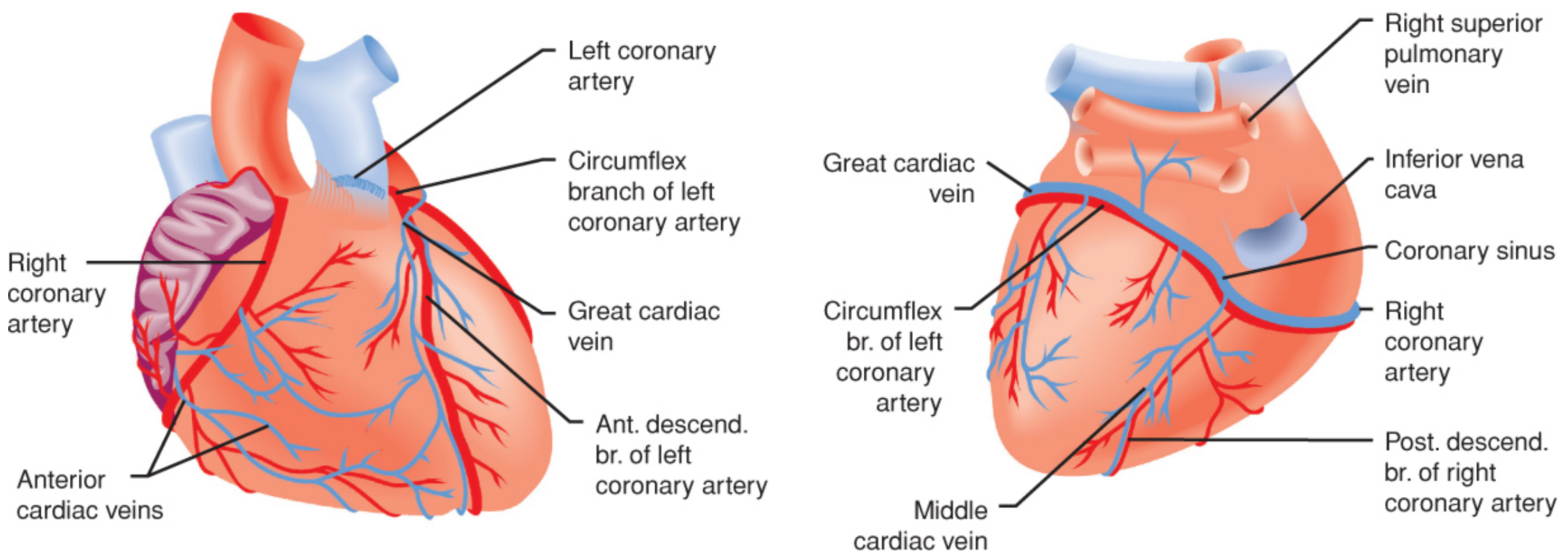
Figure: Coronary artery anatomy - anterior (left) and posterior (right) views. Barash Clinical Anesthesia, 9e, Fig. 12-3
2.1 Basic Flow Dynamics - Poiseuille's Law
Coronary blood flow follows the relationship:
Flow = Perfusion Pressure / Vascular Resistance
- Resistance varies with the 4th power of vessel radius - even tiny changes in caliber produce large changes in resistance
- Coronary blood flow is exquisitely sensitive to arteriolar vasomotor tone
- Blood viscosity (driven mainly by hemoglobin concentration) is a minor factor unless extreme polycythemia or haemodilution is present
Normal resting coronary blood flow:
- ~225-250 mL/min (~70 mL/min/100 g)
- Represents 4-5% of cardiac output
- With strenuous exercise: increases 3-4 fold (but cardiac workload increases 6-9 fold, so efficiency of oxygen utilisation must increase)
(Guyton & Hall, p. 270; Barash, p. 857)
2.2 Coronary Perfusion Pressure (CPP)
Left ventricular CPP = Aortic Diastolic Pressure - LV End-Diastolic Pressure (LVEDP)
- Coronary venous pressure is negligible
- Elevated LVEDP impedes subendocardial blood flow - critical in aortic stenosis, LVH, heart failure
- After coronary stenosis, distal vessels dilate maximally - flow becomes pressure-dependent and manipulating CPP is the primary tool for managing ischaemia
Key formula:
CPP (LV) = Aortic DBP - LVEDP
(Miller's, p. 7585; Barash, p. 857)
2.3 Phasic Nature of Coronary Flow - Systole vs Diastole
LV coronary flow:
- ~70-80% occurs during diastole because extravascular compressive forces during systole markedly impede subendocardial intramural vessel flow
- During systole: intraventricular pressure and coronary vascular resistance both rise, compressing intramural vessels
- During diastole: metabolically mediated arteriolar dilation preferentially increases subendocardial flow
Transmural perfusion ratio:
- Normal subepicardial : subendocardial flow = 1:1 (preserved by metabolic dilation during diastole)
- This ratio is compromised first by tachycardia, stenosis, or hypertrophy
RV, LA, RA:
- Intracavitary pressures remain below aortic pressure throughout the cardiac cycle
- Therefore, compressive forces do NOT significantly impede flow - RV/LA/RA flow is relatively continuous throughout the cardiac cycle
(Barash, p. 857)
2.4 Heart Rate and Coronary Flow
- Tachycardia shortens diastole disproportionately more than systole (non-linear relationship)
- Subendocardial flow is initially maintained by recruiting coronary flow reserve
- When flow reserve is exhausted (stenosis, LVH), tachycardia causes subendocardial ischaemia
- This is the mechanism behind beta-blockers as anti-ischaemic drugs - by slowing HR, they lengthen diastole and restore subendocardial perfusion time
(Miller's, p. 7586; Barash, p. 857)
2.5 Myocardial Metabolism and Oxygen Requirements
The heart is almost exclusively aerobic:
- Heart weight = 0.5% of total body weight, yet accounts for 7% of total body oxygen consumption
- ~75% of haemoglobin-bound oxygen is extracted across the myocardial capillaries
- Coronary sinus PO₂ ≈ 20 mmHg (extremely low - near-maximal extraction at rest)
Energy substrates (in order of preference):
- Fatty acids (predominant substrate, ~60-70%)
- Glucose
- Lactate (taken up and oxidised under normal conditions; produced and released during ischaemia)
In ischaemia:
- Anaerobic glycolysis is activated but ATP yield is severely limited
- Contraction ceases within 10-15 seconds of acute coronary occlusion (unless collateral flow is present)
- Lactate is released (rather than consumed) - lactate production is a marker of myocardial ischaemia
Key implication: Because O₂ extraction is near-maximal at rest, the LV has almost no O₂ extraction reserve - any increase in demand MUST be met by increased blood flow. There is no "extraction reserve" to fall back on.
(Barash, p. 857)
3. Regulation of Coronary Blood Flow
3.1 Metabolic (Local) Regulation - DOMINANT MECHANISM
- Most important regulator of coronary flow
- Coronary flow is tightly coupled to myocardial oxygen demand ("flow-metabolism coupling")
- When O₂ demand rises, the myocardium releases vasodilator mediators that dilate coronary arterioles
- Key mediators of metabolic vasodilation:
- Adenosine (most important) - produced from AMP breakdown during increased ATP turnover; potent arteriolar dilator
- Nitric oxide (NO) - produced by endothelial NOS; causes SMC relaxation
- Hypoxia - direct relaxation of vascular smooth muscle
- CO₂ / H⁺ (local acidosis) - vasodilation
- K⁺ released during repolarisation
- Prostaglandins (prostacyclin, PGI₂)
(Barash, p. 858; Guyton, p. 271)
3.2 Autoregulation
- Coronary flow is maintained constant over a CPP range of approximately 60-140 mmHg
- Below this range (CPP < 60 mmHg): flow falls (pressure-dependent zone) → ischaemia
- Above this range (CPP > 140 mmHg): flow rises (pressure breakthrough)
- Autoregulation is mediated by myogenic response of arteriolar smooth muscle + metabolic feedback
- In stenotic vessels: distal vessels are already maximally dilated to maintain resting flow; autoregulation is abolished and flow becomes entirely pressure-dependent - hence the importance of CPP in CAD patients
- Autoregulation is impaired by: volatile anaesthetics (dose-dependent), extreme tachycardia, severe hypoxia, marked acidosis
3.3 Neural (Autonomic) Regulation
- The coronary vasculature receives both sympathetic and parasympathetic innervation
- Sympathetic (α₁-adrenergic): direct vasoconstriction of coronary vessels
- Sympathetic (β₂-adrenergic): vasodilation (less prominent)
- Net effect of sympathetic stimulation: vasodilation predominates because metabolic effects (increased HR, contractility → increased O₂ demand → metabolic vasodilation) override the direct α₁ constrictive effect
- Parasympathetic (vagal): mild coronary vasodilation; acetylcholine acts via endothelial muscarinic receptors to produce NO
- Coronary conductance is regulated by arterial baroreflex: vasodilator response is induced by a rise in aortic pressure
- At low plasma fentanyl concentrations (1-2 ng/mL), baroreflex coronary control is enhanced; at higher concentrations, it is depressed (Miller's, p. 2769)
3.4 Endothelial Regulation
Vasodilator endothelial mediators:
- Nitric oxide (NO) - synthesised by eNOS; released in response to shear stress, acetylcholine, bradykinin, substance P
- Prostacyclin (PGI₂) - via endothelial COX pathway
- Endothelium-derived hyperpolarising factor (EDHF)
Vasoconstrictive endothelial mediators:
- Endothelin-1 (ET-1) - potent vasoconstrictor; upregulated in atherosclerosis, heart failure
- Thromboxane A₂ (TXA₂)
Endothelial dysfunction (atherosclerosis, diabetes, hypertension): loss of NO-mediated dilation → paradoxical vasoconstriction in response to stimuli that normally cause dilation (e.g., acetylcholine causes constriction instead of dilation when endothelium is dysfunctional)
4. Determinants of Myocardial Oxygen Supply and Demand
(Miller's, p. 7585; Barash, p. 857)
4.1 Oxygen Supply Determinants
| Factor | Components | Clinical Relevance |
|---|---|---|
| O₂ content of blood | Hb × SaO₂ × 1.34 + 0.003 × PaO₂ | Anaemia, hypoxia directly cut supply |
| Coronary blood flow | CPP / coronary vascular resistance | Tachycardia, hypotension, stenosis reduce flow |
| Coronary perfusion pressure | Aortic DBP - LVEDP | Hypotension or raised LVEDP dangerous |
| Duration of diastole | Function of heart rate | Tachycardia the most common cause of ischaemia |
| Haemoglobin level | Oxygen carrying capacity | Minimum Hb for CAD patients higher than for normal patients |
O₂ release from Hb: Leftward shift (alkalosis, hypothermia, low 2,3-DPG) decreases O₂ release to tissues - particularly relevant in cardiac surgery with hypothermia and cardioplegia.
4.2 Oxygen Demand Determinants
| Factor | Effect on MVO₂ | Notes |
|---|---|---|
| Heart rate | ↑↑↑ | Single most important determinant; increases both demand and decreases diastolic supply time |
| Contractility (inotropic state) | ↑↑ | Increased by catecholamines, Ca²⁺, digoxin |
| Wall tension (LaPlace) | ↑↑ | Wall tension = Pressure × Radius / (2 × Wall thickness) |
| Preload (LVEDV) | ↑ | Increases radius → increases wall tension |
| Afterload (SVR, aortic pressure) | ↑ | Increases systolic wall tension |
| Basal metabolism | Constant | ~15-20% of total MVO₂ |
LaPlace's Law and wall stress:
Wall Stress = (Pressure × Radius) / (2 × Wall Thickness)
- LV dilatation (increased radius) → increased wall stress → increased O₂ demand
- LV hypertrophy (increased wall thickness) → reduced wall stress per unit - but capillary density may not increase proportionately, reducing O₂ supply to the hypertrophied myocardium
"Rate-pressure product" (RPP) = HR × Systolic BP - simple bedside index of MVO₂; RPP > 12,000 associated with myocardial ischaemia in susceptible patients.
5. Coronary Flow Reserve (CFR)
- CFR = Maximum coronary blood flow (after maximal vasodilation) / Resting coronary blood flow
- Normal CFR ≈ 4-5 (flow can increase 4-5 fold from baseline)
- CFR is the physiological "buffer" that allows increased demand (exercise, tachycardia) to be met
- A stenosis of >50% diameter begins to limit maximum flow; >70-80% impairs resting flow
Factors reducing CFR:
- Coronary artery stenosis (most common)
- LV hypertrophy (increased resting flow + reduced maximum flow due to reduced capillary density)
- Microvascular disease (diabetes, hypertension)
- Tachycardia (exhausts reserve)
- Anaemia, hypotension
6. Coronary Steal Phenomenon
- Occurs when a vasodilator (e.g., adenosine, dipyridamole, isoflurane) dilates normal vessels but cannot further dilate vessels distal to a stenosis (already maximally dilated)
- Blood is "stolen" from the ischaemic territory through collateral vessels to the dilated normal territory
- Isoflurane: most implicated volatile agent; causes coronary vasodilation and can produce steal in patients with "steal-prone" anatomy (parallel coronary stenoses with collaterals)
- Opioids: do NOT produce steal phenomena and do not diminish coronary vasomotor response (Miller's, p. 2769)
- Steal-prone anatomy: requires two stenoses and a collateral vessel connecting the territories; relatively uncommon (~23% of patients with CAD have steal-prone anatomy)
7. Anaesthetic Effects on Coronary Circulation
Volatile Anaesthetics
- All volatile agents decrease MVO₂ by reducing HR, contractility, and wall tension (decrease in SVR reduces afterload)
- Isoflurane, desflurane, sevoflurane: all produce coronary vasodilation via K-ATP channel opening
- Ischaemic preconditioning by volatile agents: clinically significant myoprotective mechanism mediated through K-ATP channel opening, adenosine receptors, and PKC activation
Opioids (Miller's, p. 2769)
- No significant effect on coronary vasomotion or myocardial metabolism at clinical doses
- Do NOT produce steal phenomena
- Do not diminish large coronary arteriole response to vasoactive agents
- Baroreflex coronary control: enhanced at low fentanyl concentrations (1-2 ng/mL), depressed at high concentrations
- Opioids can mimic ischaemic preconditioning - opioid receptor stimulation (especially κ and δ receptors) reduces infarct size in animal models
- Remote preconditioning (ischaemia of intestine, kidney, limb) is mediated through myocardial κ-opioid receptors
- Postconditioning: δ-opioid receptor activation during early reperfusion protects against infarction; can be enhanced by morphine + isoflurane combination
- Late preconditioning (24 hours after morphine) has also been demonstrated in animal models
Perioperative Ischaemia Management
- Vigilant monitoring needed for supply-demand imbalance: ECG with ST-segment analysis + TEE for regional wall motion abnormalities
- Causes of reduced supply: hypotension, tachycardia, anaemia, coronary vasoconstriction
- Causes of increased demand: tachycardia, increased afterload (hypertension), increased contractility
- Beta-blockers: cornerstone - reduce HR (lengthen diastole, reduce demand), and prevent even small perioperative HR increases
(Miller's, p. 7586)
8. Myocardial Ischaemia and Infarction
Ischaemia Cascade (in order of appearance):
- Metabolic changes (lactate production, reduced ATP)
- Diastolic dysfunction (relaxation impaired)
- Systolic dysfunction (regional wall motion abnormality - detected by TEE)
- ECG changes (ST changes)
- Symptoms (chest pain) - last to appear
Consequences of Acute Myocardial Infarction (Guyton, p. 271-272):
- Decreased cardiac output - systolic stretch: ischaemic muscle passively bulges outward during systole, wasting contractile energy; cardiogenic shock if >40% LV infarcted (mortality 40-50%)
- Pulmonary oedema - reduced CO → reduced renal flow → fluid retention → elevated pulmonary venous pressure
- Ventricular fibrillation - most common cause of sudden death; two high-risk windows: within 10 minutes of infarction, and 1-several hours later
Ischaemic Preconditioning
- Brief episodes of ischaemia-reperfusion make the myocardium more resistant to subsequent prolonged ischaemia
- Mediated by: adenosine A₁ receptors, K-ATP channels, PKC, mitochondrial permeability transition pore
- Early preconditioning: within minutes, lasts 1-2 hours
- Late preconditioning: 24-72 hours after stimulus (mediated by protein synthesis, iNOS)
- Opioids and volatile anaesthetics can both pharmacologically precondition the heart
9. Quick Reference - Key Numbers
| Parameter | Value |
|---|---|
| Resting coronary blood flow | 225-250 mL/min (1 mL/min/g; ~5% CO) |
| % flow during diastole (LV) | 70-80% |
| Coronary sinus PO₂ | ~20 mmHg |
| O₂ extraction at rest | ~75% of Hb-bound O₂ |
| Normal coronary flow reserve (CFR) | 4-5 |
| Subepicardial:subendocardial flow ratio | 1:1 (normal) |
| Right coronary dominance | 80-85% of patients |
| SA node supply: RCA | 55%; LCx = 45% |
| AV node supply | Dominant artery (usually RCA) |
| Autoregulation range (CPP) | 60-140 mmHg |
| Cardiogenic shock threshold | >40% LV infarction |
| LV contraction ceases after occlusion | ~10-15 seconds |
Sources:
- Barash, Cullen & Stoelting's Clinical Anesthesia, 9e - Chapter 12 (Coronary Anatomy, pp. 850-858; Coronary Physiology, pp. 857-869)
- Miller's Anesthesia, 10e - Coronary Anatomy & Determinants of MVO₂ (pp. 7584-7586); Opioids & Coronary Circulation (pp. 2769-2770)
- Guyton & Hall Textbook of Medical Physiology - Coronary Circulation (pp. 270-272)
Concise blood transfusion chapter from miller’s anaesthesia so that I am able to learn all key concepts and points and then frame questions for OSCE on the same
Finding Sources
Finding Sources
The main blood transfusion chapter is in block18. Let me now explore its full table of contents and read the key sections in parallel.
Finding Sources
Finding Sources
The block18 file contains multiple chapters. The blood transfusion chapter starts around line 1257. Let me get the full TOC for the transfusion section and start reading simultaneously.
Finding Sources
Reading File
Finding Sources
Reading File
Finding Sources
Reading File
Reading File
Reading File
Reading File
Reading File
Reading File
Finding Sources
Reading File
Reading File
Reading File
I now have comprehensive content from the entire chapter. Let me compile the full notes and OSCE questions.
Blood Transfusion - Concise Chapter Notes
Miller's Anesthesia, 10th Edition - Chapter 45
PART 1: COMPREHENSIVE NOTES
1. Historical Evolution of Transfusion Practice
| Era | Key Shift |
|---|---|
| 1960s | Whole blood was the standard |
| 1970s-80s | Component therapy - "give only what is lacking"; concerns over HIV and hepatitis |
| 1990s-2000s | HIV screening improved 10,000-fold; focus shifted to non-infectious hazards (TRALI, TACO, hemolytic reactions); liberal vs restrictive debate; "reconstituted whole blood" for trauma |
| 2010-present | Patient Blood Management (PBM) - multipronged, evidence-based, patient-centred approach |
Patient Blood Management (PBM) - Definition (SABM):
"The timely application of evidence-based medical and surgical concepts designed to maintain hemoglobin concentration, optimize hemostasis, and minimize blood loss to improve patient outcome."
2. Blood Procurement and Transfusion-Transmitted Infections
Donor Sources
- Allogeneic (homologous): From another person
- Autologous: Patient's own blood (preoperative donation, ANH, cell salvage)
- Directed donation: From a specific designated donor (e.g., family member)
Infectious Disease Testing (All units in the US, 2021)
Tests performed on every unit:
- HIV-1/2 antibody (ELISA + NAT)
- Hepatitis B surface antigen (HBsAg)
- Hepatitis B core antibody (Anti-HBc)
- Hepatitis C antibody (Anti-HCV + NAT)
- HTLV-I/II antibody
- West Nile Virus (NAT)
- Treponema pallidum (syphilis)
- Trypanosoma cruzi (Chagas disease) - first donation only
- Babesia (endemic areas)
- Zika virus (NAT)
Risk of Transfusion-Transmitted Infection (per unit, screened blood, USA)
| Pathogen | Risk per Unit |
|---|---|
| HIV | ~1:1,500,000 |
| Hepatitis C | ~1:1,000,000 |
| Hepatitis B | ~1:1,000,000 |
| HTLV | ~1:2,000,000 |
| Bacterial contamination (PRBCs) | ~1:500,000 |
| Bacterial contamination (platelets) | ~1:75,000 (higher - stored at room temp) |
Window period: The interval between infection and antibody formation (detectable by current tests). NAT (nucleic acid testing) has dramatically shortened this.
3. Biochemical Changes in Stored Blood (Storage Lesion)
Anticoagulant-Preservative Solutions
| Solution | Contents | Shelf Life |
|---|---|---|
| CPDA-1 | Citrate (chelates Ca²⁺), Phosphate (buffer), Dextrose (energy), Adenine | 35 days |
| AS-1 (Adsol) | Adenine, Glucose, Mannitol, NaCl | 42 days |
| AS-3 (Nutricel) | Glucose, Adenine, Citrate, Phosphate, NaCl | 42 days |
| AS-5 (Optisol) | Dextrose, Adenine, NaCl, Mannitol | 42 days |
| AS-7 | (FDA approved 2015) | 56 days |
- Hct of CPDA-1 PRBCs ≈ 65%, volume ~250 mL
- Hct of AS-1 PRBCs ≈ 55-60%, volume ~310 mL (100 mL additive solution added)
- Storage regulation: At least 70% of transfused RBCs must remain in circulation for 24 hours post-transfusion
Progressive Storage Lesion Changes (RBC)
| Change | Direction | Significance |
|---|---|---|
| ATP | Decreases | Reduced RBC survival, shape change from biconcave → spherical |
| 2,3-DPG | Decreases (gone by 1 week) | Left-shift of O₂-dissociation curve → impaired O₂ delivery |
| Potassium (plasma K⁺) | Increases (19-50 mEq/L by 21 days; 45-60 by 42 days) | Hyperkalemia risk in massive transfusion, neonates, renal failure |
| pH | Decreases (7.4 → 7.1 immediately; falls to 6.9 by 21 days) | Acidosis |
| Lactate | Increases | Marker of RBC metabolism |
| pCO₂ | Rises (150-220 mmHg) | Cannot escape through plastic bag |
| Bicarbonate | Decreases | Metabolic acidosis |
| Sodium (intracellular) | Increases | Na/K pump inhibited at 1-6°C |
| Microaggregates | Form | May obstruct pulmonary microvasculature |
| Free Hb | Increases | Scavenges nitric oxide, causes vasoconstriction, renal injury |
Metabolic characteristics of PRBCs (stored blood - Table 45.5):
- Hct: 57%, pH 6.79, pCO₂ 79 mmHg, HCO₃ 11 mmol/L, Na⁺ 126 mmol/L, K⁺ 20.5 mmol/L, Glucose 24 mmol/L, Lactate 9.4 mmol/L
2,3-DPG Restoration
- 2,3-DPG is depleted within 1-2 weeks of storage
- After transfusion: 50% restored within 24 hours, complete restoration in 48-72 hours in most patients
- Clinical significance: Stored blood transfused acutely may initially deliver less O₂ at tissue level (though at normal physiologic pH, the oxyhemoglobin curve returns toward normal fairly quickly)
Duration of Storage - Clinical Evidence
- Multiple RCTs (RECESS, INFORM, TRANSFUSE, ABLE trials) have largely shown no significant difference in outcomes between fresh and older stored blood in most clinical settings
- Current evidence does not support a specific preference for fresh blood in most patients
4. Blood Components - Characteristics and Indications
4.1 Packed Red Blood Cells (PRBCs)
- Same Hb content as whole blood but plasma removed; Hct ~57-60%
- Indication: Symptomatic anaemia OR Hb below the transfusion trigger
Compatible diluents for PRBCs:
- ✅ Normal saline (0.9% NaCl) - most common
- ✅ 5% Dextrose in 0.9% or 0.45% NaCl
- ✅ Normosol-R (pH 7.4)
- ❌ Lactated Ringer - contains Ca²⁺ → can activate clotting cascade, NOT recommended
- ❌ Hypotonic solutions → RBC swelling → haemolysis
Estimating transfusion requirements:
- 1 unit PRBCs raises Hb by ~1 g/dL (Hct by ~3%) in a 70 kg adult
- Formula: Units needed = (Target Hb - Current Hb) × Body weight (kg) × 0.3
4.2 Preoperative Anaemia and Transfusion Triggers
Liberal vs Restrictive Strategy:
- Restrictive: Transfuse when Hb ≤ 7-8 g/dL
- Liberal: Transfuse when Hb ≤ 9-10 g/dL
- 2022 Cochrane Review (48 trials, >21,000 patients): Restrictive strategy reduced transfusion exposure by 41% with NO increase in mortality, MI, stroke, pneumonia, or thromboembolism
Special population triggers:
- Cardiac surgery / active CAD: Consider transfusion at Hb 8-9 g/dL
- Hip fracture repair (FOCUS trial): Restrictive (Hb 8 g/dL) equivalent to liberal
- ICU patients (TRICC trial): Restrictive (7 g/dL) equivalent or superior to liberal (10 g/dL) except in acute MI/unstable angina
Preoperative anaemia management (PBM pillars):
- Iron (oral or IV) - treat iron-deficiency anaemia ≥4 weeks preoperatively ideally
- Erythropoiesis-stimulating agents (ESAs) - epoetin alfa, darbepoetin - for renal anaemia, chemotherapy anaemia; risk: hypertension, thrombotic events
- Vitamin B12/folate if deficient
- Avoid preoperative transfusion in stable anaemia - recent data show no benefit and may increase complications
4.3 Platelet Concentrates
| Type | Content | Shelf Life | Indications |
|---|---|---|---|
| Random donor platelets (RDP) | From 1 unit whole blood; ~5.5 × 10¹⁰ platelets | 5 days at 22°C with agitation | Thrombocytopenia, platelet dysfunction with bleeding |
| Single donor apheresis platelets (SDP) | From 1 donor apheresis; ~3 × 10¹¹ platelets (equivalent to 6 RDP units) | 5 days | Preferred - reduces alloimmunisation |
1 "pool" of platelets = 4-6 random donor units = raises platelet count by ~20,000-30,000/μL
Platelet transfusion triggers (ASA guidelines):
- Prophylactic: < 50,000/μL for most invasive procedures; < 100,000/μL for CNS/ocular surgery
- Therapeutic (active bleeding): < 50,000/μL + microvascular bleeding
- Massive transfusion: Platelet count < 50,000/μL (massive transfusion dilutes platelets)
Bacterial contamination risk of platelets is higher than RBCs (~1:75,000) because stored at room temperature. Testing (bacterial culture, pathogen reduction) is performed to mitigate this.
4.4 Fresh Frozen Plasma (FFP)
- Contains all plasma proteins including labile factors V and VIII
- Processed soon after donation, frozen within 8 hours (FFP) or 24 hours (PF24)
- Thawed plasma: stored 1-6°C for up to 5 days - logistically useful for trauma centres
- PF24 is comparable to FFP except ~25% less factor VIII and slight reduction in factor V
- Dose: 10-15 mL/kg typically; 1 unit (~250 mL) raises all factors by ~3%
ASA 2015 Guidelines for FFP:
- Obtain coagulation studies first (when feasible)
- INR > 2.0 in absence of heparin with active/anticipated bleeding
- Coagulopathy during massive transfusion > 1 blood volume (~70 mL/kg) when coagulation tests unavailable
- Known factor deficiency with bleeding when specific concentrates unavailable
- Warfarin reversal with severe bleeding when prothrombin complex concentrates (PCCs) unavailable
Risks of FFP: TRALI (highest risk - high plasma volumes), TACO, allergic/anaphylactic reactions, infection
4.5 Cryoprecipitate
- Produced by slow thawing of FFP at 4°C - precipitate re-suspended in ~10-15 mL plasma
- Contents per unit: Fibrinogen (~250 mg), Factor VIII (~80 IU), vWF, Factor XIII, Fibronectin
- Stored at -18°C; once thawed, used within 4-6 hours
- 1 unit per 7-10 kg raises fibrinogen by ~50 mg/dL
Indications for Cryoprecipitate:
- Hypofibrinogenaemia (fibrinogen < 100 mg/dL with bleeding; < 150 mg/dL in obstetric haemorrhage)
- Haemophilia A (factor VIII deficiency) - if specific concentrate unavailable
- von Willebrand disease - if specific concentrate/DDAVP unavailable
- Factor XIII deficiency
- DIC with bleeding
4.6 Freeze-Dried (Lyophilised) Plasma
- Dehydrated plasma that reconstitutes with sterile water
- Advantage: Room temperature storage, long shelf life, instant availability
- Being re-evaluated for prehospital/military use
5. Massive Transfusion
Definition: Transfusion of ≥ 10 units PRBCs in 24 hours (or replacement of >1 blood volume, or >4 units PRBCs in 1 hour with ongoing haemorrhage)
Lethal Triad of Trauma
Hypothermia + Acidosis + Coagulopathy - mutually reinforce each other
Massive Transfusion Protocol (MTP) - Damage Control Resuscitation
- Reconstituted whole blood concept: PRBC : FFP : Platelets in a ratio approaching 1:1:1
- This ratio mimics whole blood and prevents dilutional coagulopathy
- Evidence: Military experience, prospective studies (PROPPR trial - 1:1:1 vs 1:1:2 ratio showed improved haemostasis and 24-hour survival with 1:1:1)
Complications of Massive Transfusion:
| Complication | Mechanism | Management |
|---|---|---|
| Dilutional thrombocytopenia | RBC transfusion dilutes platelets | 1:1:1 ratio; replace when < 50,000/μL |
| Dilutional coagulopathy | Factors V and VIII depleted in stored RBCs | FFP at 1:1 ratio; goal INR < 1.5 |
| Hypofibrinogenaemia | Fibrinogen consumed/diluted | Cryoprecipitate; goal > 150-200 mg/dL |
| Citrate intoxication → Hypocalcaemia | Citrate chelates Ca²⁺; overwhelms hepatic metabolism at >1 unit/10 min | IV Calcium chloride or calcium gluconate |
| Hyperkalaemia | K⁺ leaks from stored RBCs; stored K⁺ 19-60 mEq/L | Monitor levels; clinically significant at >120 mL/min infusion rate |
| Hypothermia | Blood stored at 4°C | Blood warmer (37-38°C); impairs coagulation; VF < 30°C |
| Metabolic acidosis | Stored blood pH 6.9-7.1; lactic acid; anaerobic metabolism | Correct perfusion; bicarbonate if severe |
| TRALI | Leukocyte antibodies in donor plasma | Stop transfusion; supportive |
| TACO | Volume overload | Slow rate; diuretics; reduce volume |
| DIC | Tissue injury, factor consumption | Treat underlying cause; replace factors |
Factors V and VIII in massive transfusion:
- Factor V: falls to 50% at 21 days, 20% at 35 days
- Factor VIII: falls to 30% at 21 days, 20% at 35 days
- However: only 5-20% of Factor V and 30% of Factor VIII are needed for surgical haemostasis - levels rarely fall below these thresholds during massive transfusion alone
6. Compatibility Testing
ABO-Rh System
ABO typing:
- Serum contains naturally occurring antibodies (anti-A, anti-B) against absent antigens
- Anti-A and anti-B are IgM antibodies - activate complement → acute intravascular haemolysis
- 15% of all transfusion-related deaths are from ABO-incompatibility haemolytic reactions
Rh(D) system:
- 85% of people are Rh(D) positive; 15% Rh(D) negative
- Anti-D antibodies are IgG - do NOT fix complement readily → primarily extravascular haemolysis
- 60-70% of Rh(D)-negative recipients given Rh(D)-positive blood will form anti-D antibodies
Blood type compatibility for PRBCs:
| Recipient | Compatible donor |
|---|---|
| O− | O− only (universal donor for PRBCs) |
| O+ | O−, O+ |
| A− | A−, O− |
| A+ | A+, A−, O+, O− |
| B− | B−, O− |
| B+ | B+, B−, O+, O− |
| AB− | AB−, A−, B−, O− |
| AB+ | All (universal recipient) |
Note: AB plasma (FFP) is the universal donor for plasma; O negative is universal donor for RBCs
Types of Compatibility Tests
| Test | What it Detects | Time Required |
|---|---|---|
| ABO-Rh Typing | Patient's blood group | Minutes |
| Antibody Screen | Unexpected alloantibodies in patient serum | 45 min |
| Crossmatch (full serologic) | Patient serum vs donor RBCs | 45-60 min |
| Electronic crossmatch | Computer-verified compatibility | Minutes (requires 2 ABO typings) |
| Type and Screen (T&S) | ABO-Rh + antibody screen; blood held but not crossmatched | If screen negative, release in <10 min |
| Emergency release (Type O−) | No testing; universal donor RBCs | Immediate |
Emergency blood strategies:
- Uncrossmatched O− PRBCs: Immediate availability; for life-threatening haemorrhage
- ABO-specific uncrossmatched: After blood group identified (~5-10 min)
- Electronic crossmatch: After 2 separate ABO typings - equivalent safety to full crossmatch
- Full serologic crossmatch: Full compatibility assured
Joint Commission requirement: Two patient identifiers + confirmation of correct blood product before ANY blood transfusion.
7. Autologous Blood Strategies
7.1 Preoperative Autologous Donation (PAD)
- Patient donates their own blood 3-5 weeks before elective surgery
- Usually 1-3 units collected weekly, last donation ≥72 hours pre-op
- Advantages: Eliminates allogeneic transfusion risks; no cross-matching needed
- Disadvantages:
- Risk of same collection/labelling errors
- Blood cannot always be used (wastage)
- Donation itself causes anaemia → may paradoxically increase transfusion rate
- Not cost-effective for low-risk surgeries
Contraindications to PAD:
- Severe aortic stenosis, unstable angina, bacteraemia/septicaemia, severe anaemia, seizure disorders, acute illness, inability to donate (very young, morbidly obese)
7.2 Acute Normovolemic Haemodilution (ANH)
Principle: Remove whole blood immediately pre-op, replace volume with crystalloid (3:1) or colloid (1:1), then re-infuse blood when major bleeding has stopped
Mechanism of benefit: When the patient bleeds during surgery, they lose blood with a LOWER haematocrit (diluted blood) → less RBC mass lost per mL of blood loss
Re-infusion order: Last unit collected first (i.e., the first unit drawn is re-infused last) - NO, wait - the first unit collected is re-infused LAST is incorrect. The correct order is: reverse order of collection - last collected first, because the first collected has the highest Hb, most factors, and best platelets and should be saved to re-infuse at the end.
Actually per Miller's: "the sequestered blood is then reinfused into the patient in the reverse order of collection because the first unit collected has the highest concentration of coagulation factors and platelets and the highest Hb level" - first collected = re-infused last (saved for maximum benefit).
Evidence: Meta-analysis of 29 RCTs in cardiac surgery - ANH patients received ~¾ fewer allogeneic blood units; also reduces platelet loss on bypass
Criteria for ANH (ideal patient):
- Expected major blood loss > 20% blood volume
- Hb ≥ 12 g/dL preoperatively
- No significant CAD, renal/hepatic impairment, hypovolaemia
7.3 Intraoperative Cell Salvage (ICS)
Principle: Shed blood collected, anticoagulated, filtered, centrifuged to wash and concentrate RBCs; returned as washed PRBCs (Hct ~55-80%)
Components of cell salvage system: Suction wand → reservoir → anticoagulant (heparin or citrate) → centrifuge/washing → reinfusion bag
Advantages:
- Provides leukocyte-reduced, washed, plasma-free blood
- No risk of allogeneic transfusion complications
- Cost-effective in procedures with >1 L expected blood loss
Relative contraindications to ICS:
- Malignancy (risk of tumour cell reinfusion) - can use leukocyte depletion filter
- Bacterial contamination of surgical field (intestinal perforation) - relative contraindication
- Sickle cell disease - may return sickled cells (though debated)
- Amniotic fluid contamination in obstetric surgery - relative; leukodepletion filters reduce risk
ICS in obstetrics: Previously contraindicated (amniotic fluid embolism risk); now acceptable with use of leukocyte depletion filter when used with appropriate precautions
Adverse reactions from ICS:
- Air embolism, haemolysis, microaggregates, coagulopathy (washing removes coagulation factors), hypovolaemia from inadequate re-infusion
8. Transfusion Reactions
8.1 Acute Haemolytic Transfusion Reaction (AHTR)
Cause: ABO incompatibility (most common cause of fatal reactions); usually a clerical/identification error (>half occur after blood is issued from the blood bank)
Mechanism: Recipient anti-A or anti-B IgM antibodies → complement activation → intravascular haemolysis → haemoglobinaemia → haemoglobinuria + renal failure + DIC
Incidence: ~1:76,000 transfusions (ABO-incompatible); fatal reactions ~1:1,800,000
Signs and Symptoms:
- In awake patient: Fever, chills, back/flank pain, chest pain, nausea/vomiting, anxiety, sense of doom, flushing, haematuria
- Under general anaesthesia (masked): Haemoglobinuria (first sign), unexplained hypotension, microvascular bleeding (oozing), DIC
- As little as 10 mL of incompatible blood can trigger a reaction
- As little as 50 mL can exceed haptoglobin binding capacity (Hb > 100 mg/100 mL plasma) → free Hb in plasma
Treatment of AHTR (Box 45.6):
- Stop the transfusion immediately
- Keep IV access open with normal saline
- Notify blood bank and return blood bag + tubing
- Maintain urine output ≥ 1 mL/kg/hr (goal 75-100 mL/hr)
- Administer furosemide or mannitol to force diuresis
- Treat hypotension with IV fluids ± vasopressors
- Send blood and urine to blood bank for examination (DAT, repeat crossmatch)
- Monitor for DIC (PT, aPTT, fibrinogen, D-dimer, platelet count)
- Prevent/treat acute renal failure (avoid nephrotoxins, maintain BP)
8.2 Delayed Haemolytic Transfusion Reaction (DHTR)
- Occurs 2-21 days post-transfusion
- Mechanism: Prior sensitisation to minor RBC antigens (Rh, Kidd system most common) → anamnestic IgG response after re-exposure → extravascular haemolysis (reticuloendothelial system)
- More common in: females, multiply-transfused patients
- Signs: Unexplained fall in Hb, jaundice, haemoglobinuria (mild), rarely renal failure
- NOT preventable by pre-transfusion testing (antibody level too low to detect at time of transfusion)
- Clinical pearl: Consider DHTR in any patient with unexplained Hb drop 2-21 days post-transfusion before returning to OR for "bleeding"
8.3 Transfusion-Related Acute Lung Injury (TRALI)
- Leading cause of transfusion-related mortality (2012-2016, FDA data)
- Definition: New acute lung injury (ALI/ARDS) within 6 hours of transfusion in absence of other ALI risk factor; bilateral infiltrates on CXR, PaO₂/FiO₂ < 300, no left atrial hypertension
Mechanism (two-hit model):
- First hit: Patient risk factors (surgery, critical illness, inflammation)
- Second hit: Donor antibodies (anti-HLA class I/II or anti-neutrophil antibodies) or biologically active lipids in stored blood → neutrophil activation → pulmonary capillary leak
- All blood components implicated; FFP highest risk (large plasma volume, female multiparous donors have higher anti-HLA antibody prevalence)
- Mitigation strategy: Male or never-transfused female donors for plasma/platelets
Clinical features: Fever, dyspnea, hypoxia (SpO₂ drop), pulmonary oedema on CXR, often within 1-2 hours; no evidence of fluid overload (LAP/LVEDP normal - this distinguishes from TACO)
Management:
- Stop transfusion immediately
- Supportive care (O₂, ventilatory support - ARDS protocol)
- Notify blood bank; quarantine all units from same donor
- No specific therapy; most recover within 96 hours
8.4 Transfusion-Associated Circulatory Overload (TACO)
- Hydrostatic pulmonary oedema from volume overload
- Presentation: Dyspnoea, hypertension, elevated JVP, bilateral crackles, pulmonary oedema
- Distinguishes from TRALI by: Evidence of fluid overload (raised BNP/NT-proBNP, elevated LAP), hypertension, responds to diuretics
- Management: Slow or stop transfusion; diuretics; upright positioning; O₂
- Risk factors: Elderly, small body weight, cardiac dysfunction, renal failure, rapid infusion rate
8.5 Comparison: TRALI vs TACO
| Feature | TRALI | TACO |
|---|---|---|
| Onset | ≤ 6 hours | During or within 6-12 hours |
| Mechanism | Immunologic / non-cardiogenic | Volume overload / cardiogenic |
| BP | Low/normal | Elevated |
| JVP | Normal | Elevated |
| Response to diuretics | Poor | Good |
| BNP | Normal or mildly elevated | Markedly elevated |
| CXR | Bilateral infiltrates (normal heart size) | Cardiomegaly, Kerley B lines |
| Treatment | Supportive; stop transfusion | Diuretics; stop/slow transfusion |
8.6 Febrile Non-Haemolytic Transfusion Reaction (FNHTR)
- Most common transfusion reaction
- Cause: Recipient antibodies against donor leukocyte antigens; cytokines accumulated in stored components
- Temperature rise ≥ 1°C during or within 4 hours of transfusion
- Management: Stop transfusion, rule out haemolytic reaction; antipyretics (paracetamol)
- Prevention: Leukoreduction of blood products (filtration to remove >99.9% WBCs)
8.7 Allergic and Anaphylactic Reactions
- Mild allergic: Urticaria, pruritus - caused by donor plasma proteins
- Management: Antihistamines; can restart transfusion slowly if symptoms resolve
- Anaphylaxis: IgA-deficient patients with anti-IgA antibodies
- Management: Stop transfusion; epinephrine; standard anaphylaxis treatment
- Prevention: Use IgA-deficient donor blood or washed RBCs
8.8 Other Adverse Effects
- Transfusion-associated graft-versus-host disease (TA-GvHD): Donor T-lymphocytes attack immunocompromised recipient; near 100% fatal; prevented by irradiation of cellular blood products for at-risk patients
- Post-transfusion purpura: Sudden severe thrombocytopenia 5-10 days post-transfusion; anti-HPA-1a antibodies; treated with IVIG
- Immunomodulation (TRIM): Transfusion-related immunomodulation - transfusion may suppress immunity; associated with increased infections, cancer recurrence, and possibly worse outcomes in some settings (data mixed)
- Iron overload: In chronically transfused patients (e.g., sickle cell, thalassaemia); treated with chelation therapy
9. Citrate, Hyperkalaemia, Hypothermia, Acid-Base
Citrate Intoxication
- Citrate chelates ionised Ca²⁺ → hypocalcaemia → myocardial depression, hypotension, dysrhythmias, coagulopathy
- Risk: Infusion > 1 unit every 10 minutes, liver disease, liver transplantation, hypothermia, hyperventilation (increases citrate binding), neonates
- Even at these rates, ionised Ca²⁺ may not fall enough to cause bleeding in healthy adults
- Treatment: IV Calcium chloride (preferred in cardiac surgery - faster onset) or calcium gluconate
Hyperkalaemia
- Stored blood K⁺: ~20 mEq/L at 21 days; up to 60 mEq/L at 42 days and in irradiated units
- Net K⁺ gain per unit is modest (~10 mEq when accounting for blood lost)
- Clinically significant: Transfusion rate > 120 mL/min
- High risk: Neonates, renal failure, massive transfusion of irradiated blood
- Use the freshest available blood for at-risk patients
Hypothermia
- Stored blood at 4°C can rapidly drop patient's core temperature
- Hypothermia impairs coagulation factors and platelet function even at small decreases
- < 30°C: Ventricular irritability, cardiac arrest
- Prevention: Blood warmers (37-38°C water bath or plate warmers); never microwave blood; upper temperature limit ~43°C to prevent haemolysis
Acid-Base
- CPD preservation pH 5.5; immediately drops blood pH from 7.4 → 7.1
- After 21 days: pH ~6.9, pCO₂ ~150-220 mmHg, lactate elevated
- With adequate ventilation: The respiratory component (high pCO₂) has little consequence
- Post-transfusion: Often a metabolic alkalosis develops (citrate metabolised to bicarbonate by liver) rather than expected acidosis
10. Measurement of Blood Loss
| Method | Principle | Notes |
|---|---|---|
| Visual estimation | Clinical observation of sponges, suction, field | Notoriously inaccurate (underestimates by 30-50%) |
| Gravimetric (weighing) | Weight of used sponges vs dry weight | Most accurate method intraoperatively |
| Haematocrit measurement | Serial Hct/Hb monitoring | Reflects status after fluid shifts; may lag actual loss |
| Colorimetric | Spectrophotometric Hb measurement in suction | Research use |
PART 2: OSCE QUESTIONS ON BLOOD TRANSFUSION
OSCE Station 1: Emergency Transfusion Scenario
Scenario: A 42-year-old male is undergoing emergency laparotomy for trauma. He is haemodynamically unstable and has lost an estimated 2 litres of blood. The blood bank has been called.
Q1: What is the immediate blood product you would request while awaiting crossmatch?
A: Uncrossmatched O Rhesus-negative PRBCs (universal donor; immediately available; no crossmatching needed)
Q2: What ratio of blood components would you ideally request as part of massive transfusion protocol?
A: 1:1:1 ratio (PRBCs : FFP : Platelets) - "damage control resuscitation" to reconstitute whole blood and prevent coagulopathy
Q3: What is the "lethal triad" in trauma and why is it important?
A: Hypothermia + Acidosis + Coagulopathy - mutually reinforcing; hypothermia impairs coagulation factor function, acidosis impairs enzyme activity, coagulopathy causes further bleeding and worsening perfusion. Recognising and breaking this cycle is the cornerstone of massive haemorrhage management.
OSCE Station 2: Transfusion Reaction Under Anaesthesia
Scenario: During a general anaesthetic for a total hip replacement, 20 minutes into transfusion of the first unit of PRBCs, the patient develops unexplained hypotension, and you notice dark brown urine in the urinary catheter bag.
Q1: What is the most likely diagnosis and what has caused it?
A: Acute haemolytic transfusion reaction (AHTR), most likely due to ABO incompatibility from a clerical/identification error. Intravascular haemolysis caused by complement activation from recipient anti-A or anti-B IgM antibodies against donor RBC antigens.
Q2: What is the immediate management?
A: 1. Stop the transfusion immediately 2. Maintain IV access with normal saline 3. Return blood bag + tubing to blood bank 4. Maintain urine output ≥ 1 mL/kg/hr (furosemide/mannitol + IV fluids) 5. Treat hypotension (fluids ± vasopressors) 6. Send blood and urine samples to blood bank 7. Monitor for DIC (coagulation screen) 8. Recheck patient identity and blood product labelling
Q3: How does the presentation differ from what you would expect in an awake patient?
A: In an awake patient: fever, chills, back/flank pain, chest pain, nausea, anxiety, "sense of doom". Under GA, all symptoms are masked except: haemoglobinuria, unexplained hypotension, and microvascular bleeding/oozing (signs of DIC).
OSCE Station 3: TRALI vs TACO
Scenario: 2 hours into transfusion of FFP for a patient post-cardiac surgery, they develop sudden dyspnoea, SpO₂ falls from 98% to 84%, and chest X-ray shows bilateral white-out.
Q1: What are the two diagnoses to differentiate and what are the distinguishing features?
| Feature | TRALI | TACO |
|---|---|---|
| BP | Low/normal | Elevated (hypertension) |
| JVP | Normal | Raised |
| Response to diuretics | Poor | Good |
| BNP | Near normal | Markedly elevated |
| Mechanism | Non-cardiogenic (immunologic) | Cardiogenic (fluid overload) |
Q2: What is the management of TRALI?
A: Stop transfusion; notify blood bank; quarantine donor units; supportive care (O₂, ARDS ventilation strategy - low tidal volume); most recover within 96 hours; no specific therapy.
Q3: Which blood component carries the highest risk for TRALI and why?
A: FFP - highest plasma volume; female multiparous donors have higher rates of anti-HLA antibodies. Mitigation: Use male or never-transfused female donors for plasma products.
OSCE Station 4: Blood Storage and Compatibility
Q1: A unit of blood in CPDA-1 was collected 36 days ago. Can it be transfused?
A: No. CPDA-1 extends shelf life to only 35 days. It would need to have been collected in an additive solution (AS-1/3/5) which extends shelf life to 42 days.
Q2: Which IV fluid would you NOT co-administer with PRBCs and why?
A: Lactated Ringer solution - contains calcium which can chelate the citrate anticoagulant and re-activate the coagulation cascade, causing clot formation in the tubing/blood bag.
Q3: A patient urgently needs blood but their blood group is unknown. Which group do you give and why?
A: O Rhesus-negative PRBCs - universal donor; O group lacks A and B antigens so no ABO reaction; Rh-negative to prevent alloimmunisation (especially in females of childbearing age).
Q4: What is the "type and screen" and when would you use it instead of a full crossmatch?
A: T&S = ABO-Rh typing + antibody screen; blood held but NOT formally crossmatched. Used for elective procedures with a low probability of transfusion (e.g., elective cholecystectomy). If screen is negative and emergency arises, blood can be released quickly (<10 min). Saves resources vs crossmatching blood that is unlikely to be needed.
OSCE Station 5: Blood Components and Indications
Q1: What are the contents of cryoprecipitate and when would you use it?
A: Contents: Fibrinogen (~250 mg), Factor VIII, vWF, Factor XIII, fibronectin. Indications: Hypofibrinogenaemia (< 100-150 mg/dL with bleeding), DIC, Haemophilia A (if concentrate unavailable), vWD, Factor XIII deficiency.
Q2: A patient has a platelet count of 45,000/μL and is about to have a lumbar puncture. What would you do?
A: Transfuse platelets preoperatively. The threshold for neuraxial procedures (spinal, lumbar puncture) is generally ≥ 50,000/μL (CNS/eye surgery requires ≥ 100,000/μL). After 1 adult therapeutic dose (pool) of platelets, expect an increment of ~20,000-30,000/μL.
Q3: When is FFP indicated? List the ASA 2015 criteria.
A: (1) INR > 2.0 with active/anticipated bleeding; (2) Coagulopathy during massive transfusion > 1 blood volume when coag tests unavailable; (3) Known factor deficiency with bleeding when specific concentrate unavailable; (4) Warfarin reversal with severe bleeding when PCC unavailable.
OSCE Station 6: Autologous Blood / Patient Blood Management
Q1: Describe acute normovolemic haemodilution (ANH). What is the principle and in which order is blood re-infused?
A: Pre-operatively, whole blood is withdrawn and volume replaced with crystalloid (3:1) or colloid (1:1). During surgery, surgical bleeding has a lower Hct, so less RBC mass is lost per unit volume. Blood is re-infused in reverse order of collection (last unit drawn is given back first); the first unit collected (highest Hb, most factors, best platelets) is saved and given last.
Q2: What are three contraindications/concerns with intraoperative cell salvage?
A: (1) Malignancy - risk of tumour cell reinfusion (mitigated by leukodepletion filter); (2) Bacterial contamination of field (bowel perforation); (3) Amniotic fluid contamination in obstetrics - relative contraindication; leukodepletion filters now used to allow use in obstetric haemorrhage.
Q3: Define Patient Blood Management (PBM) and its three pillars.
A: PBM = evidence-based, multidisciplinary approach to preserve and optimise the patient's own blood. Three pillars: (1) Optimise red cell mass preoperatively (treat anaemia with iron, ESAs, vitamins); (2) Minimise blood loss (meticulous haemostasis, antifibrinolytics - TXA, surgical techniques); (3) Optimise/harness anaemia tolerance (restrictive transfusion thresholds, optimise O₂ delivery and consumption).
Source: Miller's Anesthesia, 10th Edition, Chapter 45 - Blood Therapy
This is a shared conversation. Sign in to Orris to start your own chat.