
Chronic inflammation pathology
chronic inflammation histology pathology macrophages lymphocytes
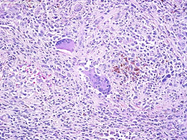
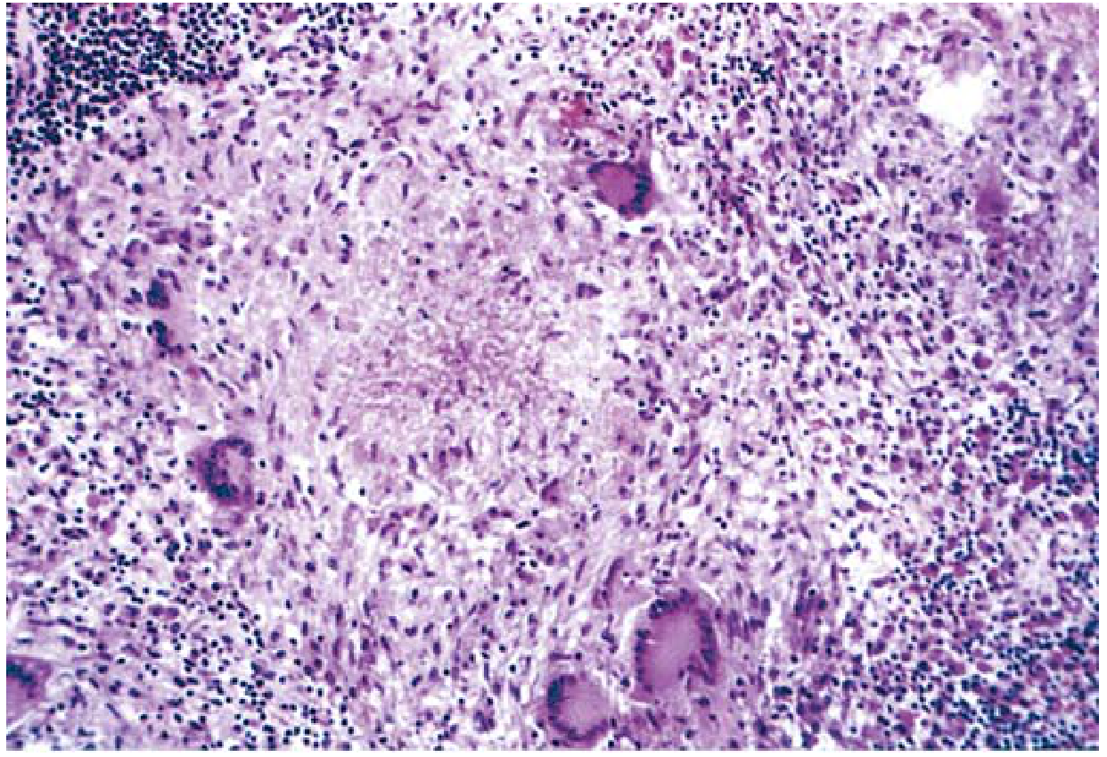
This is a light microscopy image of a tissue biopsy stained with hematoxylin and eosin (H&E). The slide shows granulomatous inflammation with multinucleated giant cells and scattered hemosiderin-laden macrophages embedded in a dense inflammatory background. Epithelioid histiocytes predominate, forming nodular aggregates with elongated nuclei, abundant cytoplasm, and indistinct cell borders. Multinucleated giant cells are evident within the granulomas, sometimes with a peripheric arrangement suggestive of Langhans-type morphology. Intermixed small lymphocytes and plasma cells contribute to the chronic inflammatory milieu. Brown to golden-brown hemosiderin pigment within macrophages is conspicuous and suggests prior hemorrhage or vascular congestion; occasional clusters of iron-laden macrophages are dispersed in the interstitium. Mild edema and vascular congestion may be present; necrosis is not a prominent feature in this field. The tissue architecture reflects organized granulomatous reaction rather than acute suppurative inflammation. No overt microbial organisms are visible at this magnification; however, special stains or molecular tests are required to identify potential infectious etiologies. Clinically, these findings raise differential diagnoses including mycobacterial and fungal infections, sarcoidosis, foreign body granulomatous reaction, or hemorrhagic granulomatous response. This image is valuable for diagnostic pathology review, education on granulomatous histology, and research into macrophage activation and iron handling in chronic inflammation process.
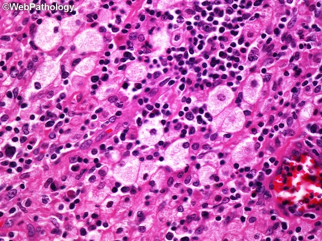
Light microscopy on hematoxylin and eosin (H&E) stained renal tissue illustrating the characteristic xanthogranulomatous pyelonephritis pattern. Renal parenchyma with focal involvement of the renal pelvis and calyces, reflecting chronic suppurative infection of the kidney. The image shows numerous foamy (lipid-laden) histiocytes arranged in aggregates within a background of mixed chronic inflammatory infiltrate comprising lymphocytes and plasma cells; scattered multinucleated giant cells are present with areas of necrosis and fibrous scarring. Lipid-rich macrophages impart a yellow-white appearance at gross inspection, corresponding to the xanthogranulomatous reaction. Additional features may include cholesterol clefts, dilated tubules, and focal nephron destruction; conspicuous vascular congestion and pigment deposition are not prominent. This histology is highly suggestive of Xanthogranulomatous Pyelonephritis; differential considerations include malakoplakia and renal abscess, but the combination of foamy histiocytes with granulomatous inflammation is characteristic. Clinical correlation with chronic obstruction, nephrolithiasis, and infection by uropathogens such as Proteus or Escherichia coli supports diagnosis and informs management, which frequently involves nephrectomy. Clinically, this pattern aids decision-making in chronic kidney infection workups, educational histology resources, and radiologic-pathologic correlation studies for renal masses and obstructive disease. This specimen illustrates the diagnostic hallmark of XGP and underscores the importance of correlating gross appearance with microscopic granulomatous lipid-laden macrophages for accurate pathology reporting.
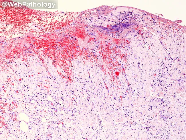
This histopathology image depicts gallbladder wall tissue from chronic calculous cholecystitis evaluated by light microscopy after Hematoxylin and Eosin (H&E) staining. The mucosal lining shows variability in integrity with focal atrophy and scarring, while the underlying layers display transmural chronic inflammation involving the mucosa, muscularis propria, and extending toward the serosa. The inflammatory infiltrate is dominated by lymphocytes and plasma cells, with macrophages and rare neutrophils, consistent with a long-standing inflammatory process. Fibrosis and architectural distortion of the gallbladder wall are evident, reflecting chronic injury and healing. The serosal surface demonstrates hemorrhagic exudate, indicating serositis and superimposed vascular leakage. Although gallstones are classically associated with chronic cholecystitis, this histological field focuses on wall pathology and may not clearly display calculi. These features help distinguish chronic cholecystitis from acute inflammatory changes, which would show predominant neutrophilic infiltration and edema, and from cholesterolosis or adenomyomatosis, which feature mucosal lipid-laden macrophages or mucosal gland proliferation, respectively. Clinically, such histology correlates with chronic biliary pain, prior gallstone disease, cholecystitis risk for gallbladder dysfunction, and potential complications such as cholestasis or gallbladder empyema if untreated. This image is valuable for pathology education, differential diagnosis practice, and radiologic–pathologic correlation in hepatobiliary diseases.
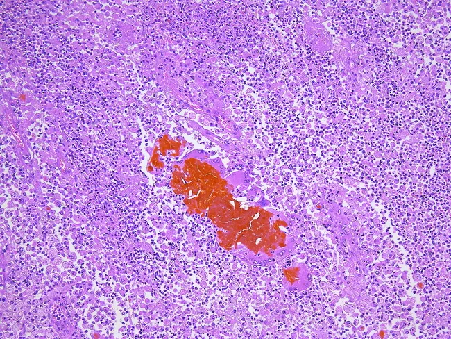
Imaging modality: bright-field light microscopy of a hematoxylin and eosin–stained gallbladder wall section, illustrating xanthogranulomatous inflammation. Anatomy: gallbladder wall with mucosa, submucosa, muscularis, and Rokitansky-Aschoff sinuses. Visual features: dense inflammatory infiltrate composed of lipid-laden foamy macrophages (xanthomatous histiocytes), plasma cells, lymphocytes, scattered neutrophils, and multinucleated giant cells; cholesterol clefts and bile pigments embedded within fibrotic stroma; focal ulceration and scattered calcifications are possible; perivascular and interstitial distribution with intralesional granulomas. Pathology: xanthogranulomatous cholecystitis (XC) characterized by foam cells, cholesterol crystallization, cholesterol clefts, foreign-body-type granulomas, and bile extravasation into subepithelial mesenchyme that provokes a vigorous inflammatory response; secondary bacterial superinfection may contribute. Diagnostic significance: essential to differentiate XC from gallbladder neoplasia; recognition prevents misdiagnosis of carcinoma and guides surgical management; histology may reflect chronic biliary obstruction and pigment deposition. Differential considerations: chronic cholecystitis without XC; cholesterolosis; adenomyomatosis; gallbladder adenocarcinoma or other gallbladder neoplasms with desmoplasia; infectious cholecystitis. Clinical correlation: commonly accompanies gallstones and chronic biliary disease; post-surgical histology confirms diagnosis and influences prognosis, follow-up, and patient counseling. Potential educational uses: pathology atlas references, diagnostic pattern recognition, and research into inflammatory gallbladder diseases. This image exemplifies XC histology for teaching, reference libraries, and computational indexing.
"chronic inflammation" pathology mechanisms
chronic inflammation fibrosis tissue repair lymphocyte plasma cell histology
Chronic Inflammation - Pathology
Definition
- Robbins & Kumar Basic Pathology (Robbins Pathology), p. 61
- Robbins, Cotran & Kumar Pathologic Basis of Disease, p. 103
Causes
| Category | Examples |
|---|---|
| Persistent infections | Mycobacteria (TB), certain viruses, fungi, parasites - difficult to eradicate; evoke delayed-type hypersensitivity |
| Hypersensitivity/Immune diseases | Autoimmune diseases (rheumatoid arthritis, multiple sclerosis, IBD); allergic diseases (bronchial asthma) |
| Toxic/exogenous agents | Silica dust (silicosis) |
| Endogenous agents | Excess cholesterol deposition driving atherosclerosis |
| Metabolic/neurodegenerative | Alzheimer disease, metabolic syndrome, type 2 diabetes (role of low-grade chronic inflammation) |
Morphologic Features (The Classic Triad)
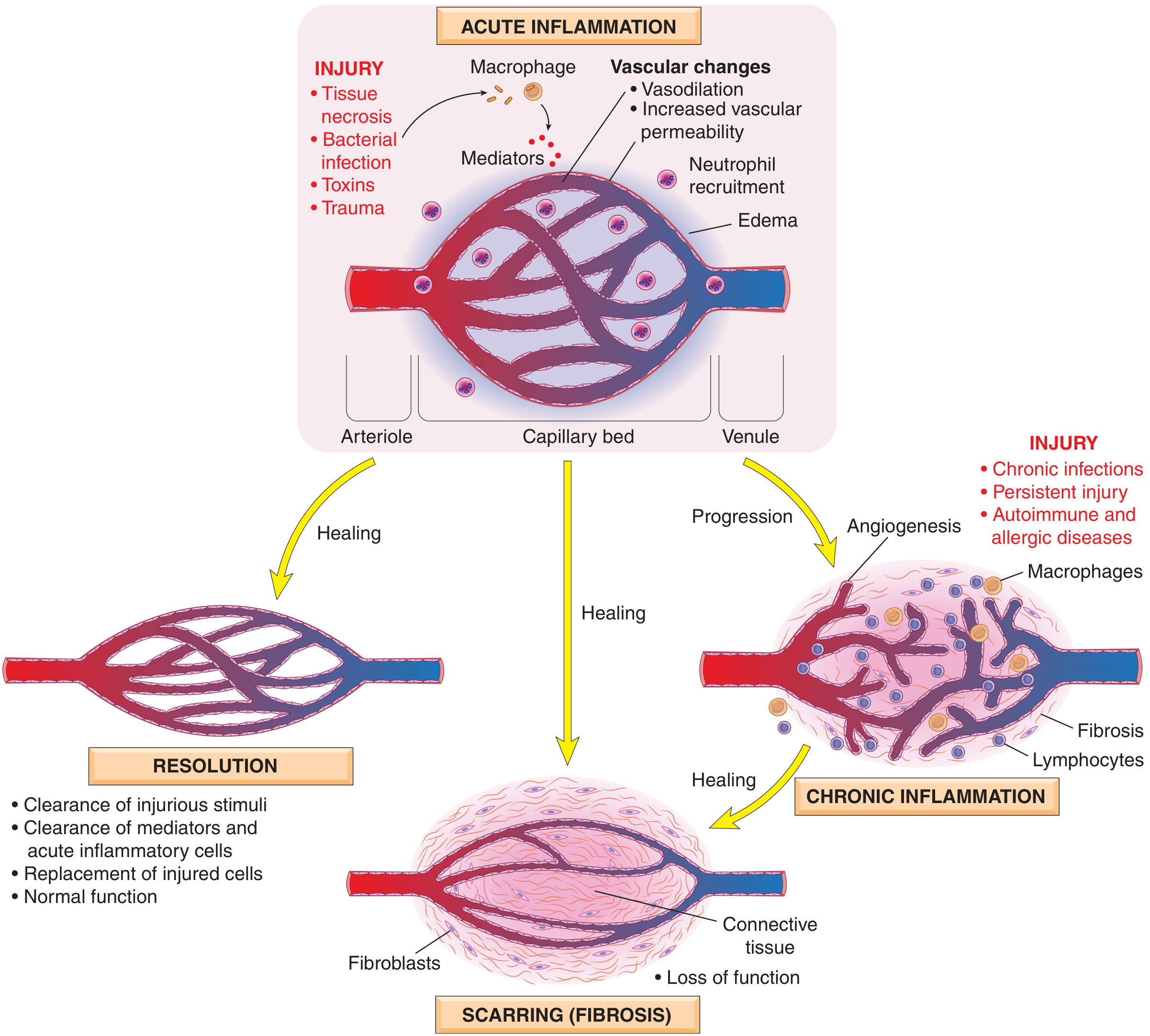
- Mononuclear cell infiltration - macrophages, lymphocytes, and plasma cells (rather than neutrophils)
- Tissue destruction - induced by the persistent offending agent or by the inflammatory cells themselves
- Attempts at healing - by connective tissue replacement via angiogenesis and fibrosis
Cells and Mediators
1. Macrophages (dominant cells)
| Activation Pathway | Stimulus | Actions |
|---|---|---|
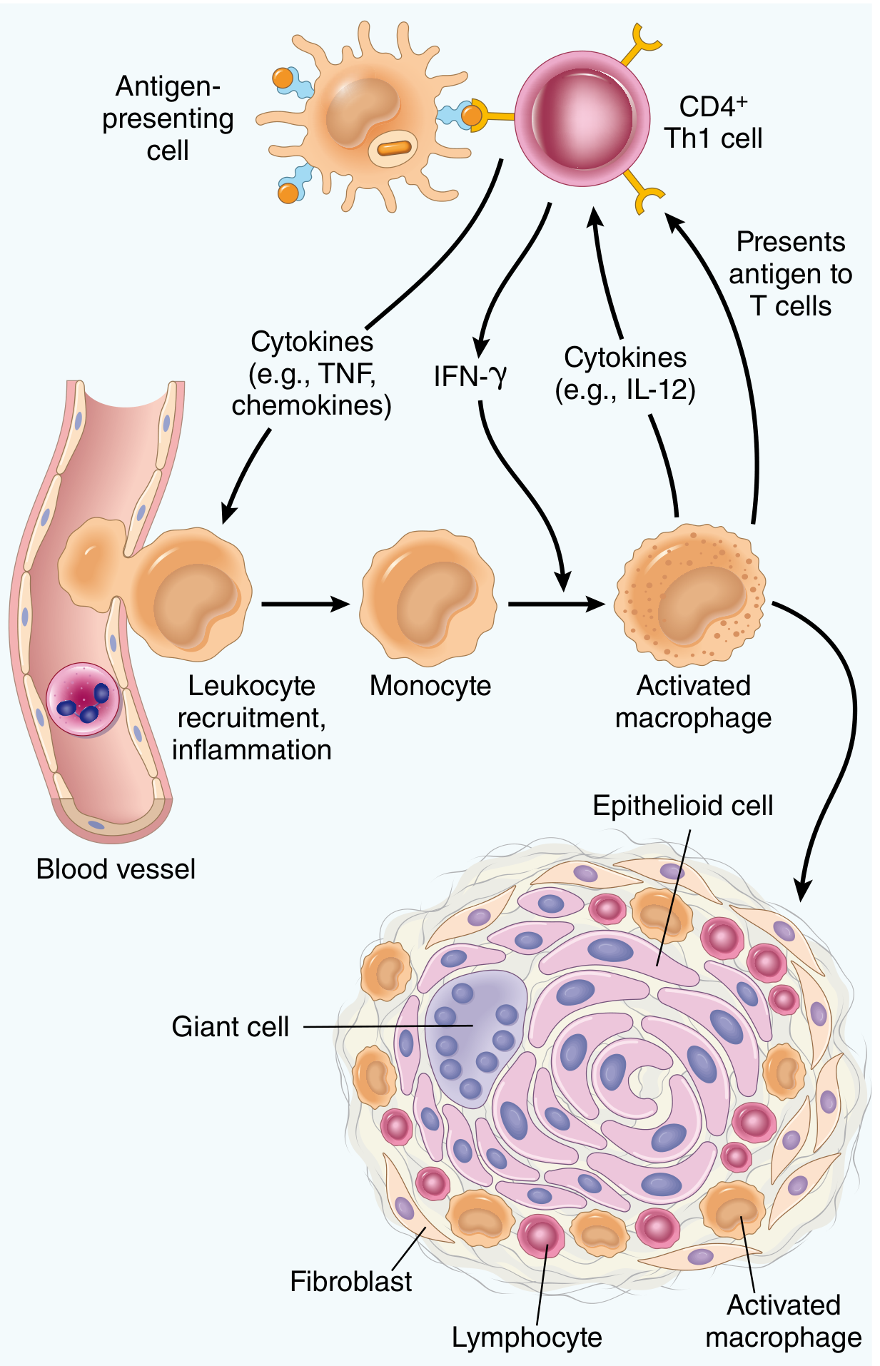
| Classical activation (M1) | Microbial products, IFN-γ | Phagocytose/kill microbes and dead tissue; secrete TNF, IL-1, IL-12, ROS, NO; can cause tissue damage |
| Alternative activation (M2) | IL-4, IL-13 | Tissue repair, anti-inflammatory, stimulate fibrosis via TGF-β |
2. Lymphocytes
| Subset | Cytokine secreted | Effect |
|---|---|---|
| Th1 | IFN-γ | Classical macrophage activation |
| Th2 | IL-4, IL-5, IL-13 | Alternative macrophage activation; eosinophil recruitment; IgE |
| Th17 | IL-17 | Neutrophil (and monocyte) recruitment via chemokines |
- Th1 and Th17 drive autoimmune diseases (RA, psoriasis, IBD)
- Th2 drives helminthic defense and allergic inflammation
3. Other Cells
- Eosinophils: Abundant in Th2-driven reactions (parasitic infections, allergies). Contain major basic protein - toxic to helminths but also injures host epithelium
- Mast cells: Express FcεRI (IgE receptor); involved in allergic and chronic inflammatory reactions
- Neutrophils: May persist in chronic reactions driven by bacterial infections or Th17 responses
Granulomatous Inflammation
Two Types:
| Type | Pathogenesis | Examples |
|---|---|---|
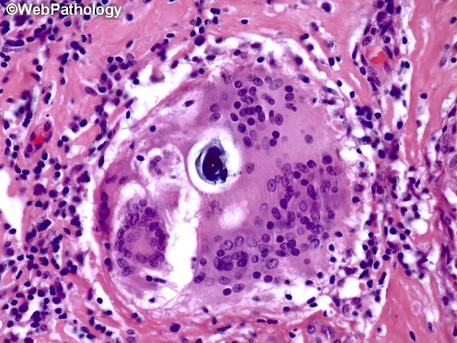
| Foreign body granulomas | Reaction to inert, non-immunogenic material too large to be phagocytosed; no T cell-mediated response | Talc (IV drug use), sutures, fibers - foreign material often visible under polarized light |
| Immune granulomas | Persistent T cell-mediated (Th1) immune response; IFN-γ drives macrophage activation | TB, sarcoidosis, Crohn disease, syphilis, fungal infections, schistosomiasis (Th2/eosinophils) |
Histologic Features (H&E):
- Epithelioid cells: Activated macrophages with pink granular cytoplasm and indistinct cell borders (resemble epithelium)
- Langhans giant cells: 40-50 μm multinucleated giant cells formed by fusion of multiple activated macrophages; nuclei arranged at periphery in horseshoe pattern
- Collar of lymphocytes surrounding the macrophage aggregate
- Rim of fibroblasts and connective tissue in older granulomas
- Caseous necrosis: Central zone of necrosis (amorphous, structureless, eosinophilic debris) - characteristic of M. tuberculosis infection; caused by hypoxia + free radical injury
- Noncaseating granulomas: No necrotic center - seen in Crohn disease, sarcoidosis, foreign body reactions
Diseases With Granulomatous Inflammation:
| Disease | Key Feature |
|---|---|
| Tuberculosis | Caseating tubercles; prototype; always exclude first |
| Sarcoidosis | Noncaseating; Schaumann bodies, asteroid bodies |
| Crohn disease | Noncaseating; in bowel wall |
| Fungal infections | (Histoplasma, Coccidioides) |
| Syphilis | Gummas |
| Schistosomiasis | Th2/eosinophil-associated |
| Foreign body | Talc, sutures |
Clinically important: Recognition of granulomas is significant because the differential is relatively limited, some causes are life-threatening (TB), and identification guides workup - special stains (acid-fast for TB), cultures, PCR, and serology.
Systemic Effects of Chronic Inflammation
| Manifestation | Mechanism |
|---|---|
| Fever | IL-1 and TNF (endogenous pyrogens) → upregulate cyclooxygenase → PGE2 in hypothalamus → raises temperature setpoint |
| Leukocytosis | Cytokine-driven accelerated release of granulocytes from bone marrow; shift to the left; lymphocytosis in viral infections; eosinophilia in parasites/allergy |
| Acute-phase response | Liver synthesizes acute-phase proteins (CRP, SAA, fibrinogen) - CRP acts as opsonin; SAA is precursor of AA amyloid; fibrinogen increases ESR |
| Weight loss / cachexia | TNF and IL-1 mobilize fats and proteins; suppress appetite |
Outcome of Chronic Inflammation
- Fibrosis and scarring - the most common end result, causing loss of function (e.g., hepatic cirrhosis, pulmonary fibrosis, renal scarring)
- Resolution - if the causative agent is eliminated and repair is effective
- Persistence and disease - autoimmune or unresolvable stimulus leads to ongoing tissue destruction
Recent Evidence (2025)
Prepare a otee for me in pdf
Prepare a note for me in pdf
ls -la /tmp/workspace/ 2>/dev/null || mkdir -p /tmp/workspace && echo "workspace ready"
mkdir -p /tmp/workspace/chronic-inflammation-pdf && python3 << 'PYEOF' from reportlab.lib.pagesizes import A4 from reportlab.lib import colors from reportlab.lib.styles import getSampleStyleSheet, ParagraphStyle from reportlab.lib.units import cm, mm from reportlab.platypus import (SimpleDocTemplate, Paragraph, Spacer, Table, TableStyle, HRFlowable, KeepTogether, ListFlowable, ListItem) from reportlab.lib.enums import TA_LEFT, TA_CENTER, TA_JUSTIFY from reportlab.platypus import PageBreak OUTPUT = "/tmp/workspace/chronic-inflammation-pdf/Chronic_Inflammation_Pathology.pdf" doc = SimpleDocTemplate( OUTPUT, pagesize=A4, rightMargin=2*cm, leftMargin=2*cm, topMargin=2.5*cm, bottomMargin=2*cm, title="Chronic Inflammation – Pathology Notes", author="Orris Medical Library" ) W, H = A4 styles = getSampleStyleSheet() # ── Custom Styles ────────────────────────────────────────────────── BLUE = colors.HexColor("#1a3a5c") ACCENT = colors.HexColor("#2e7bcf") LIGHT_BG = colors.HexColor("#eaf3fb") TEAL = colors.HexColor("#1a6b6b") GREEN_BG = colors.HexColor("#e8f6f3") YELLOW_BG = colors.HexColor("#fffbe6") RED_SOFT = colors.HexColor("#c0392b") GRAY_LINE = colors.HexColor("#bdc3c7") title_style = ParagraphStyle("Title2", parent=styles["Title"], fontSize=22, textColor=BLUE, spaceAfter=4, alignment=TA_CENTER, fontName="Helvetica-Bold") subtitle_style = ParagraphStyle("Sub", parent=styles["Normal"], fontSize=11, textColor=ACCENT, spaceAfter=12, alignment=TA_CENTER, fontName="Helvetica-Oblique") h1_style = ParagraphStyle("H1", parent=styles["Heading1"], fontSize=14, textColor=colors.white, spaceAfter=4, spaceBefore=14, fontName="Helvetica-Bold", backColor=BLUE, leftIndent=-8, rightIndent=-8, borderPad=6) h2_style = ParagraphStyle("H2", parent=styles["Heading2"], fontSize=12, textColor=BLUE, spaceAfter=3, spaceBefore=10, fontName="Helvetica-Bold", borderPad=2, borderWidth=0, leftIndent=0) h3_style = ParagraphStyle("H3", parent=styles["Heading3"], fontSize=11, textColor=TEAL, spaceAfter=2, spaceBefore=6, fontName="Helvetica-Bold") body_style = ParagraphStyle("Body2", parent=styles["Normal"], fontSize=10, leading=15, spaceAfter=4, alignment=TA_JUSTIFY, fontName="Helvetica") bullet_style = ParagraphStyle("Bullet2", parent=styles["Normal"], fontSize=10, leading=14, spaceAfter=2, leftIndent=16, bulletIndent=4, fontName="Helvetica") note_style = ParagraphStyle("Note", parent=styles["Normal"], fontSize=9, leading=13, spaceAfter=2, fontName="Helvetica-Oblique", textColor=colors.HexColor("#555555")) bold_body = ParagraphStyle("BoldBody", parent=body_style, fontName="Helvetica-Bold", textColor=BLUE) important_style = ParagraphStyle("Important", parent=styles["Normal"], fontSize=10, leading=14, fontName="Helvetica-Bold", backColor=YELLOW_BG, textColor=RED_SOFT, borderPad=6, leftIndent=8, rightIndent=8, spaceAfter=6, spaceBefore=4) def h1(text): return Paragraph(f" {text}", h1_style) def h2(text): return Paragraph(text, h2_style) def h3(text): return Paragraph(text, h3_style) def body(text): return Paragraph(text, body_style) def note(text): return Paragraph(text, note_style) def sp(h=6): return Spacer(1, h) def hr(): return HRFlowable(width="100%", thickness=0.5, color=GRAY_LINE, spaceAfter=4, spaceBefore=4) def bullet(items): return ListFlowable( [ListItem(Paragraph(i, bullet_style), bulletColor=ACCENT, leftIndent=16) for i in items], bulletType='bullet', bulletFontSize=8, leftIndent=0 ) def table(data, col_widths, header=True): t = Table(data, colWidths=col_widths, repeatRows=1 if header else 0) style_cmds = [ ('BACKGROUND', (0,0), (-1,0), ACCENT if header else colors.white), ('TEXTCOLOR', (0,0), (-1,0), colors.white if header else colors.black), ('FONTNAME', (0,0), (-1,0), 'Helvetica-Bold'), ('FONTSIZE', (0,0), (-1,-1), 9), ('ROWBACKGROUNDS', (0,1), (-1,-1), [colors.white, LIGHT_BG]), ('GRID', (0,0), (-1,-1), 0.4, GRAY_LINE), ('LEFTPADDING', (0,0), (-1,-1), 6), ('RIGHTPADDING', (0,0), (-1,-1), 6), ('TOPPADDING', (0,0), (-1,-1), 4), ('BOTTOMPADDING', (0,0), (-1,-1), 4), ('VALIGN', (0,0), (-1,-1), 'TOP'), ('WORDWRAP', (0,0), (-1,-1), True), ] t.setStyle(TableStyle(style_cmds)) return t def info_box(text, bg=GREEN_BG, border_color=TEAL): data = [[Paragraph(text, ParagraphStyle("IB", parent=body_style, backColor=bg, fontSize=9.5, leading=14, leftIndent=4))]] t = Table(data, colWidths=[doc.width]) t.setStyle(TableStyle([ ('BACKGROUND', (0,0), (-1,-1), bg), ('LEFTPADDING', (0,0), (-1,-1), 10), ('RIGHTPADDING', (0,0), (-1,-1), 10), ('TOPPADDING', (0,0), (-1,-1), 7), ('BOTTOMPADDING', (0,0), (-1,-1), 7), ('LINEWIDTH', (0,0), (0,-1), 3), ('LINECOLOR', (0,0), (0,-1), border_color), ])) return t # ══════════════════════════════════════════════════════════════════ story = [] # TITLE PAGE BLOCK story += [ sp(20), Paragraph("CHRONIC INFLAMMATION", title_style), Paragraph("Pathology Notes · Based on Robbins & Kumar", subtitle_style), HRFlowable(width="80%", thickness=2, color=ACCENT, spaceAfter=4, spaceBefore=4), sp(4), Paragraph("Robbins & Kumar Basic Pathology | Robbins, Cotran & Kumar Pathologic Basis of Disease", note_style.__class__("NC", parent=note_style, alignment=TA_CENTER, fontSize=9, textColor=colors.HexColor("#888888"))), sp(30), ] # ── 1. DEFINITION ────────────────────────────────────────────────── story += [h1("1. DEFINITION"), sp(6)] story.append(body( "Chronic inflammation is a response of <b>prolonged duration (weeks to months)</b> in which " "inflammation, tissue injury, and attempts at repair coexist simultaneously, in varying combinations. " "It may follow unresolved acute inflammation, <i>or</i> it may begin insidiously as a smoldering " "process <b>without</b> a preceding acute reaction." )) story.append(sp(4)) story.append(info_box( "<b>Key concept:</b> Unlike acute inflammation (dominated by vascular changes, edema, neutrophils), " "chronic inflammation involves mononuclear cell infiltration, tissue destruction, and concurrent attempts " "at repair — all three processes coexisting at the same time.", bg=LIGHT_BG, border_color=ACCENT )) # ── 2. CAUSES ────────────────────────────────────────────────────── story += [sp(8), h1("2. CAUSES OF CHRONIC INFLAMMATION"), sp(6)] causes_data = [ [Paragraph("<b>Category</b>", bullet_style), Paragraph("<b>Examples</b>", bullet_style)], [Paragraph("Persistent infections", bullet_style), Paragraph("Mycobacteria (TB), viruses, fungi, parasites — difficult to eradicate; evoke T-cell (delayed-type hypersensitivity) responses", bullet_style)], [Paragraph("Hypersensitivity / Immune diseases", bullet_style), Paragraph("Autoimmune: rheumatoid arthritis, multiple sclerosis, IBD; Allergic: bronchial asthma", bullet_style)], [Paragraph("Exogenous toxic agents", bullet_style), Paragraph("Inhaled silica → silicosis (nondegradable particulate)", bullet_style)], [Paragraph("Endogenous toxic agents", bullet_style), Paragraph("Excess cholesterol deposition → atherosclerosis", bullet_style)], [Paragraph("Metabolic / Neurodegenerative", bullet_style), Paragraph("Alzheimer disease, metabolic syndrome, type 2 diabetes (low-grade chronic inflammation)", bullet_style)], ] story.append(table(causes_data, [5.5*cm, 11.5*cm])) # ── 3. MORPHOLOGY ────────────────────────────────────────────────── story += [sp(10), h1("3. MORPHOLOGIC FEATURES — THE CLASSIC TRIAD"), sp(6)] story.append(body("Three processes coexist and define chronic inflammation:")) story.append(sp(4)) triad_data = [ [Paragraph("<b>#</b>", bullet_style), Paragraph("<b>Feature</b>", bullet_style), Paragraph("<b>Details</b>", bullet_style)], [Paragraph("1", bullet_style), Paragraph("<b>Mononuclear cell infiltration</b>", bullet_style), Paragraph("Macrophages, lymphocytes, plasma cells (NOT neutrophils as in acute)", bullet_style)], [Paragraph("2", bullet_style), Paragraph("<b>Tissue destruction</b>", bullet_style), Paragraph("Caused by the persistent agent or by the inflammatory cells themselves", bullet_style)], [Paragraph("3", bullet_style), Paragraph("<b>Attempts at healing</b>", bullet_style), Paragraph("Angiogenesis + fibrosis (connective tissue replacement of damaged tissue)", bullet_style)], ] story.append(table(triad_data, [1.2*cm, 5.5*cm, 10.3*cm])) # ── 4. CELLS ─────────────────────────────────────────────────────── story += [sp(10), h1("4. CELLS AND MEDIATORS"), sp(6)] # Macrophages story += [h2("4.1 Macrophages (Dominant Cells)"), sp(3)] story.append(body( "Macrophages are the principal effector cells of chronic inflammation, derived from blood monocytes " "(circulating monocytes → tissue macrophages). They are activated via two distinct pathways:" )) story.append(sp(4)) mac_data = [ [Paragraph("<b>Pathway</b>", bullet_style), Paragraph("<b>Stimulus</b>", bullet_style), Paragraph("<b>Actions</b>", bullet_style)], [Paragraph("Classical (M1)", bullet_style), Paragraph("Microbial products, IFN-γ", bullet_style), Paragraph("Phagocytose/kill microbes; secrete TNF, IL-1, IL-12, ROS, NO; <b>tissue damage</b>", bullet_style)], [Paragraph("Alternative (M2)", bullet_style), Paragraph("IL-4, IL-13 (Th2 cytokines)", bullet_style), Paragraph("Tissue repair; anti-inflammatory; stimulate fibrosis via TGF-β", bullet_style)], ] story.append(table(mac_data, [3.5*cm, 5*cm, 8.5*cm])) story.append(sp(4)) story.append(body( "Macrophage functions: <b>(a)</b> ingest/destroy microbes & debris; <b>(b)</b> secrete cytokines " "(TNF, IL-1, IL-6, chemokines, eicosanoids); <b>(c)</b> initiate tissue repair/fibrosis; " "<b>(d)</b> present antigens to T lymphocytes — setting up the critical macrophage-lymphocyte feedback loop." )) # Lymphocytes story += [sp(6), h2("4.2 Lymphocytes"), sp(3)] story.append(body( "T and B lymphocytes amplify and perpetuate chronic inflammation. Their activation generates <b>long-lived " "memory cells</b>, explaining why chronic inflammatory reactions are so persistent and severe." )) story.append(sp(4)) thelper_data = [ [Paragraph("<b>Subset</b>", bullet_style), Paragraph("<b>Key Cytokine</b>", bullet_style), Paragraph("<b>Effect</b>", bullet_style), Paragraph("<b>Diseases</b>", bullet_style)], [Paragraph("Th1", bullet_style), Paragraph("IFN-γ", bullet_style), Paragraph("Classical macrophage activation (M1)", bullet_style), Paragraph("RA, IBD, psoriasis, autoimmunity", bullet_style)], [Paragraph("Th2", bullet_style), Paragraph("IL-4, IL-5, IL-13", bullet_style), Paragraph("Alternative macrophage activation; eosinophil recruitment; IgE production", bullet_style), Paragraph("Helminth infection, allergic inflammation", bullet_style)], [Paragraph("Th17", bullet_style), Paragraph("IL-17", bullet_style), Paragraph("Chemokines → neutrophil/monocyte recruitment", bullet_style), Paragraph("RA, psoriasis, IBD, autoimmunity", bullet_style)], ] story.append(table(thelper_data, [1.8*cm, 3*cm, 6*cm, 6.2*cm])) story.append(sp(4)) story.append(info_box( "<b>Bidirectional macrophage–lymphocyte loop:</b> Macrophages present antigens to T cells + secrete IL-12 " "→ T cells produce IFN-γ + chemokines → further macrophage activation → more antigen presentation. " "This self-perpetuating cycle fuels and sustains chronic inflammation.", bg=GREEN_BG, border_color=TEAL )) story.append(sp(3)) story.append(body( "<b>Plasma cells:</b> Activated B lymphocytes differentiate into plasma cells that produce antibodies " "against persistent foreign/self antigens at the site of inflammation." )) story.append(sp(3)) story.append(body( "<b>Tertiary lymphoid structures:</b> In prolonged reactions (e.g., RA synovium, Hashimoto thyroiditis), " "accumulated lymphocytes and APCs organize into follicle-like structures resembling lymph nodes, potentially " "perpetuating the local immune response." )) # Other cells story += [sp(6), h2("4.3 Other Cells"), sp(3)] other_data = [ [Paragraph("<b>Cell</b>", bullet_style), Paragraph("<b>Stimulus</b>", bullet_style), Paragraph("<b>Role / Key Mediator</b>", bullet_style)], [Paragraph("Eosinophils", bullet_style), Paragraph("Th2/eotaxin", bullet_style), Paragraph("<b>Major basic protein</b> — toxic to helminths; also injures host epithelium in allergy", bullet_style)], [Paragraph("Mast cells", bullet_style), Paragraph("IgE (FcεRI receptor)", bullet_style), Paragraph("Degranulate → histamine, prostaglandins; allergic and chronic inflammatory reactions", bullet_style)], [Paragraph("Neutrophils", bullet_style), Paragraph("Bacterial / Th17", bullet_style), Paragraph("Persist in certain chronic bacterial infections and Th17-driven reactions", bullet_style)], ] story.append(table(other_data, [3.2*cm, 3.5*cm, 10.3*cm])) # ── 5. GRANULOMATOUS INFLAMMATION ────────────────────────────────── story += [PageBreak(), h1("5. GRANULOMATOUS INFLAMMATION"), sp(6)] story.append(info_box( "<b>Definition:</b> A specialized form of chronic inflammation characterized by collections of " "<b>activated macrophages (epithelioid cells)</b>, often with T lymphocytes, and sometimes associated " "with central necrosis. It represents a cellular attempt to contain an offending agent that is difficult to eradicate.", bg=YELLOW_BG, border_color=RED_SOFT )) story.append(sp(6)) story += [h2("5.1 Two Types of Granulomas"), sp(3)] gran_data = [ [Paragraph("<b>Type</b>", bullet_style), Paragraph("<b>Pathogenesis</b>", bullet_style), Paragraph("<b>Examples</b>", bullet_style)], [Paragraph("Foreign body granuloma", bullet_style), Paragraph("Reaction to inert, non-immunogenic material too large to phagocytose; NO T-cell response", bullet_style), Paragraph("Talc (IV drug use), suture material, fibres — foreign material visible under polarized light", bullet_style)], [Paragraph("Immune granuloma", bullet_style), Paragraph("Persistent T cell-mediated (Th1) immune response; IFN-γ drives macrophage activation", bullet_style), Paragraph("TB, sarcoidosis, Crohn disease, syphilis, fungal infections, schistosomiasis (Th2/eosinophil)", bullet_style)], ] story.append(table(gran_data, [4*cm, 6*cm, 7*cm])) story += [sp(6), h2("5.2 Histologic Features (H&E)"), sp(3)] story.append(bullet([ "<b>Epithelioid cells:</b> Activated macrophages with pink granular cytoplasm and indistinct cell borders (resemble epithelium — hence the name)", "<b>Langhans giant cells:</b> 40–50 μm multinucleated giant cells formed by fusion of multiple activated macrophages; nuclei arranged at periphery in horseshoe/ring pattern", "<b>Collar of lymphocytes</b> surrounding the epithelioid macrophage aggregate", "<b>Rim of fibroblasts and connective tissue</b> in older/healing granulomas", "<b>Caseous necrosis:</b> Central zone of amorphous, structureless, eosinophilic debris — characteristic of <i>M. tuberculosis</i>; caused by hypoxia + free radical injury", "<b>Noncaseating granulomas:</b> No necrotic centre — seen in Crohn disease, sarcoidosis, foreign body reactions", ])) story.append(sp(6)) story += [h2("5.3 Diseases With Granulomatous Inflammation"), sp(3)] disease_data = [ [Paragraph("<b>Disease</b>", bullet_style), Paragraph("<b>Key Feature</b>", bullet_style), Paragraph("<b>Notes</b>", bullet_style)], [Paragraph("Tuberculosis", bullet_style), Paragraph("Caseating (tubercles)", bullet_style), Paragraph("<b>Prototype</b> — always exclude first; acid-fast stain", bullet_style)], [Paragraph("Sarcoidosis", bullet_style), Paragraph("Noncaseating", bullet_style), Paragraph("Schaumann bodies, asteroid bodies; serum ACE elevated", bullet_style)], [Paragraph("Crohn disease", bullet_style), Paragraph("Noncaseating", bullet_style), Paragraph("In bowel wall; transmural inflammation", bullet_style)], [Paragraph("Fungal infections", bullet_style), Paragraph("Variable", bullet_style), Paragraph("Histoplasma, Coccidioides; special stains, culture", bullet_style)], [Paragraph("Syphilis", bullet_style), Paragraph("Gummas", bullet_style), Paragraph("Central necrosis; serologic diagnosis", bullet_style)], [Paragraph("Schistosomiasis", bullet_style), Paragraph("Th2/eosinophil-associated", bullet_style), Paragraph("Granulomas around schistosome eggs", bullet_style)], [Paragraph("Foreign body", bullet_style), Paragraph("Non-immune", bullet_style), Paragraph("Talc, sutures; birefringent material on polarized light", bullet_style)], ] story.append(table(disease_data, [3.8*cm, 4*cm, 9.2*cm])) story.append(sp(6)) story.append(info_box( "<b>Clinical importance:</b> Recognition of granulomas is significant because the differential is " "relatively limited and some causes are life-threatening. Always exclude TB first. " "Workup includes: acid-fast stains, cultures, PCR, serologic studies.", bg=YELLOW_BG, border_color=RED_SOFT )) story.append(sp(4)) story.append(body( "Healing of granulomas is accompanied by <b>extensive fibrosis</b> in involved organs, which can lead to " "significant loss of function." )) # ── 6. SYSTEMIC EFFECTS ──────────────────────────────────────────── story += [sp(10), h1("6. SYSTEMIC EFFECTS OF INFLAMMATION"), sp(6)] story.append(body( "Even localized chronic inflammation produces systemic effects through cytokine release — principally " "<b>TNF, IL-1, and IL-6</b> (the 'triad of systemic mediators')." )) story.append(sp(6)) sys_data = [ [Paragraph("<b>Manifestation</b>", bullet_style), Paragraph("<b>Mechanism</b>", bullet_style)], [Paragraph("<b>Fever</b>", bullet_style), Paragraph("IL-1 and TNF (endogenous pyrogens) → upregulate COX → PGE2 in hypothalamus → raises temperature set-point. NSAIDs reduce fever by inhibiting prostaglandin synthesis.", bullet_style)], [Paragraph("<b>Leukocytosis</b>", bullet_style), Paragraph("Cytokine-driven accelerated granulocyte release from bone marrow; 'shift to the left' (immature neutrophils). Neutrophilia (bacteria), lymphocytosis (viral), eosinophilia (parasites/allergy), leukopenia (typhoid, rickettsia).", bullet_style)], [Paragraph("<b>Acute-phase response</b>", bullet_style), Paragraph("Liver synthesizes acute-phase proteins: CRP (opsonin), SAA (precursor of AA amyloid), fibrinogen (↑ESR). Plasma levels increase several hundred-fold.", bullet_style)], [Paragraph("<b>Weight loss / Cachexia</b>", bullet_style), Paragraph("TNF and IL-1 mobilize fats and proteins; suppress appetite.", bullet_style)], [Paragraph("<b>Hypotension / Shock</b>", bullet_style), Paragraph("In severe/systemic inflammation — TNF-mediated vasodilation (septic shock).", bullet_style)], ] story.append(table(sys_data, [5*cm, 12*cm])) # ── 7. OUTCOMES ──────────────────────────────────────────────────── story += [sp(10), h1("7. OUTCOMES OF CHRONIC INFLAMMATION"), sp(6)] out_data = [ [Paragraph("<b>Outcome</b>", bullet_style), Paragraph("<b>When / Mechanism</b>", bullet_style), Paragraph("<b>Examples</b>", bullet_style)], [Paragraph("Resolution", bullet_style), Paragraph("If causative agent is eliminated and repair is effective", bullet_style), Paragraph("Mild infections after antibiotic treatment", bullet_style)], [Paragraph("<b>Fibrosis / Scarring</b> (most common)", bullet_style), Paragraph("Persistent damage → connective tissue replacement → loss of function", bullet_style), Paragraph("Hepatic cirrhosis, pulmonary fibrosis, renal scarring", bullet_style)], [Paragraph("Continued tissue destruction", bullet_style), Paragraph("Persistent, unresolvable stimulus (autoimmune, undrained abscess)", bullet_style), Paragraph("RA joint destruction, ongoing IBD", bullet_style)], [Paragraph("Neoplastic transformation", bullet_style), Paragraph("Chronic mucosal inflammation → dysplasia → carcinoma", bullet_style), Paragraph("Gastric carcinoma (H. pylori), colorectal carcinoma (IBD)", bullet_style)], ] story.append(table(out_data, [4.5*cm, 6.5*cm, 6*cm])) # ── 8. COMPARISON TABLE ──────────────────────────────────────────── story += [sp(10), h1("8. ACUTE vs. CHRONIC INFLAMMATION — COMPARISON"), sp(6)] comp_data = [ [Paragraph("<b>Feature</b>", bullet_style), Paragraph("<b>Acute</b>", bullet_style), Paragraph("<b>Chronic</b>", bullet_style)], [Paragraph("Duration", bullet_style), Paragraph("Minutes to days", bullet_style), Paragraph("Weeks to months (or years)", bullet_style)], [Paragraph("Dominant cells", bullet_style), Paragraph("Neutrophils", bullet_style), Paragraph("Macrophages, lymphocytes, plasma cells", bullet_style)], [Paragraph("Vascular changes", bullet_style), Paragraph("Prominent — vasodilation, edema", bullet_style), Paragraph("Less prominent", bullet_style)], [Paragraph("Tissue injury", bullet_style), Paragraph("Usually mild / reversible", bullet_style), Paragraph("More severe; tissue destruction prominent", bullet_style)], [Paragraph("Fibrosis", bullet_style), Paragraph("Absent", bullet_style), Paragraph("Present (hallmark outcome)", bullet_style)], [Paragraph("Granulomas", bullet_style), Paragraph("Absent", bullet_style), Paragraph("Present in granulomatous type", bullet_style)], [Paragraph("Resolution potential", bullet_style), Paragraph("High", bullet_style), Paragraph("Lower; often ends in fibrosis", bullet_style)], [Paragraph("Systemic effects", bullet_style), Paragraph("Variable", bullet_style), Paragraph("Often present (fever, acute-phase proteins, cachexia)", bullet_style)], ] story.append(table(comp_data, [4.5*cm, 5.5*cm, 7*cm])) # ── FOOTER REFERENCE ─────────────────────────────────────────────── story += [ sp(16), hr(), Paragraph( "<i>Sources: Robbins & Kumar Basic Pathology (Robbins Pathology), pp. 61–67 | " "Robbins, Cotran & Kumar Pathologic Basis of Disease, pp. 103–108 | " "Low-Grade Chronic Inflammation: Cifuentes et al., Physiology 2025 (PMID 39078396)</i>", note_style ), ] # Build doc.build(story) print("PDF created:", OUTPUT) PYEOF
Chronic Inflammation Pathology
PDF Document · PDF
| Section | Content |
|---|---|
| 1. Definition | What chronic inflammation is, how it differs from acute |
| 2. Causes | Persistent infections, hypersensitivity, toxic agents, metabolic |
| 3. Morphologic Features | The classic triad (mononuclear infiltrate, tissue destruction, repair) |
| 4. Cells & Mediators | Macrophages (M1/M2), T-cell subsets (Th1/Th2/Th17), plasma cells, eosinophils, mast cells |
| 5. Granulomatous Inflammation | Types, histologic features, disease table (TB, sarcoid, Crohn, etc.) |
| 6. Systemic Effects | Fever, leukocytosis, acute-phase response, cachexia |
| 7. Outcomes | Resolution, fibrosis, neoplastic transformation |
| 8. Comparison Table | Acute vs. chronic inflammation side-by-side |