

Is this a wart
What's Visible in the Image
1. Row of bumps along the corona (ridge of the glans)
- Not a wart
- Not an STI
- Not contagious
- Not harmful
- No treatment needed (cosmetic removal only if desired)
2. Isolated bump on the penile shaft (just below the corona)
| Possibility | Key Features | Contagious? |
|---|---|---|
| Genital wart (Condyloma acuminatum) | HPV-caused, flesh-colored, can be single or multiple, rough or smooth | Yes (HPV) |
| Molluscum contagiosum | Viral, firm, dome-shaped, often has a central dimple | Yes |
| Skin tag (acrochordon) | Benign overgrowth of skin, soft, painless | No |
| Fordyce spot / sebaceous gland | Small, yellowish, usually multiple | No |
| PPP extension | Part of the normal papule ring | No |
Bottom Line
- The bump has grown, changed shape, or new ones have appeared
- You've had unprotected sexual contact
- It bleeds, itches, or causes pain
- You're unsure about your STI status
Important disclaimer: This is not a medical diagnosis. Only an in-person clinical examination can confirm what this lesion is. Please consult a healthcare professional.
Look on the shaft clearly that is a small bump
genital wart condyloma acuminatum penile shaft small single papule
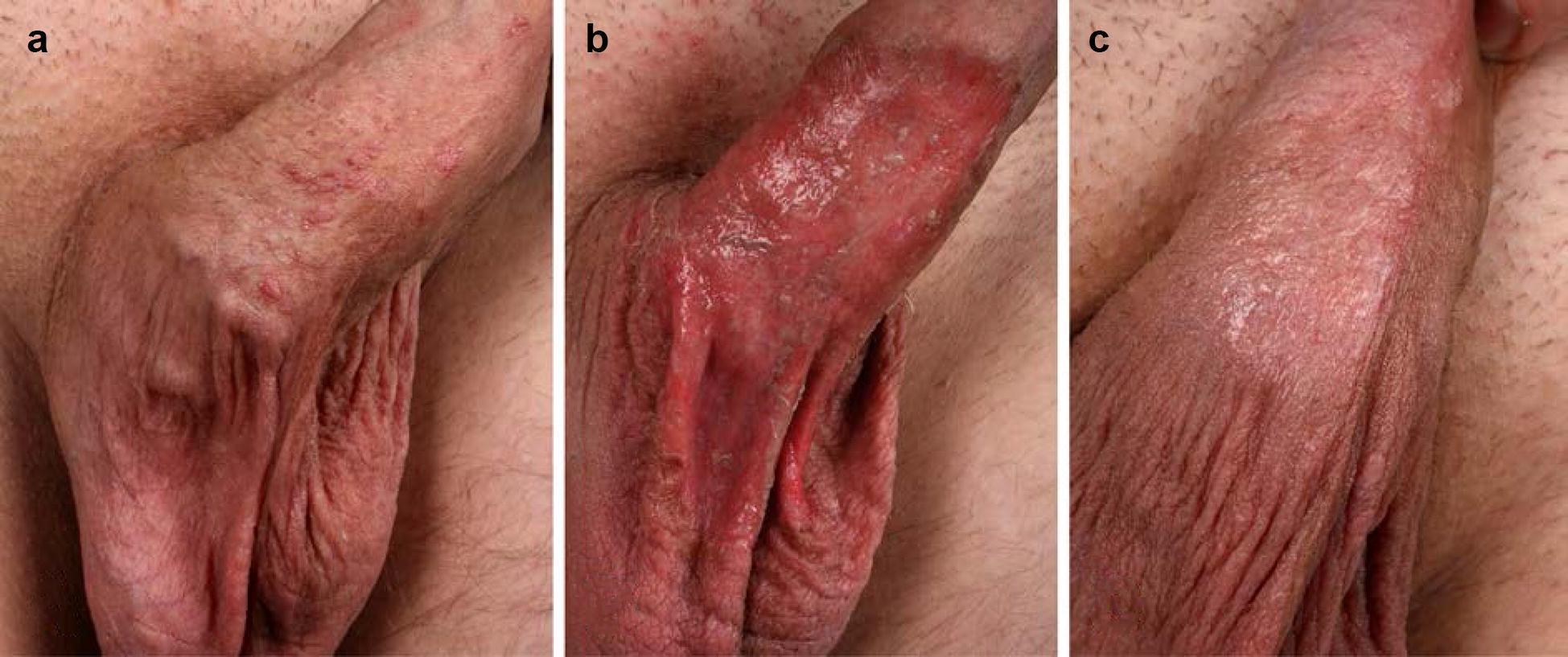
This clinical photograph sequence depicts the therapeutic progression and inflammatory response of anogenital warts (AGW) on the penile shaft treated with ingenol mebutate (IM) 0.05% gel. Panel (a) shows the baseline presentation with multiple, small, discrete, flesh-colored to slightly erythematous raised papules consistent with condyloma acuminatum. Panel (b) illustrates a severe local skin reaction 48 hours post-application, characterized by confluent, extensive erosive dermatitis. The area exhibits intense erythema, superficial tissue desquamation, and moist erosions without evidence of purulent superinfection. Panel (c) shows the clinical status four days later, demonstrating rapid wound healing and resolution of the acute inflammation. The skin displays significant reduction in erythema with early re-epithelialization and partial clearance of the original wart lesions. This series demonstrates the typical lesion-directed inflammatory response and subsequent uncomplicated healing process associated with topical ingenol mebutate treatment for genital viral lesions.
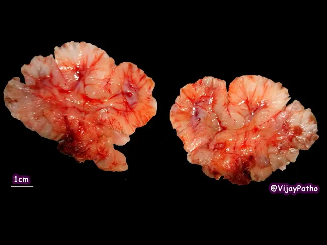
Gross pathology photograph of a bisected genital condyloma acuminatum lesion, showing a soft, fleshy exophytic mass with cauliflower-like papillary architecture. The specimen comprises two symmetrical halves laid side-by-side against a dark background; each fragment measures approximately 1–2 cm in greatest dimension as suggested by a 1 cm scale bar. The surface is pink-tan with irregular, frond-like projections and subtle keratotic grooves. The cut surfaces reveal a fibrous to loosely arranged stroma and thickened epidermis with pronounced papillary infoldings. The lesions are consistent with condyloma acuminatum, most commonly HPV types 6 and 11; they arise on mucocutaneous genital skin including corona of glans, urethral meatus, fossa navicularis, penile shaft, and scrotal skin; similar lesions may occur in vulvar region and perineum. In males, lesions may extend proximally toward the urethra and bladder in extensive disease; symptoms can include itching, bleeding, pain; sexual transmission risk is high; co-infection with other STIs is possible; recurrence is common after treatment. This image highlights the characteristic wart-like architecture: soft, friable, papillary projections with a cauliflower appearance. Understanding gross morphology aids clinical diagnosis and guides subsequent histopathologic confirmation and treatment planning (cryotherapy, topical agents, surgical excision).
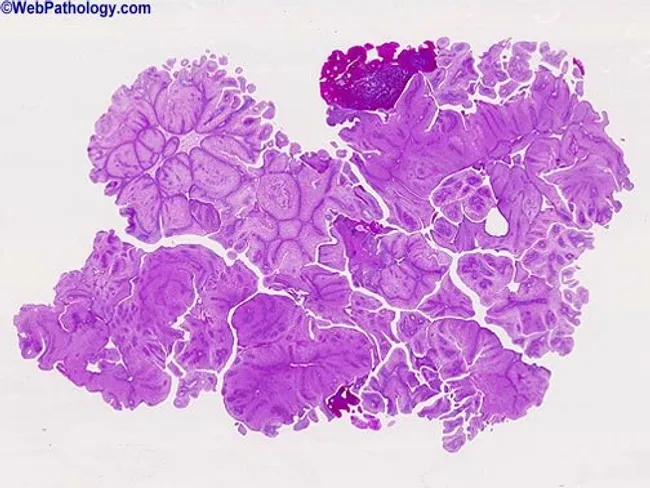
Imaging modality and technique: Light microscopy of fixed tissue stained with Hematoxylin and Eosin (H&E). Specimen: genital mucosa biopsy from a cauliflowers-like neoplastic lesion on the penile skin, viewed at low magnification. Location: genital mucosa of penis, corona glandis region, a typical site for condyloma acuminatum. Morphology: hallmark papillary or polypoid fronds with warty, cauliflower-like architecture; marked papillomatosis and acanthosis producing thickened, undulating epithelium with elongated rete ridges; hyperkeratosis and parakeratosis at the surface. Cellular features: koilocytosis in the upper epithelial layers, characterized by nuclear enlargement with perinuclear halos and irregular nuclear membranes, consistent with HPV infection. The dermal stroma shows mild chronic inflammatory infiltrate and fibrovascular cores within the papillary projections; cytologic atypia is absent, indicating benign HPV-related lesion. Diagnostic impression: condyloma acuminatum (genital wart) most commonly caused by low-risk HPV types 6 and 11; presence of koilocytic change and papillomatous growth supports HPV-induced lesion. Clinical significance: differentiates from malignant or precancerous processes; important for STI counseling and management; differential diagnoses include verruca vulgaris, penile intraepithelial neoplasia, and Buschke-Lowenstein tumor in rare cases. Potential uses: educational reference, correlating histology with gross morphology, and guiding patient counseling and treatment planning. Correlation with clinical presentation strengthens diagnostic confidence and informs management.
The Shaft Bump - Detailed Analysis
| Feature | What's Seen |
|---|---|
| Size | ~2-3 mm |
| Color | Flesh-colored to slightly brownish-grey |
| Shape | Raised, broad-based (sessile), slightly lobulated |
| Surface | Rough and verrucous (warty texture) |
| Shine | Dull, not shiny or pearly |
| Borders | Well-defined, distinct from surrounding skin |
Most Likely: Genital Wart (Condyloma Acuminatum)
Why it's NOT likely to be:
- Molluscum - those are smooth, pearly, with a central pit/dimple. This is rough.
- Skin tag - those are smooth, soft, hanging. This is sessile and rough.
- PPP - only occur on the corona ridge, not the shaft.
What You Should Do
- See a doctor - a sexual health clinic, dermatologist, or urologist can confirm this with a quick physical exam. They may use a dermatoscope (magnifying tool) for clarity.
- Do not pick or scratch it - this can spread HPV to other areas.
- Inform recent sexual partners - if confirmed as a wart, HPV is easily transmitted through skin-to-skin contact.
- Treatment options (if confirmed as genital wart) include topical creams (imiquimod, podophyllotoxin), cryotherapy (freezing), or minor procedures - all done by a doctor.
Disclaimer: This is an assessment based on a photo, not a medical diagnosis. Only an in-person examination can confirm the diagnosis. Please see a healthcare professional.
But I haven't done sex ever
If You've Never Had Sex
More Likely Possibilities Now
| Condition | Why it fits |
|---|---|
| Fordyce spot | Enlarged sebaceous (oil) gland - extremely common, harmless, present from puberty, no cause needed |
| Perifollicular papule | A blocked hair follicle causing a small bump - rough texture, flesh-colored |
| Ingrown hair / folliculitis | Especially common on the shaft, can look rough and raised |
| Tyson's gland | Small sebaceous glands naturally present on the penis, can become visible/raised |
| Benign fibrous papule | Random benign overgrowth of skin tissue, no infectious cause |
| Common wart (verruca vulgaris) | Caused by non-genital HPV strains that CAN spread non-sexually - from touching warts on your own hands/body and then touching the skin there |