
Quadriceps tendinosis
"quadriceps tendinosis" OR "quadriceps tendinopathy" treatment
quadriceps tendinosis MRI ultrasound pathology
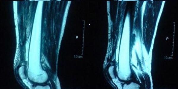
This diagnostic image consists of two sagittal T2-weighted MRI views of the bilateral knees, demonstrating a bilateral quadriceps tendon rupture. The pathology is characterized by a complete midsubstance disruption of the quadriceps tendon approximately 1.5 cm proximal to its insertion into the superior pole of the patella. The right knee shows a more extensive retraction and disruption compared to the left. Key findings include visible thickening and abnormal signal intensity in the remaining tendon segments, indicative of pre-existing tendinosis or degeneration. While the sagittal views highlight the tendon gap and associated soft tissue edema/hemarthrosis, clinical context suggests associated lateral patellar tilt and retinacular injury. This case is an educational example of simultaneous bilateral quadriceps rupture, a rare clinical entity often associated with systemic metabolic conditions or chronic tendon degeneration. The imaging is critical for surgical planning to differentiate between osteotendinous avulsion and midsubstance tears.
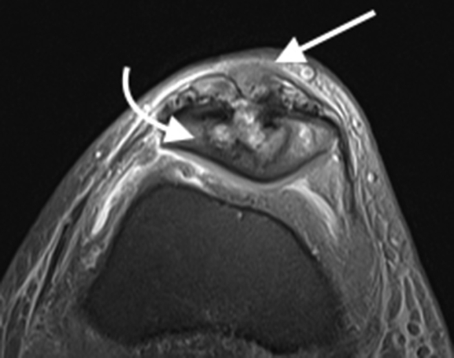
**Imaging Modality:** Magnetic Resonance Imaging (MRI), axial Proton Density-weighted (PD) with fat suppression. **Anatomical Region:** Anterior compartment of the knee at the level of the patellofemoral joint. **Observed Pathology:** Chronic tendinopathy and secondary osseous changes. The distal quadriceps tendon exhibits significant pathological thickening with a heterogeneous, nodular internal signal. Adjacent to the tendon insertion, there is prominent cortical irregularity and erosive change involving the anterior aspect of the patella. **Characteristic Visual Features:** * **Quadriceps Tendon:** Loss of normal thin, low-signal morphology; replaced by an expanded, intermediate-signal nodular mass (indicated by straight arrow). * **Patella:** Focal osseous destruction and subchondral irregularities (indicated by curved arrow) suggestive of pressure necrosis or inflammatory erosion. * **Joint Space:** The underlying trochlear groove and patellofemoral articulation are visible, with mild reactive fluid signal in the peritendinous tissues. **Diagnostic Significance:** These findings are characteristic of severe, chronic quadriceps tendinosis or intratendinous tophaceous deposition (e.g., gout), which can lead to secondary bone erosion. The image serves as a reference for distinguishing tendinous hypertrophy from acute traumatic rupture.
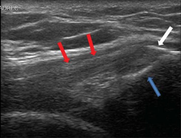
**Imaging Modality:** Ultrasound (Grey-scale, longitudinal view) **Anatomical Region:** Anterior knee, specifically the quadriceps tendon and the superior pole of the patella. **Observed Pathology:** Calcific tendinopathy of the distal quadriceps tendon. **Characteristic Visual Features:** * **Quadriceps Tendon:** The image displays a thickened quadriceps tendon with a loss of the normal organized fibrillar pattern, appearing heterogeneously hypoechoic. * **Calcification:** A focal, linear hyperechoic focus is visible at the distal aspect of the tendon near its insertion site. * **Bony Landmark:** The superior cortex of the patella is visualized, showing a distinct interface with the tendon insertion. * **Annotated Features:** Red arrows delineate the body of the quadriceps tendon; the blue arrow indicates the patellar insertion site; the white arrow highlights the intratendinous calcification. **Diagnostic Differentiating Features:** The presence of focal hyperechoic material within a thickened, hypoechoic tendon at the insertion site is highly characteristic of calcific insertional tendinopathy, distinguishing it from simple tendinosis or an acute partial tear.
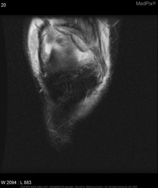
**Imaging Modality:** Magnetic Resonance Imaging (MRI), T2-weighted or STIR sequence. **Anatomical Region:** Anterior knee, axial or coronal cross-section of the extensor mechanism. **Observed Pathology:** Evidence of a high-grade or complete quadriceps tendon rupture. There is a significant disruption of the normal low-signal tendinous fibers proximal to the superior pole of the patella. **Characteristic Visual Features:** - **Signal Abnormality:** Presence of extensive high-signal intensity (edema/hemorrhage) occupying the expected location of the quadriceps tendon. - **Tendon Morphology:** Loss of continuity and wavy, retracted appearance of the remaining tendon fibers. - **Associated Findings:** Evidence of a medial retinacular tear, indicated by signal hyperintensity and fiber discontinuity along the medial aspect of the patellofemoral joint. Significant soft tissue swelling and joint effusion are visible surrounding the injury site. **Differentiating Features:** The total disruption of the trilaminar appearance of the quadriceps tendon and the associated retraction distinguish this from simple tendinosis or partial-thickness tears. The localization proximal to the patella differentiates it from patellar tendon rupture.
"quadriceps tendinopathy" diagnosis management
Quadriceps Tendinosis
Definition and Pathology
Epidemiology and Risk Factors
- More common in older athletes (the quadriceps tendon is the dominant rupture site after age 40, compared to the patellar tendon before 40) - Miller's Review of Orthopaedics
- Part of the "jumper's knee" spectrum, but located at the superior pole of the patella (vs. patellar tendinosis at the inferior pole)
- Sports: elite volleyball (40-50%), basketball (35-40%), track and field athletes
- Systemic predispositions include: SLE, renal failure, diabetes, endocrinopathies - Firestein & Kelley's Textbook of Rheumatology, p. 943
Clinical Features
| Feature | Detail |
|---|---|
| Location | Pain and tenderness at the superior border of the patella |
| Quality | Aching, activity-related; may include painful clicking |
| Aggravation | Worse with extension than flexion (quadriceps loading) |
| Onset | Insidious, repetitive microtrauma; occasionally a single eccentric overload event |
Staging (Blazina Classification, modified by Ferretti)
| Phase | Symptoms |
|---|---|
| 1 | Pain only after activity |
| 2 | Pain during and after activity; no functional impairment |
| 3 | Pain during and after activity with progressive difficulty in performance |
| 4 (added later) | End-stage: stress fracture of patella or extensor mechanism disruption |
- Campbell's Operative Orthopaedics, 15th Ed. 2026
Imaging
- MRI findings: focal thickening, abnormal (increased) signal intensity within the tendon, correlating with angiofibroblastic degeneration at surgery
- A partial-thickness tear >50% of tendon width, or tendon thickness >11.45 mm, predicts poor response to non-operative treatment
- Campbell's Operative Orthopaedics, 15th Ed. 2026, p. 2968
Treatment
Conservative (Phases 1-2, and initial trial for Phase 3)
| Modality | Notes |
|---|---|
| Activity modification & rest | First-line; jumping and repetitive eccentric loading discouraged initially |
| NSAIDs | Adjunct for pain relief |
| Physical therapy | Eccentric quadriceps strengthening (results are conflicting in RCTs - one RCT showed results comparable to surgery; another showed no benefit at 12 weeks) |
| Hamstring stretching | Reduces load on extensor mechanism |
| Counterforce brace (Cho-Pat strap) | Can reduce symptoms |
| Ultrasound therapy | Used as adjunct |
| ESWT (shockwave) | Option for refractory cases; 74% return to sport at ~6 weeks; two studies showed PRP was superior at 6 and 12 months |
| PRP injection | Growing evidence; outperformed ESWT in recent comparative studies |
| Cortisone injection | Contraindicated - significantly increases risk of complete tendon rupture |
- Textbook of Family Medicine, 9th Ed.; Campbell's Operative Orthopaedics, 15th Ed. 2026
Surgical (Phase 3 refractory, Phase 4)
- Longitudinal incision of the tendon sheath
- Identification and excision of the degenerated area using longitudinal tendon incisions
- The patellar pole can be curetted or drilled to stimulate a healing response
- Defect repaired with side-to-side interrupted 2-0 Vicryl sutures
- Peritenon closure; hinged brace or immobilizer applied
-
Brace worn 3-4 weeks; weight-bearing with knee locked straight permitted
-
Early passive ROM to 90°
-
Stage 1: ROM + isometric strengthening
-
Stage 2: Closed-chain kinetics (when swelling/tenderness resolved)
-
Stage 3: Activity-specific exercises, avoiding eccentric overload
-
Return to full activity when 85-90% strength and full ROM are achieved
-
Campbell's Operative Orthopaedics, 15th Ed. 2026
Key Distinctions
| Quadriceps Tendinosis | Patellar Tendinosis ("Jumper's Knee") | |
|---|---|---|
| Location | Superior pole of patella | Inferior pole of patella |
| Common age | > 40 years | Younger athletes |
| Tenderness | Above patella | Below patella |
| Prevalence | Less common | More common |
| Rupture risk after 40 | Higher | Lower |
Summary Points
- Quadriceps tendinosis = degenerative overuse condition, not acute inflammation
- Core pathology: angiofibroblastic degeneration with collagen disorganization
- Key warning: never inject corticosteroids - predisposes to complete rupture
- Eccentric exercise is the cornerstone of conservative rehab (evidence is mixed but favored)
- PRP is emerging as the preferred injection therapy over corticosteroids or ESWT
- Surgery (debridement/tenotomy) is reserved for phase 3-4 or conservative failure