
important questions in ent viva regarding retromolar trigone and how to examine it clinically
Reading File
Now let me search the medical library for retromolar trigone content in parallel.
Finding Sources
Finding Sources
Searching the Web
retromolar trigone ENT viva important questions clinical examination anatomy malignancy
Good results. Now let me read the key textbook sections in parallel.
Reading File
Reading File
Reading File
Now let me get more detailed content - from Scott-Brown's on epidemiology/presentation and KJ Lee's on carcinoma, plus the Cummings block for more detail, and the Bailey & Love examination section.
Reading File
Reading File
Reading File
Now let me get a bit more from Scott-Brown's on workup/management and the Schwartz Surgery section on RMT, and also fetch the CT image of the RMT tumor.
Reading File
Reading File
Loading Image
I now have comprehensive information from multiple authoritative ENT textbooks. Let me compile the complete viva answer.
Retromolar Trigone (RMT) - ENT Viva: Important Questions & Clinical Examination
1. What is the Retromolar Trigone? (Definition & Anatomy)
The retromolar trigone is a triangular mucosal area posterior to the last lower molar tooth, overlying the ascending ramus of the mandible toward the coronoid process. Its boundaries are:
| Boundary | Structure |
|---|---|
| Anterior | Posterior aspect of the last mandibular molar (2nd or 3rd) |
| Superior (apex) | Maxillary tuberosity / coronoid process |
| Medial | Anterior tonsillar pillar (palatoglossal arch) |
| Lateral | Buccal mucosa |
| Inferior | Posterior mandibular alveolus |
The mucosa here is extremely thin with very little submucosa between the overlying mucosa and the underlying mandibular periosteum. This is clinically important because early bone invasion is common.
- Cummings Otolaryngology, block 19 | Scott-Brown's Otorhinolaryngology, block 2 | Sabiston Surgery, block 15
2. Why is the RMT Clinically Important? (The Viva "Trap")
Examiners love this because the RMT is:
- A small, anatomically difficult-to-visualize site - tumors are detected late
- Intimately related to several critical structures simultaneously (mandible, pterygomandibular raphe, masticator space, oropharynx)
- Often misclassified due to early spread to adjacent subsites
3. Nerve Supply (High-Yield for Viva)
| Nerve | Supply |
|---|---|
| Lesser palatine nerve (CN V2) | Sensation to RMT |
| Glossopharyngeal nerve (CN IX) | Also supplies RMT region |
| Inferior alveolar nerve (CN V3) | Runs in mandibular canal nearby |
Key viva point: The presence of CN IX explains referred otalgia in RMT lesions - pain referred to the ear via Jacobson's nerve (tympanic branch of CN IX). This is a classic ENT viva question.
Perineural invasion (PNI): Lower lip paresthesia indicates PNI at the mandibular foramen via the inferior alveolar nerve.
4. Lymphatic Drainage
- Primary drainage: Level II (upper jugular/jugulodigastric) lymph nodes
- Regional metastases present in 26-56% at diagnosis
- Occult cervical nodal involvement: 8-15%
5. How to Examine the RMT Clinically
Equipment Needed:
- Good light source (headlight preferred)
- Two tongue depressors/spatulas
- Gloves for bimanual palpation
Systematic Oral Cavity Examination Sequence (Bailey & Love):
- Lips
- Teeth, gums, gingival sulci
- Buccal mucosa + parotid duct opening (opposite upper 2nd molar)
- Floor of mouth + submandibular duct openings
- Hard and soft palate
- Retromolar trigone region (specifically named in the sequence)
- Anterior and posterior faucial pillars, tonsils
- Posterior pharyngeal wall
- Tongue (all movements observed)
Specific RMT Examination Steps:
Inspection:
- Ask patient to open mouth maximally
- Use two spatulas - one to retract the cheek laterally and the other to depress the tongue
- Look posteriorly behind the last lower molar on each side
- Identify the triangular mucosa overlying the ascending ramus
- Look for: ulceration, induration, erythema (erythroplakia), white patches (leukoplakia), or exophytic lesion
Palpation (CRITICAL):
- Bimanual palpation with a gloved finger intraorally pressing against the region
- Assess: induration, fixity to underlying bone, trismus (inability to open mouth >35 mm suggests masticator space involvement)
- Check mouth opening - trismus is a red flag for masticator space invasion
- Feel the pterygomandibular raphe (runs from hamulus of medial pterygoid plate to mylohyoid line)
Assess for spread:
- Buccal mucosa (lateral spread - most common, ~84%)
- Anterior tonsillar pillar and soft palate (medial/posterior)
- Floor of mouth
- Check neck nodes - level II, III bilaterally
Cranial nerve assessment:
- CN V3: Test lower lip sensation (inferior alveolar nerve involvement)
- CN IX: Ask about referred otalgia
- Mouth opening: measure inter-incisal distance (trismus = masticator space involvement)
6. Common Presentations / Why Patients Present Late
Patients typically present with:
- Pain (most common)
- Trismus - jaw stiffness; suggests masticator space involvement (T4b)
- Referred otalgia - via CN IX (Jacobson's nerve)
- Lingual paresthesia - tongue numbness
- Lower lip paresthesia - PNI of inferior alveolar nerve
The RMT is anatomically small and posteriorly located - tumors are hard to see and patients ignore early symptoms. At presentation, 55-73% have Stage III/IV disease.
7. Pathology
- Most common: Squamous cell carcinoma (SCC)
- Histology: usually well or moderately differentiated
- Incidence: 6-7% of all oral cavity carcinomas (relatively uncommon)
- Risk factors: Tobacco (smoking, chewing), alcohol - as with other oral cavity sites
- More common in males and older individuals
8. Spread Pattern (Routes of Extension - Very High Yield)
The pterygomandibular raphe is the key highway for tumor spread. It runs from the hook of the hamulus of the medial pterygoid to the mylohyoid line on the medial border of the mandible. Because the buccinator, superior constrictor, and orbicularis oris all insert into it, RMT tumors can spread to:
| Direction | Structure involved | Frequency |
|---|---|---|
| Lateral | Buccal mucosa | ~84% |
| Posterior | Oropharynx (tonsillar fossa, soft palate) | ~14% |
| Deep | Masticator space (medial pterygoid, masseter, temporalis, V3) | ~22% |
| Inferior | Mandible (bone invasion) | 12-75% |
| Superior | Maxilla/posterior maxilla | More common than mandibular |
T4b disease: Extension into masticator space or pterygoid plates = T4b (unresectable in most systems).
9. Imaging
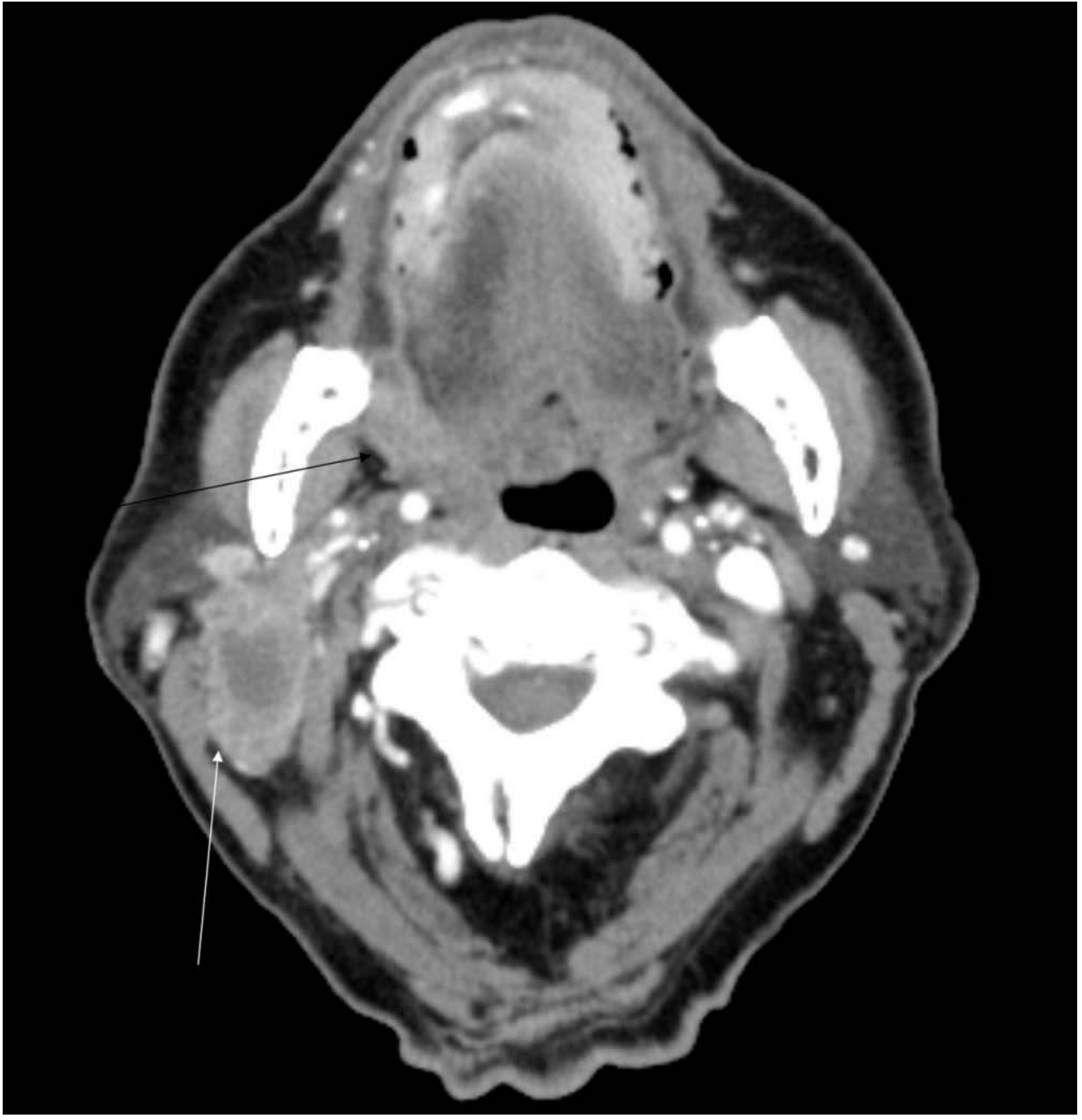
- MRI is the modality of choice for RMT tumors - best for soft tissue detail, perineural spread, and accurate staging
- CT: High specificity but low sensitivity for mandibular bone invasion - do not rely on CT alone to exclude bone involvement
- Clinical examination alone is unreliable for determining bone involvement
- Principle: Maintain a low threshold for bone resection given CT's poor negative predictive value
CT image of right RMT SCC with necrotic level II node:
10. Treatment (Viva Points)
| Stage | Treatment |
|---|---|
| Stage I/II | Surgery OR radiotherapy equally effective; surgery preferred (avoids osteoradionecrosis) |
| Advanced (III/IV) | Surgery + adjuvant radiotherapy/chemoradiotherapy |
Surgical approach:
- Small tumors: transoral resection
- Posterior location/adjacent spread: mandibulotomy (lip-splitting + lateral mandibulotomy with "mandibular swing") for access
- Extensive masticator space involvement: cervicofacial incision + parotidectomy ± zygomatic osteotomy
Mandibular management:
- Marginal mandibulectomy (posterior marginal, including coronoid): acceptable if no cortical involvement on imaging and no prior radiotherapy
- Segmental mandibulectomy: required if cortical bone invaded on imaging or after prior radiotherapy
- Reconstruction: fibula osteocutaneous free flap for segmental defects
Neck dissection: Levels I-III elective neck dissection for N0 neck if significant risk of nodal disease
Reconstruction of soft tissue defect:
- Small mucosal defects: secondary intention healing
- Larger defects: split-skin graft, buccal fat pad, local flaps (tongue flap, masseteric flap), or free flap
Aim for 1 cm clear margins in all planes. Note: RMT has higher rates of positive margins than other oral sites.
11. Prognosis (5-year outcomes after surgery + radiotherapy)
| Stage | Locoregional control | Disease-specific survival |
|---|---|---|
| Stages I-III | 87% | 83% |
| Stage IV | 62% | 61% |
| Overall | 71% | 69% |
12. High-Yield Viva Q&A Summary
Q: Why does RMT carcinoma cause referred otalgia?
A: Via Jacobson's nerve (tympanic branch of CN IX), which supplies the RMT region.
Q: Why does bone invasion occur early in RMT tumors?
A: The mucosa overlying the RMT is extremely thin with minimal soft tissue between it and the mandibular periosteum, allowing early periosteal and cortical invasion.
Q: What does trismus signify in an RMT tumor?
A: Involvement of the masticator space (medial pterygoid, masseter, temporalis muscles), classifying it T4b in most staging systems. However, if resectable, outcomes are comparable to T4a.
Q: What nerve causes lower lip numbness in RMT cancer?
A: Inferior alveolar nerve (V3) due to perineural invasion near the mandibular foramen.
Q: What is the preferred imaging for RMT tumors and why?
A: MRI - superior soft tissue resolution for perineural spread, masticator space involvement, and accurate staging. CT has low sensitivity for mandibular invasion.
Q: What is the pterygomandibular raphe and why is it important in RMT cancer?
A: A fibrous band from the hamulus of the medial pterygoid plate to the mylohyoid line on the mandible. Tumor extension along this raphe allows spread to buccal space, tonsillar fossa, floor of mouth, and nasopharynx.
Q: What is the RMT as a subsite of?
A: The oral cavity (not oropharynx). This is important for TNM staging.
Q: What type of elective neck dissection is performed for cN0 RMT cancer?
A: Unilateral elective neck dissection, levels I-III.
Sources: Cummings Otolaryngology Head and Neck Surgery (block 19) | Scott-Brown's Otorhinolaryngology Head & Neck Surgery (block 2, 6) | Sabiston Textbook of Surgery (block 15) | Schwartz's Principles of Surgery (block 7) | KJ Lee's Essential Otolaryngology (block 4) | Bailey and Love's Short Practice of Surgery (block 6)
This is a shared conversation. Sign in to Orris to start your own chat.