
Air emoblism and how to treat mechanism and management symptoms Postion
Reading File
Now I'll search the medical library and web in parallel for comprehensive information on air embolism.
Finding Sources
Finding Sources
Searching the Web
air embolism mechanism symptoms treatment position management 2024
Now let me read the most comprehensive textbook sections in parallel:
Reading File
Reading File
Reading File
Reading File
Searching Images
air embolism venous arterial mechanism diagram treatment position
Now let me read the nephrology and Miller's Anesthesia sections for more management detail:
Reading File
Finding Sources
Finding Sources
I have rich information from multiple authoritative textbooks. Let me also fetch the management flowchart image:
Loading Image
Reading File
I now have comprehensive information from multiple authoritative sources. Here is the complete answer:
Air Embolism
Definition
Air (gas) embolism occurs when gas bubbles enter the vascular system, obstructing blood flow and causing ischemic injury. It is divided into two major types:
- Venous Air Embolism (VAE) - air enters the venous circulation and affects the right heart/pulmonary vasculature
- Arterial (Systemic) Air Embolism (AAE) - air enters the left side of the circulation and reaches systemic organs (brain, heart)
Causes / Portals of Entry
| Category | Examples |
|---|---|
| Iatrogenic | Central venous catheter insertion, disconnection, or removal; IV infusions with glass bottles; hemodialysis circuits; laparoscopic/obstetric procedures; neurosurgery in sitting position |
| Trauma | Penetrating chest injuries, bronchovenous fistulas; blunt trauma with lung injury |
| Positive-pressure ventilation | Creates air-blood gradient that drives air across bronchovenous fistulas - the most important factor in traumatic air embolism |
| Scuba diving / decompression | Rapid ascent causes dissolved nitrogen to bubble out ("decompression sickness" / the bends) |
| Head and neck surgery | Air sucked into open veins |
| Illegal abortion / Fallopian tube insufflation | Historically documented causes |
Mechanism (Pathophysiology)
Venous Air Embolism
Air enters the venous system → travels to right heart → two main consequences:
- Airlock effect: A large bolus accumulates in the right ventricle or pulmonary outflow tract, forming foam that blocks right ventricular output → acute right heart failure, obstructive shock, and sudden death. Lethal volume is approximately 5 to 8 mL/kg.
- Microvascular obstruction: Smaller bubbles lodge in pulmonary capillary beds → abrupt rise in pulmonary artery pressure → noncardiogenic pulmonary edema + reduced lung compliance → hypoxemia.
- Platelet-fibrin aggregates form around bubbles → diffuse microthrombi → thrombocytopenia.
- Bubbles can pass through pulmonary microvascular shunts → enter left-sided (arterial) circulation → paradoxical air embolism.
Arterial (Systemic) Air Embolism
Air in the left heart/arteries is far more dangerous per volume:
- As little as 0.5 mL in the left anterior descending coronary artery can cause ventricular fibrillation.
- 2 mL injected into the cerebral circulation can be fatal.
- Entry occurs via bronchovenous fistulas (lung trauma) or paradoxical embolism through a patent foramen ovale.
Decompression Sickness (Special Form)
Rapid ascent from depth → dissolved nitrogen expands and bubbles out of solution in blood/tissues → gas emboli in joints (the bends), lungs (respiratory distress), and CNS (memory loss, ataxia, coma). Chronic form = caisson disease with avascular necrosis of femoral/humeral/tibial heads.
Symptoms
Symptoms vary by volume of air, rate of entry, and site of involvement. All presentations are potentially life-threatening emergencies.
Cardiovascular
- Sudden drop in blood pressure / circulatory collapse
- Irregular heartbeat / cardiac arrest
- "Millwheel murmur" - a churning sound audible on cardiac auscultation (classic finding)
Respiratory
- Rapid, shallow breathing / dyspnea
- Continuous coughing
- Chest pain
- Coughing up blood (hemoptysis)
- Respiratory arrest
Neurological (if cerebral involvement)
- Headache, confusion, dizziness
- Vision changes
- Seizures
- Focal neurologic deficits / hemiparesis
- Loss of consciousness / coma
- Altered mental status
Systemic
- Sense of impending doom
- Paralysis in one area of the body
Key diagnostic clue: In trauma patients, cardiac arrest after intubation and initiation of positive-pressure ventilation + hemoptysis is highly suggestive. Focal neurological changes without head injury also raise suspicion. Confirmed at thoracotomy by needle aspiration of foamy air-blood mixture from the ventricle or visualization of air in coronary arteries.
Position (Durant's Maneuver + Trendelenburg)
Patient positioning is a critical, immediate intervention:
Venous Air Embolism
Left Lateral Decubitus (Durant's Maneuver) + Trendelenburg (head-down)
- Left lateral decubitus: Encourages the air bubble to float to the right ventricular apex, away from the pulmonary outflow tract - relieves the "airlock" and prevents outflow obstruction.
- Trendelenburg (head-down): Encourages air to enter veins in the lower body; reduces air migrating to the cerebral circulation.
- Together these two positions allow air to be trapped in the right atrium and prevent further cardiovascular and cerebral migration.
Arterial Air Embolism
- Keep the patient in the flat supine position.
- The head-down position is avoided in arterial embolism because it may worsen cerebral edema.
Management
Immediate Steps (Stop the Source)
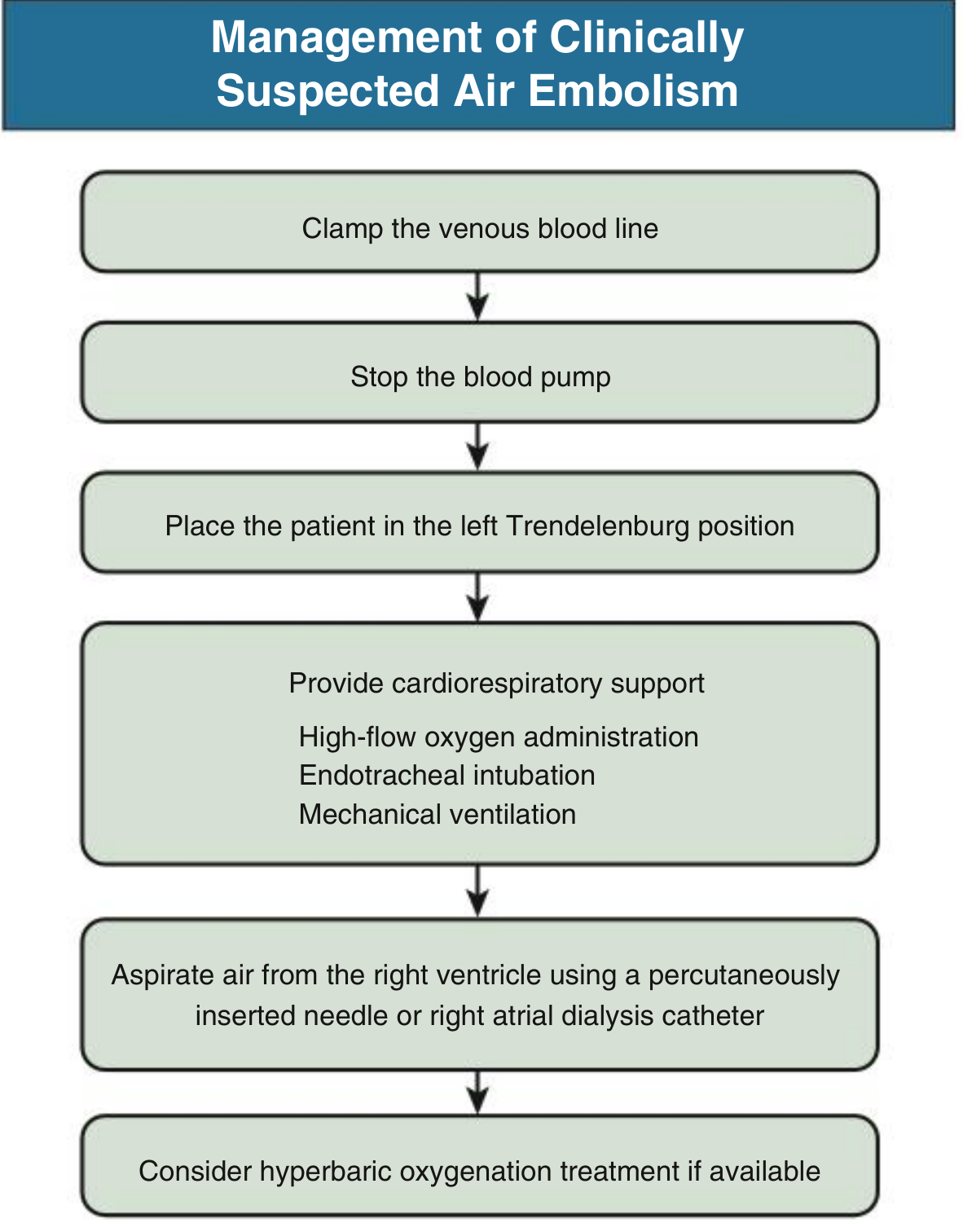
- Clamp or disconnect the venous line / IV circuit immediately to stop further air entry
- Stop blood pump (in dialysis setting)
- Position the patient: Left lateral decubitus + Trendelenburg (VAE)
Cardiorespiratory Support
- 100% high-flow oxygen - accelerates bubble reabsorption (oxygen replaces nitrogen in bubbles, which are then absorbed faster)
- Secure airway: endotracheal intubation + mechanical ventilation if needed
- Hemodynamic support: maintain blood pressure and perfusion
Air Removal
- Aspiration through central venous catheter or right atrial catheter
- In extreme cases: needle aspiration of air from the right ventricle (needle introduced below the left costal margin, through the cardiac apex)
- Closed-chest cardiac massage if in arrest (helps break up air bubbles)
Definitive Therapy
- Hyperbaric oxygen therapy (HBO) - the treatment of choice for arterial gas embolism and severe venous events. Forces gas back into solution under high pressure; subsequent slow decompression allows gradual reabsorption without re-formation of bubbles. Also used for decompression sickness.
Management Flowchart (from Comprehensive Clinical Nephrology)
Summary Table
| Feature | Venous Air Embolism | Arterial Air Embolism |
|---|---|---|
| Entry site | Veins → right heart | Left heart / arteries |
| Lethal volume | ~5-8 mL/kg | 0.5 mL (LAD) / 2 mL (cerebral) |
| Key mechanism | Right heart airlock, pulmonary HTN | Coronary/cerebral ischemia |
| Position | Left lateral decubitus + Trendelenburg | Flat supine |
| Key treatment | O2, aspiration, HBO | HBO (first choice) |
| Classic auscultation finding | Millwheel murmur | - |
Prevention
- Air detection alarms on dialysis machines (never run dialysis without functioning alarm)
- Occlusive dressings over central line insertion sites; clamp lines during connection/disconnection
- Avoid glass IV bottles (create vacuum effect)
- Avoid sitting/head-elevated position in at-risk surgeries; use TEE monitoring in neurosurgery
- Aspirate and flush catheters with saline before connecting
Sources: Roberts and Hedges' Clinical Procedures in Emergency Medicine; Murray & Nadel's Textbook of Respiratory Medicine; Fishman's Pulmonary Diseases and Disorders; Robbins & Kumar Basic Pathology; Comprehensive Clinical Nephrology 7th Ed; Bailey and Love's Surgery 28th Ed; Pye's Surgical Handicraft.
This is a shared conversation. Sign in to Orris to start your own chat.