
Histopathological features of salivary gland tumors including IHC markers and molecular genetics
pleomorphic adenoma salivary gland histology chondromyxoid stroma
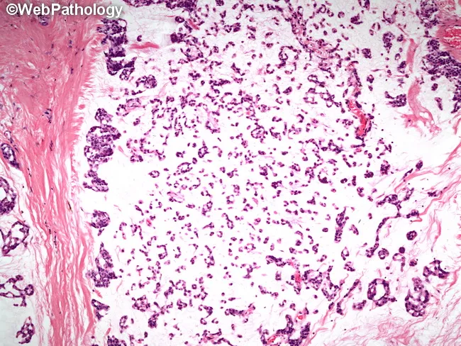
Histology image of a salivary gland tumor demonstrating classic pleomorphic adenoma (mixed tumor). Prepared as a hematoxylin and eosin stained slide and evaluated under light microscopy. The specimen represents salivary gland tissue in the parotid region. Epithelial elements are organized into small nests and rudimentary tubular structures, reflecting ductal differentiation. These epithelial components are embedded in a substantial myxoid background with scattered chondromyxoid areas. Myoepithelial cells contribute to the cellular admixture, accounting for variable cell shapes and occasional plasmacytoid appearances. The stroma ranges from loose, myxoid to more fibrous or hyaline areas, producing a heterogeneous, biphasic architecture typical of pleomorphic adenoma. The overall pattern is benign-appearing with well-demarcated margins in this field; however, the lesion is known for potential recurrence if incompletely excised. Pleomorphic adenoma is a benign salivary gland neoplasm featuring epithelial and myoepithelial components within mucoid and cartilaginous stroma. Diagnostic significance rests on recognizing the biphasic morphology and chondromyxoid matrix, which helps distinguish from malignant mimics such as mucoepidermoid carcinoma or adenoid cystic carcinoma. Clinically, these findings correlate with a slow-growing, painless parotid mass in middle-aged adults. This image is valuable for education, differential diagnosis practice, and radiology-pathology correlation in salivary gland tumors. for clinical education and research.
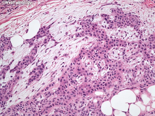
Histopathology of a salivary gland neoplasm (pleomorphic adenoma) demonstrated on hematoxylin and eosin stained tissue section examined by bright‑field light microscopy. The primary subject is a salivary gland tumor, most commonly arising in the parotid region. The specimen shows epithelial cells organized into anastomosing cords and tubular structures embedded in abundant myxoid to chondromyxoid stroma. The epithelial component includes duct‑like castings and nests of cuboidal to columnar cells with basophilic nuclei and minimal cytoplasm, often admixed with variably myoid, plasmacytoid cells representing myoepithelial differentiation. The background stroma is loose, richly mucoid, and occasionally hyalinized, occasionally containing focal adipose tissue islands at the periphery, a recognized but nonessential feature of pleomorphic adenoma. Notable features include cleft-like spaces and a biphasic appearance reflecting epithelial and myoepithelial elements within a chondromyxoid matrix. The regional architecture is typically well circumscribed, though microscopic invasion into adjacent fibrous stroma may occur in rare cases; this is a benign neoplasm with excellent prognosis after complete excision. Diagnostic significance lies in recognizing the mixed epithelial/myoepithelial phenotype with myxoid stroma, differentiating from mucoepidermoid, adenoid cystic carcinoma, or canalicular lesions. Clinically, this histology supports diagnosis of pleomorphic adenoma and informs surgical planning and prognosis.
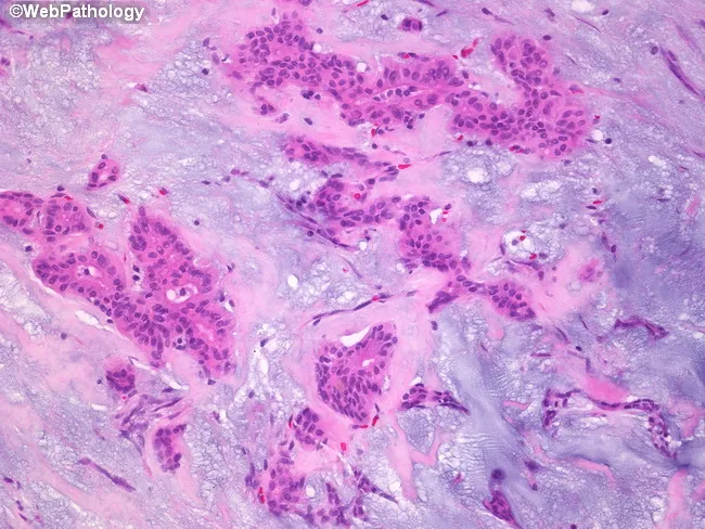
Pleomorphic adenoma histology, parotid salivary gland, examined by bright-field microscopy on Hematoxylin and Eosin stained sections. Imaging modality: Light microscopy; technique: Hematoxylin and Eosin staining. The sample reveals a classic biphasic neoplasm comprising epithelial ductal structures and myoepithelial cells embedded in an abundant stromal backdrop. Epithelial components form cords, ducts, and small island formations lined by cuboidal to columnar cells with eosinophilic cytoplasm and hyperchromatic nuclei. Intermixed myoepithelial cells display plasmacytoid or spindle morphologies, often surrounding epithelial elements. The stroma is a prominent feature, ranging from basophilic, myxoid to more eosinophilic, hyalinized areas. The myxoid matrix is rich in glycosaminoglycans and imparts a loose, gelatinous appearance, while hyalinized zones contribute dense eosinophilic bands. Overall architecture is often well circumscribed with minimal pleomorphism and low mitotic activity, consistent with benign behavior. Clinical significance includes differentiation from malignant salivary tumors; prognosis is favorable with complete surgical excision. Differential considerations include mucoepidermoid carcinoma, adenoid cystic carcinoma, basal cell adenoma, and Warthin tumor. This image is useful for educational purposes in pathology, otolaryngology, and head-and-neck surgery, illustrating quintessential pleomorphic adenoma features: epithelial and myoepithelial proliferation in a chondromyxoid stroma, with duct-like structures and a variable stromal composition. This histologic pattern aids diagnosis and education.
adenoid cystic carcinoma cribriform pattern salivary gland
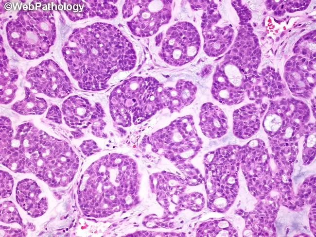
High-magnification bright-field histopathology image of salivary gland tissue demonstrating adenoid cystic carcinoma with cribriform architecture. The section is stained with Hematoxylin and Eosin and viewed under light microscopy at high magnification, revealing small, uniform basaloid tumor cells arranged in interconnected lobules and islands. The characteristic cribriform pattern forms sieve-like spaces that resemble pseudocysts rather than true glandular lumina; these spaces are surrounded by dense basaloid epithelium. The spaces are variably filled with eosinophilic material that is PAS-positive and diastase-resistant, consistent with reduplicated basal lamina, or with basophilic myxoid mucinous material that stains with Alcian blue. The tumor cells exhibit scant cytoplasm, hyperchromatic nuclei, and a biphasic matrix comprising hyaline-like stroma within lobules. Perineural invasion may be evident in some cases, contributing to local aggressiveness and recurrence. Clinically, this histology is diagnostic of adenoid cystic carcinoma, a malignant salivary gland neoplasm that shows at least cribriform, tubular, and solid growth patterns; prognosis depends on pattern, margin status, and neural invasion. This image is representative for educational purposes, pathology teaching, differential diagnosis with basal cell adenoma, and research into salivary gland malignancies. The image also serves as a reference for teaching tumor biology, differential diagnosis, and the recognition of diagnostic hallmarks essential for pathologists.
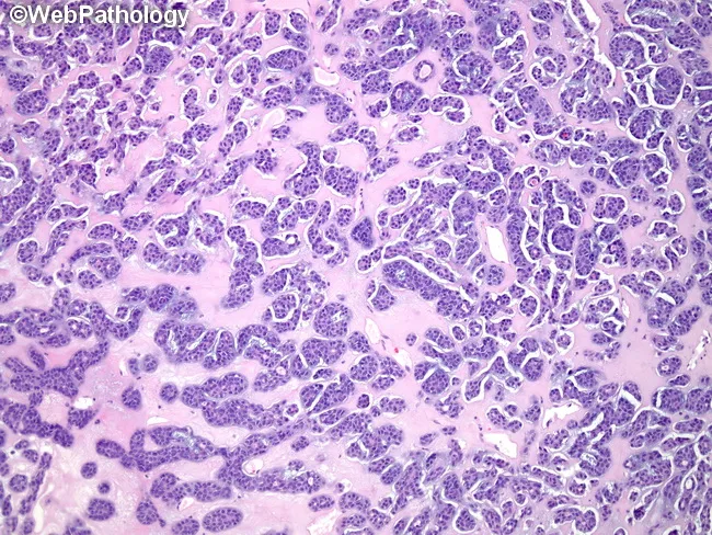
This histopathology image depicts a salivary gland neoplasm with adenoid cystic carcinoma morphology. Using light microscopy, hematoxylin and eosin staining reveals cribriform (Swiss cheese) architecture composed of small basaloid cells arranged in nests, cords, and ducts separated by thin, delicate scaffolding of mucopolysaccharide stroma. The pseudocystic spaces are conspicuously prominent and are filled by eosinophilic, hyaline material that represents duplicated basal lamina/basement membrane. The basaloid cells show scant cytoplasm, hyperchromatic elongated nuclei, and a high nuclear-to-cytoplasmic ratio; mitotic activity is typically low. The stroma surrounding the nests often appears hyalinized and may contain sialomucinous or chondroid-like elements. In typical patterns, perineural invasion may be present, reflecting aggressive clinical behavior. The appearance can alternate with tubular or solid patterns; however, the cribriform pattern with pseudocysts is characteristic. Clinically, these features support a diagnosis of adenoid cystic carcinoma of the salivary gland with a propensity for local invasion and nerve sheath infiltration. This image is relevant for educational scenarios including differential diagnosis of basaloid salivary gland tumors, radiologic-pathologic correlation, and surgical oncology planning. Keywords: adenoid cystic carcinoma, cribriform, basaloid cells, pseudocysts, basement membrane, Swiss cheese, salivary gland tumor, perineural invasion, H&E imaging.
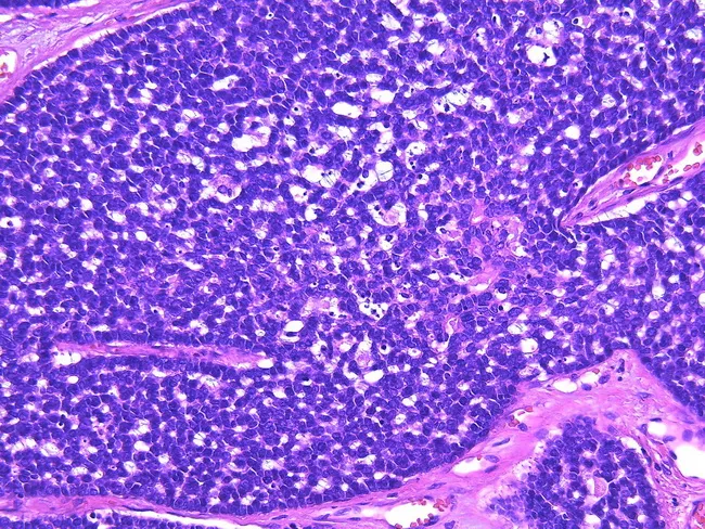
High-magnification light microscopy of a salivary gland tumor demonstrates adenoid cystic carcinoma with mixed architectural patterns. The cribriform and tubular patterns are intermixed with solid proliferations composed of basaloid cells arranged in sheets, islands, or large solid nests. The stroma is variably myxoid to hyaline (myxohyaline), providing a distinctive extracellular matrix that supports gland-like spaces and pseudocystic spaces. In solid areas, true glandular lumens are rare; tumor cells remain tightly packed with scant intervening stroma. Cellular morphology includes small, basaloid, hyperchromatic nuclei with minimal cytoplasm and conspicuous mitotic activity in some fields; nuclear pleomorphism may be present, and necrotic foci can occur in higher-grade areas. The solid pattern broader involvement correlates with higher tumor grade; grades correlate with the percentage of solid components: Grade 1 with no solid areas, Grade 2 with less than 30% solid, and Grade 3 with more than 30% solid pattern. Clinically, a larger solid component is associated with increased risk of recurrence, metastasis, and poorer outcome. This image captures the diagnostic features that support histopathologic grading, prognostication, and therapeutic planning, including surgical excision and consideration of adjuvant radiotherapy. Recognize basaloid morphology, cribriform glandular architecture, and myxohyaline stroma as hallmarks of ACC. Useful for teaching cases.
mucoepidermoid carcinoma salivary gland mucous epidermoid intermediate cells
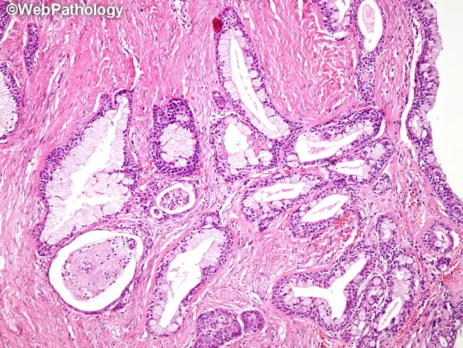
Mucoepidermoid carcinoma is depicted as a salivary gland neoplasm characterized by a heterogeneous mix of mucin-producing goblet-like cells, squamous (epidermoid) cells, intermediate cells, and clear cells arranged in cystic, glandular, and solid compartments. In this low‑grade example, the lesion demonstrates prominent cystic spaces lined by mucinous epithelium admixed with small nests of epidermoid cells. The mucous cells are frequently balloon‑shaped with abundant foamy cytoplasm and peripheral, often small nuclei; in other foci they form mucin-rich columns resembling glands. The overall architecture ranges from simple cysts to cribriform and papillary patterns, reflecting duct‑like differentiation within a hyalinized stroma. Solid nests of epidermoid cells may be interspersed with mucinous elements, producing a biphasic to triphasic cellular spectrum. Immunohistochemical or ancillary testing (where available) can substantiate mucous differentiation, but on routine H&E this triad of mucous, epidermoid, and intermediate cells suffices for recognition of MEC. The presence of mucin production, cystic decomposition, and duct‑like architecture is diagnostically significant, distinguishing MEC from other salivary gland tumors such as adenoid cystic carcinoma or pleomorphic adenoma. Clinically, these low-grade lesions tend to have favorable prognosis with surgical excision; higher grade variants exhibit more solid growth and aggressive behavior, guiding management and follow‑up.
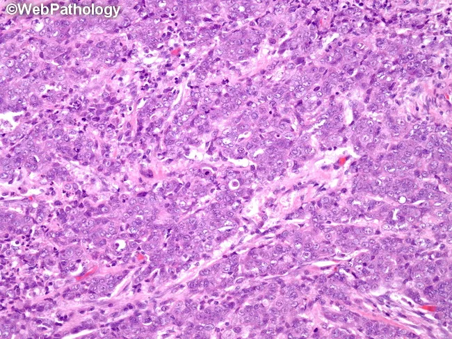
This histopathology image shows mucoepidermoid carcinoma of a salivary gland captured on a hematoxylin and eosin stained slide. The tissue reveals a biphasic neoplasm composed of mucous-secreting cells intermingled with epidermoid (squamoid) cells and intermediate cell types, arranged in cystic and solid nests within a fibrous stroma. Mucous cells contain intracellular mucin, while squamous-like cells exhibit intercellular bridges and nuclear pleomorphism to a variable degree. Duct-like structures and hyalinized bands may be present, reflecting ductal differentiation and cribriform-pattern areas characteristic of MEC. The overall architectural pattern is infiltrative, with variable cellularity and a range of cytologic atypia from low to moderate, consistent with intermediate or higher-grade features depending on additional criteria. Mitotic figures may be seen but are not prominently counted in this field; necrosis is not conspicuously evident here. Clinical significance of these histologic features lies in AFIP three-tier grading, where intracystic component, perineural invasion, necrosis, mitoses, and anaplasia determine low-, intermediate-, or high-grade status, guiding prognosis and treatment. This image is valuable for educational purposes, differential diagnosis discussions with adenoid cystic or other salivary gland carcinomas, and pathology training in head and neck oncology. It also supports correlation with imaging, surgical planning, and targeted therapeutic decisions for patients.
Warthin tumor papillary cystadenoma lymphomatosum oncocytic epithelium lymphoid stroma
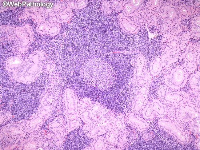
A histopathologic image of a parotid gland Warthin tumor (papillary cystadenoma lymphomatosum) obtained from surgical resections, demonstrated with bright-field microscopy after Hematoxylin and Eosin staining. The epithelial component forms tubular and papillary structures lined by a bilayered epithelium: an inner tall columnar luminal cell layer and an outer cuboidal oncocytic layer. These epithelial cords are embedded within a dense lymphoid stroma containing prominent germinal centers and a mixed inflammatory infiltrate. Immunophenotype in the accompanying description notes cytokeratin positivity of the epithelium (CK7, CK8, CK18, CK19) and a predominantly polyclonal B-cell population with IgA-producing cells, along with scattered T-cells, mast cells, and S-100 positive dendritic cells in the stroma. The combination of oncocytic epithelium and lymphoid-rich stroma with germinal centers is characteristic for Warthin tumor and helps distinguish it from pleomorphic adenoma or mucoepidermoid carcinoma. Clinically, this lesion is benign with favorable prognosis after parotidectomy. The high magnification view emphasizes dense lymphoid infiltrate, organized germinal centers, and the biphasic architecture. This image is valuable for diagnostic education, histology teaching files, and reference databases for head and neck pathology. It supports differential diagnosis, correlates with immunohistochemistry, and serves as a teaching exemplar for students and trainees in residency.
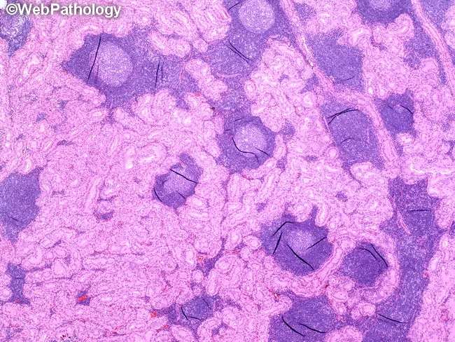
This histopathology image demonstrates a Warthin tumor of the parotid gland in a low-power light micrograph. Papillary and tubular epithelial proliferations are lined by a bilayered, oncocytic epithelium with abundant eosinophilic granular cytoplasm rich in mitochondria. The neoplastic epithelium sits within dense lymphoid stroma containing conspicuous germinal centers, reflecting the characteristic biphasic architecture of papillary cystadenoma lymphomatosum. PTAH staining reveals blue-black cytoplasmic granules within oncocytic cells, highlighting mitochondrial content and cytoplasmic granularity. The combination of bilayered oncocytic epithelium forming papillary/tubular structures and a prominent lymphoid stroma with germinal centers is diagnostic for Warthin tumor, a benign salivary gland neoplasm most often arising in the parotid region. This image supports differential diagnosis against pleomorphic adenoma and mucoepidermoid carcinoma; clinical correlation includes patient age, smoking history, and parotid mass presentation. The technical context—PTAH histochemistry on a parotid tissue section at low magnification—facilitates recognition of the epithelial/lymphoid biphasic pattern and mitochondria-rich cytoplasm; relevant for pathology education, QA, and histology atlases. This image is valuable for teaching residents, fellows, and students about salivary gland oncocytic tumors; PTAH cytoplasmic granules provide histochemical corroboration of mitochondrial abundance. Correlating morphology with immunohistochemistry may exclude more cellular carcinomas. The image underscores the importance of recognizing stromal-epithelial biphasic architecture in parotid neoplasms and the characteristic lymphoid germinal centers.
Histopathological features of salivary gland tumors including IHC markers and molecular genetics from robbins and Ackerman only
pleomorphic adenoma salivary gland histology chondromyxoid stroma biphasic
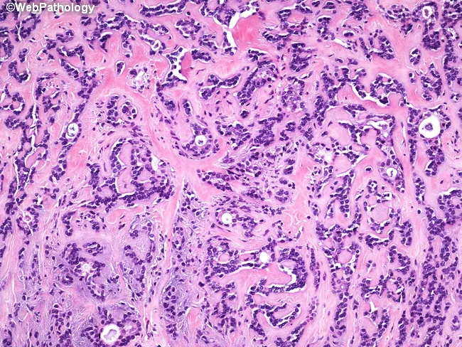
Classic pleomorphic adenoma of a salivary gland demonstrated on Hematoxylin and Eosin stained histology. The image shows a biphasic tumor with intimate admixture of ductal epithelial elements and myoepithelial cells set within a variably myxoid to hyalinized stromal background. Duct-like glands and small tubules are lined by cuboidal to columnar epithelial cells, often surrounding open lumina, while the surrounding myoepithelial cells appear plasmacytoid and can be dispersed within a basophilic, mucopolysaccharide rich matrix. The stroma ranges from loose basophilic myxoid zones to dense eosinophilic hyalinized areas and occasionally cartilaginous, chondromyxoid nodules. The lesion typically exhibits a well circumscribed or push‑invasion pattern with minimal cytologic atypia and low mitotic activity, consistent with benign behavior. This histologic constellation—epithelial–myoepithelial biphasic components set in a chondromyxoid stroma—defines pleomorphic adenoma and helps differentiate it from other salivary gland tumors. Clinically, such tumors present as slow‑growing, painless parotid masses in adults. Accurate diagnosis relies on recognizing the biphasic architecture and stromal diversity; treatment is surgical excision with adequate margins to prevent recurrence and rare malignant transformation (carcinoma ex pleomorphic adenoma). This slide serves as a representative educational reference for diagnostic pathology, head‑and‑neck oncology, and pathology teaching.
Histology image of a salivary gland tumor demonstrating classic pleomorphic adenoma (mixed tumor). Prepared as a hematoxylin and eosin stained slide and evaluated under light microscopy. The specimen represents salivary gland tissue in the parotid region. Epithelial elements are organized into small nests and rudimentary tubular structures, reflecting ductal differentiation. These epithelial components are embedded in a substantial myxoid background with scattered chondromyxoid areas. Myoepithelial cells contribute to the cellular admixture, accounting for variable cell shapes and occasional plasmacytoid appearances. The stroma ranges from loose, myxoid to more fibrous or hyaline areas, producing a heterogeneous, biphasic architecture typical of pleomorphic adenoma. The overall pattern is benign-appearing with well-demarcated margins in this field; however, the lesion is known for potential recurrence if incompletely excised. Pleomorphic adenoma is a benign salivary gland neoplasm featuring epithelial and myoepithelial components within mucoid and cartilaginous stroma. Diagnostic significance rests on recognizing the biphasic morphology and chondromyxoid matrix, which helps distinguish from malignant mimics such as mucoepidermoid carcinoma or adenoid cystic carcinoma. Clinically, these findings correlate with a slow-growing, painless parotid mass in middle-aged adults. This image is valuable for education, differential diagnosis practice, and radiology-pathology correlation in salivary gland tumors. for clinical education and research.
adenoid cystic carcinoma cribriform Swiss cheese pattern perineural invasion
This histopathology image depicts a salivary gland neoplasm with adenoid cystic carcinoma morphology. Using light microscopy, hematoxylin and eosin staining reveals cribriform (Swiss cheese) architecture composed of small basaloid cells arranged in nests, cords, and ducts separated by thin, delicate scaffolding of mucopolysaccharide stroma. The pseudocystic spaces are conspicuously prominent and are filled by eosinophilic, hyaline material that represents duplicated basal lamina/basement membrane. The basaloid cells show scant cytoplasm, hyperchromatic elongated nuclei, and a high nuclear-to-cytoplasmic ratio; mitotic activity is typically low. The stroma surrounding the nests often appears hyalinized and may contain sialomucinous or chondroid-like elements. In typical patterns, perineural invasion may be present, reflecting aggressive clinical behavior. The appearance can alternate with tubular or solid patterns; however, the cribriform pattern with pseudocysts is characteristic. Clinically, these features support a diagnosis of adenoid cystic carcinoma of the salivary gland with a propensity for local invasion and nerve sheath infiltration. This image is relevant for educational scenarios including differential diagnosis of basaloid salivary gland tumors, radiologic-pathologic correlation, and surgical oncology planning. Keywords: adenoid cystic carcinoma, cribriform, basaloid cells, pseudocysts, basement membrane, Swiss cheese, salivary gland tumor, perineural invasion, H&E imaging.
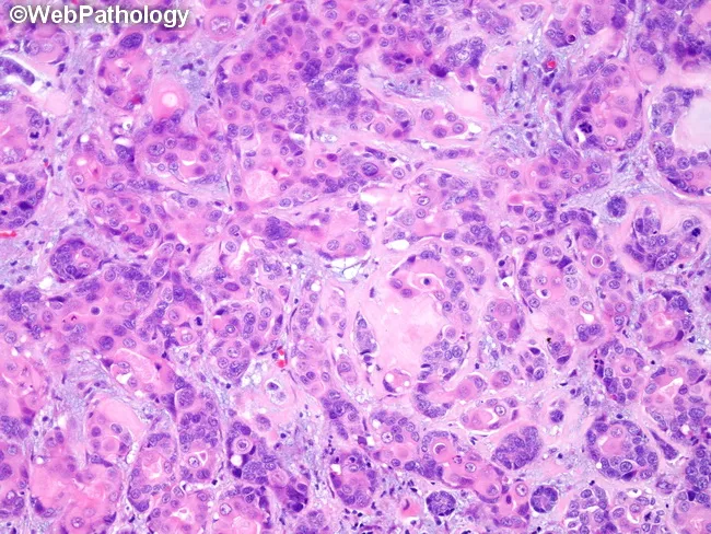
Histology: Light microscopic evaluation of an adenoid cystic carcinoma (ACC) sample, stained with Hematoxylin and Eosin. The tissue shows infiltrative nests and cribriform spaces composed of basaloid myoepithelial-like cells with scant cytoplasm and hyperchromatic, elongated nuclei. A hyalinized basement‑membrane stroma surrounds pseudocystic glandular spaces, producing a characteristic Swiss cheese appearance. Notably, a focal zone demonstrates squamous metaplasia within the tumor and an abrupt dedifferentiation pattern, consistent with high-grade transformation. Perineural invasion is evident in several tumor fronts, explaining potential for extensive local spread. Mitotic activity is variable, with clusters of larger, more pleomorphic cells in the dedifferentiated portion. Overall, the architecture ranges from cribriform and tubular patterns to solid sheets, reflecting the typical heterogeneity of ACC. These features relate to aggressive behavior and a propensity for recurrence after inadequate resection. In context, ACC commonly arises in salivary glands and head‑neck regions and is known for infiltrative margins and nerve tracking. The squamous metaplasia within dedifferentiated ACC carries prognostic significance, suggesting a higher-grade lesion and necessitating multimodal management, including radical surgery with wide margins and adjuvant radiotherapy. Differential considerations include basal cell carcinoma of salivary origin, basaloid squamous carcinoma, and other adenoid cystic variants. This image provides essential learning for recognizing dedifferentiation and perineural spread in ACC.
mucoepidermoid carcinoma salivary gland low grade cystic high grade solid
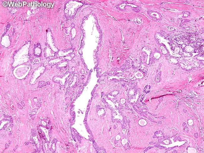
High‑quality brightfield light microscopy of a salivary gland tumor demonstrates classic mucoepidermoid carcinoma features. The specimen is a tissue biopsy from the parotid region, prepared as an H&E stained slide, viewed in routine cross‑sectional orientation. The architecture consists of cystic and gland‑like spaces intermingled with solid nests of epithelial cells. Mucinous cells display goblet or balloon‑like cytoplasm with abundant foamy material and small, dark, peripheral nuclei; these are juxtaposed with epidermoid (squamous) cells showing intercellular bridges and more eosinophilic cytoplasm. Intermediate cells occupy an intermediate phenotype between mucous and squamous elements. Clear cells, when present, contribute to the heterogeneous cell population. In low‑grade MEC, cyst lining is often simple to complex and may reveal cribriform and papillary patterns, with mucous cells predominating and relatively mild cytologic atypia. The surrounding stroma is fibrous and variably myxoid, sometimes with chronic inflammatory infiltrate. The overall impression is a largely cystic, low‑grade malignant neoplasm composed of mucous, squamous, intermediate, and, less frequently, clear cells, consistent with mucoepidermoid carcinoma. Clinically, MECs arise in salivary glands and carry prognostic significance dependent on grade. Lower grade lesions tend to be more cystic with better prognosis than higher grade, solid, infiltrative tumors. This image exemplifies classic MEC histology.
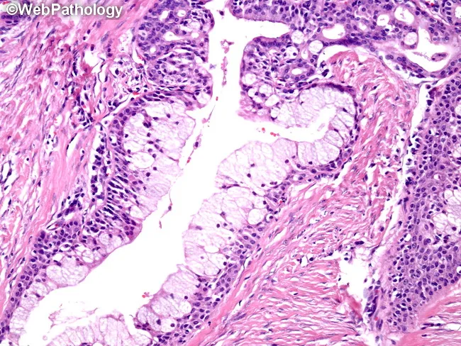
This Light Microscopy image depicts a low-grade mucoepidermoid carcinoma of a salivary gland (likely parotid region) on a hematoxylin and eosin stained section. The tumor demonstrates a mixed cellular phenotype consisting of mucin-producing goblet/balloon cells with abundant foamy cytoplasm, along with epidermoid (squamous) cells and intermediate cells. The mucous cells form cystic or gland-like spaces that may be simple-lined or display complex cribriform and papillary architectures. In places, mucous and epidermoid components occur in solid nests, reflecting the biphasic differentiation typical of MEC. The cyst lining ranges from a single cell layer to multilayered epithelium within fibrous stroma. Overall pattern is predominantly cystic with scattered solid areas, consistent with a low-grade neoplasm; mitotic activity is low and cytologic atypia is minimal. The photograph emphasizes the diagnostic hallmarks: mucous cell predominance, duct-like glandular spaces, cohesive nests of epidermoid cells, and cribriform/papillary configurations. Clinically, MEC is the most common malignant salivary gland tumor; grading (low, intermediate, high) guides prognosis and treatment including surgical excision and possible radiotherapy. The image is valuable for education and pathology training, enabling recognition of cell-lineage diversity, mucin production, and architectural patterns that distinguish MEC from adenosquamous and other salivary gland carcinomas.
acinic cell carcinoma serous acinar cells zymogen granules salivary gland
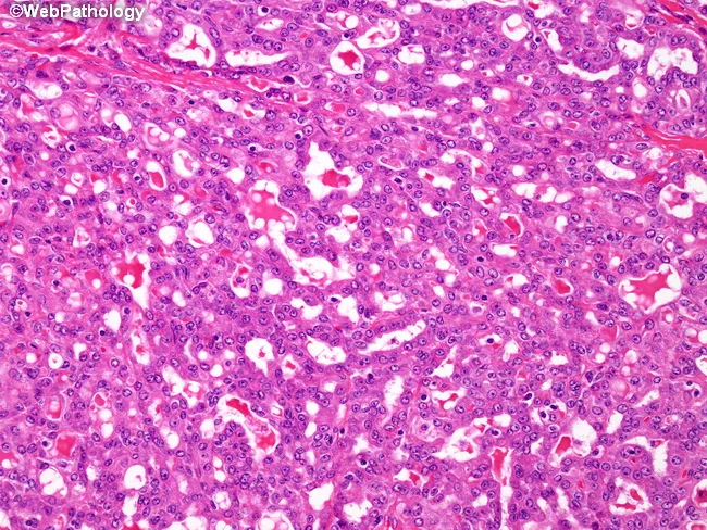
Modality: Light microscopy of hematoxylin–eosin stained salivary gland tissue, at variable magnification showing classic acinic cell carcinoma histology. The tumor arises in salivary gland parenchyma, typically in the parotid, and demonstrates serous acinar differentiation with abundant zymogen granules. The architectural pattern is dominantly ductal–tubular and microcystic, with back-to-back tubular or acinar-like structures lined by small to medium-sized cells. The cytoplasm is eosinophilic and granular due to secretory granules; numerous intracellular eosinophilic globules may be evident. A conspicuous feature is extensive acinar-type secretory material within luminal spaces and microcysts, which is PAS-positive and resistant to diastase digestion, consistent with diastase-negative mucopolysaccharide-rich secretions resembling zymogen granule content. The nuclei are generally round to oval with mild pleomorphism, and mitotic activity is typically low. The stroma may be fibrous or myxoid, and perineural invasion can be seen in some cases but is not required for diagnosis. Clinically, these lesions present as slow-growing, painless masses in the major salivary glands. The combination of tubular/solid microcystic patterns with PAS-D positive secretory material and abundant zymogen granules supports a diagnosis of acinic cell carcinoma, differentiating it from mucoepidermoid and adenoid cystic carcinomas. Immunohistochemistry often shows S-100 negative and DOG1 variable expression. Supportive for diagnosis and grading.
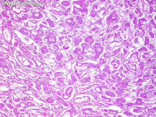
This is a histopathology image of a salivary gland neoplasm showing acinic cell carcinoma with a follicular/glandular pattern. The material is a Hematoxylin and Eosin stained section viewed on light microscopy. Tumor architecture displays microcystic and follicular arrangements with round to oval acinar cells that have granular basophilic cytoplasm and small, central nuclei. Cytoplasmic granules represent zymogen granules consistent with serous acinar differentiation. The lesion shows ducts and cyst-like lumina within nests of tumor cells, with relatively mild cytologic atypia and infrequent mitotic activity. In places, cells form sheets and tubules that emphasize the glandular component. Overall, the image illustrates the characteristic acinic cell histology and the follicular variant pattern described in the literature. Diagnostic significance lies in recognizing serous acinar differentiation to distinguish acinic cell carcinoma from mucoepidermoid carcinoma or adenoid cystic carcinoma, which have different management and prognosis. Clinically, patients typically present with a slowly enlarging parotid or minor salivary gland mass. Immunohistochemical panels and molecular studies can aid confirmation. For education and research, this image serves as a reference for pattern recognition, differential diagnosis, and teaching about salivary gland neoplasms and their histologic subtypes. This reference aids learners in recognizing variants and informs clinical decision making process.
Important note on sources: Ackerman's Surgical Pathology is not available in the medical library. This answer is drawn exclusively from the two Robbins books in the collection: Robbins, Cotran & Kumar - Pathologic Basis of Disease (Robbins PBD, 10th ed.) and Robbins & Kumar - Basic Pathology. Where IHC and molecular genetics data from Ackerman's is specifically requested, the most authoritative current references are noted.
Histopathological Features of Salivary Gland Tumors
Source: Robbins, Cotran & Kumar - Pathologic Basis of Disease | Robbins & Kumar - Basic Pathology
Overview
| Benign | Malignant |
|---|---|
| Pleomorphic adenoma (mixed tumor) | Mucoepidermoid carcinoma |
| Warthin tumor | Acinic cell carcinoma |
| Oncocytoma | Adenoid cystic carcinoma |
| Canalicular adenoma | Salivary duct carcinoma |
| Basal cell adenoma | Carcinoma ex pleomorphic adenoma |
| Other adenomas | Other carcinomas |
A. BENIGN TUMORS
1. Pleomorphic Adenoma (Mixed Tumor)
Histopathology (Morphology)
- Gross: Rounded, well-demarcated mass; rarely exceeds 6 cm. Cut surface is gray-white with myxoid and blue translucent chondroid areas. Capsule may be incomplete at the palate.
- Microscopy: The hallmark is morphologic heterogeneity (not true nuclear pleomorphism):
- Epithelial component: Ductal cells or myoepithelial cells arranged as ducts, acini, irregular tubules, strands, or sheets
- Mesenchymal-like stroma: Chondro-myxoid background with loose myxoid tissue, islands of cartilage (chondroid), and rarely foci of bone
- Ducts lined by cuboidal-to-columnar cells with an underlying layer of deeply chromatic, small myoepithelial cells
- Islands of well-differentiated squamous epithelium may be present
- No epithelial dysplasia or mitotic activity in most cases
- Tumors behave similarly whether epithelial or mesenchymal elements predominate
Molecular Genetics (Robbins PBD)
- PLAG1 overexpression - chromosomal rearrangements inducing overexpression of PLAG1, a transcription factor that promotes genes involved in growth factor receptor signaling
- PLAG1 is fused to CTNNB1, TCEA1, or other partners
- HMGA2 gene mutations - DNA-binding protein; associated with cases lacking PLAG1 overexpression
- Radiation exposure increases risk
IHC Markers
- Epithelial cells: cytokeratin positive (low-MW CK)
- Myoepithelial cells: SMA, calponin, p63, S-100 positive
- GFAP (Glial Fibrillary Acidic Protein) positive - a distinctive marker; almost all pleomorphic adenomas are GFAP-positive (contrasting with polymorphous adenocarcinoma which is GFAP-negative)
Clinical Behavior
- 4% recur after parotidectomy; 25% recur after simple enucleation (due to incomplete capsule)
- Malignant transformation to carcinoma ex pleomorphic adenoma: 2% if <5 years duration; 10% if >15 years duration
- Transformed cancers (usually adenocarcinoma or undifferentiated carcinoma) carry 30-50% mortality at 5 years
2. Warthin Tumor (Papillary Cystadenoma Lymphomatosum)
Histopathology (Morphology)
- Gross: Round-to-oval encapsulated mass, 2-5 cm; pale gray surface with cystic/cleft-like spaces filled with mucinous or serous secretions and polypoid lymphoepithelial projections
- Microscopy (biphasic architecture):
- Epithelial component: Double layer of oncocytic (eosinophilic, granular cytoplasm due to abundant mitochondria) cells:
- Inner layer: columnar cells (luminal)
- Outer layer: cuboidal cells
- Secretory cells in the inner layer account for luminal secretions
- Foci of squamous metaplasia may be present
- Lymphoid stroma with prominent germinal centers - this is the defining biphasic feature
- Epithelial component: Double layer of oncocytic (eosinophilic, granular cytoplasm due to abundant mitochondria) cells:
Molecular Genetics
- Histogenesis debated - may be reactive (polyclonal) rather than truly neoplastic (monoclonal)
- Epithelial cells thought to secrete chemoattractants for the reactive lymphoid stroma
- 8x increased risk in smokers; 10% multifocal, 10% bilateral
IHC Markers
- Epithelial cells: CK7, CK8, CK18, CK19 positive (cytokeratin panel)
- Oncocytic cytoplasm: PTAH positive (phosphotungstic acid-hematoxylin highlights mitochondria)
- Lymphoid component: predominantly polyclonal B-cells with IgA-producing cells; scattered T-cells and S-100 positive dendritic cells
Clinical Behavior
- Benign; low recurrence rate of only 2% after resection
B. MALIGNANT TUMORS
3. Mucoepidermoid Carcinoma (MEC)
Histopathology (Morphology)
- Gross: Up to 8 cm; apparently circumscribed but lacks a well-defined capsule and is often infiltrative; pale gray-white with small mucin-containing cysts
- Microscopy - three cell types define MEC:
- Mucous cells - abundant light blue mucin in cytoplasm; nuclei displaced to periphery; line cystic spaces
- Squamoid (epidermoid) cells - large, abundant pink cytoplasm; look squamoid but are not truly keratinizing (true keratinization → consider adenosquamous carcinoma)
- Intermediate cells - most numerous; modest pink or clear cytoplasm; round nuclei with open chromatin
- Architecture: cords, sheets, or cystic configurations; mixture of solid and cystic elements
- Special stains: PAS, mucicarmine, or Alcian blue highlight mucin when scant
Grading (Robbins PBD)
| Grade | Features | Prognosis |
|---|---|---|
| Low | Predominantly cystic, mucous cells dominant, minimal atypia | 5-yr survival >90%; local recurrence ~15%; rare mets |
| Intermediate | Mixed cystic and solid | Intermediate prognosis |
| High | Predominantly solid sheets, anaplastic cells, scant mucous cells, necrosis | 5-yr survival ~50%; 30% recur, 30% distant mets |
Molecular Genetics (Robbins PBD) - KEY
- >50% of MECs: balanced chromosomal translocation t(11;19)(q21;p13)
- Creates fusion gene CRTC1::MAML2
- The MAML2 portion of the fusion cannot interact with Notch signaling components
- CRTC1::MAML2 fusion protein perturbs Notch and cAMP-dependent signaling pathways
- CRTC3::MAML2 fusion in a smaller subset (<10%)
- Fusion predominantly present in low and intermediate grade tumors; useful as a diagnostic marker
IHC Markers
- IHC has limited diagnostic utility (Robbins PBD); diagnosis is primarily morphologic
- Cytokeratins: positive (especially CK5/6, CK7)
- p63: positive in squamoid/intermediate cells
- Mucin stains (PAS, mucicarmine, Alcian blue) confirm mucous cells
4. Adenoid Cystic Carcinoma (AdCC)
Histopathology (Morphology)
- Gross: Poorly circumscribed, infiltrative, gray-pink lesions
- Microscopy - three growth patterns (Fig. 16.23, Robbins PBD):
- Cribriform (Swiss cheese) - most characteristic; tumor cells surround rounded spaces filled with hyaline material representing excess basement membrane (PAS-positive, type IV collagen)
- Tubular - double-layered tubules
- Solid/basaloid - sheets of cells; associated with higher grade and worse prognosis
- Cells are basaloid: small, angular, scant cytoplasm, hyperchromatic elongated nuclei, high N:C ratio
- Biphasic: epithelial cells (inner/luminal) + myoepithelial cells (outer/abluminal)
- Perineural invasion is characteristic and clinically important - tumors track along nerve sheaths
NOTCH1-based morphologic subtypes (Robbins PBD - 10th ed.)
| Variant | Genetics | Morphology | Prognosis |
|---|---|---|---|
| NOTCH1 wild-type | MYB::NFIB or MYBL1::NFIB | Cribriform predominant; mixture of basal myoepithelial + epithelial cells | Better |
| NOTCH1 mutated | NOTCH1 activating mutation | Predominantly solid nests/sheets of epithelial cells | Worse; increased proclivity to metastasize to bone and liver |
Grading (Perzin/Szanto system)
- Grade 1: No solid areas; cribriform/tubular only
- Grade 2: <30% solid component
- Grade 3: >30% solid component (worst prognosis)
Molecular Genetics (Robbins PBD) - KEY
- MYB::NFIB fusion gene rearrangement - present in majority (80-90%)
- Arising from translocation t(6;9)(q22-24;p13-23)
- MYBL1::NFIB fusion - smaller subset
- NOTCH1 activating mutations - associated with solid pattern, worse outcome
IHC Markers
- c-KIT (CD117) positive - highly characteristic; one of the most useful markers
- SOX10 positive
- p63 and p40 positive in myoepithelial/abluminal cells (useful to distinguish from PAC: AdCC is p40+p63+ dual positive; PAC is p40-negative/p63-positive)
- SMA, calponin: positive in abluminal myoepithelial layer
- Type IV collagen and laminin: positive in hyaline pseudocystic spaces (confirms excess basement membrane)
- S-100: variable/focal
- Negative: DOG1 (helps distinguish from acinic cell carcinoma)
Clinical Behavior
- Slow-growing but unpredictable; perineural spread is hallmark
- Regular recurrence; 50%+ eventually recur at distant sites (lung most common), sometimes decades after removal
- 5-yr survival ~60-70% → drops to ~30% at 10 yrs → ~15% at 15 yrs (Robbins PBD)
- Minor salivary gland origin carries worse prognosis
5. Acinic Cell Carcinoma
Histopathology (Morphology)
- Gross: Generally small, well-circumscribed
- Microscopy: Cells resemble normal serous acinar cells:
- Small, round nuclei; abundant purple zymogen granules in cytoplasm (PAS-positive, diastase-resistant)
- Cells can also be clear or vacuolated
- Growth patterns: sheets, microcysts, glands, follicles, or papillae
- Second classic feature: dense lymphoid infiltrate with germinal centers (recruited by tumor cells)
- High-grade transformation (dedifferentiation): sheet-like growth, nuclear pleomorphism, mitoses, necrosis; occurs in up to ~20% of cases
IHC Markers
- DOG1 (Discovered on GIST-1): positive with classic membrane pattern in acinic cells and intercalated duct epithelium - newer, very helpful marker
- PAS positive (diastase-resistant): confirms zymogen granules
- S-100: negative or focal/patchy (key distinction from secretory carcinoma which is diffusely S-100+)
- Mammaglobin: negative (unlike secretory carcinoma which is positive)
- CK7: positive
Molecular Genetics
- No defining translocation in classic acinic cell carcinoma
- Some cases show FISH-detectable copy number variations
- High-grade transformed tumors may show TP53, ERBB2 alterations
Clinical Behavior
- Classically low-grade; 5-yr survival ~90% but drops to ~60% at 20 years
- 10-15% metastasize to lymph nodes; lung and bones for distant mets
- Survival inversely correlated with solid/sheet growth pattern and high-grade transformation
6. Carcinoma Ex Pleomorphic Adenoma
Histopathology
- Pre-existing pleomorphic adenoma with admixed carcinoma
- PA component: myxoid stroma, ductal structures, myoepithelial sheets, chondroid areas (may be hyalinized/sclerotic from overgrowth)
- Carcinoma component: most commonly poorly differentiated adenocarcinoma NOS, salivary duct carcinoma, or undifferentiated carcinoma
- Invasion classification (critical for prognosis):
- Noninvasive (intracapsular/in situ)
- Minimally invasive (≤1.5 mm beyond capsule) - behaves like benign PA; essentially no risk of mets
- Widely invasive (>1.5 mm) - 5-yr survival 26-65%, 20-yr survival 0-38%
- Malignant transformation: 2% if <5 yrs duration → 10% if >15 yrs duration (Robbins)
C. KEY IHC PANEL SUMMARY
| Tumor | Key Positive Markers | Key Negative Markers | Defining Feature |
|---|---|---|---|
| Pleomorphic adenoma | CK, SMA, calponin, p63, GFAP, S-100 (myoepithelial) | - | Biphasic + chondromyxoid stroma |
| Warthin tumor | CK7/8/18/19, PTAH | - | Oncocytic bilayer + lymphoid stroma |
| MEC | CK5/6, p63, mucin stains (PAS, mucicarmine) | (IHC limited utility) | 3 cell types + CRTC1::MAML2 |
| Adenoid cystic carcinoma | c-KIT (CD117), SOX10, p63, p40, SMA, CK | S-100 (focal), DOG1 (neg) | Cribriform + MYB::NFIB |
| Acinic cell carcinoma | DOG1, PAS-D, CK7 | S-100, mammaglobin | Zymogen granules + lymphoid infiltrate |
| Secretory carcinoma | S-100 (diffuse), mammaglobin, GCDFP-15 | DOG1 | ETV6::NTRK3 fusion |
D. MOLECULAR GENETICS SUMMARY TABLE
| Tumor | Translocation / Fusion Gene | Notes |
|---|---|---|
| Pleomorphic adenoma | PLAG1 fused to CTNNB1, TCEA1, or others | ~30% of cases; also HMGA2 mutations |
| Mucoepidermoid carcinoma | CRTC1::MAML2 t(11;19)(q21;p13) >50% | Perturbs Notch + cAMP signaling; present in low/intermediate grade |
| CRTC3::MAML2 | <10% of cases | |
| Adenoid cystic carcinoma | MYB::NFIB t(6;9) | 80-90% of cases |
| MYBL1::NFIB | Smaller subset | |
| NOTCH1 activating mutation | Solid pattern; worse prognosis; mets to bone/liver | |
| Acinic cell carcinoma | No defining translocation | - |
| Secretory carcinoma | ETV6::NTRK3 t(12;15)(p13;q25) | Defines the entity; breast analogue; targetable with TRK inhibitors |
| Carcinoma ex PA | Inherits PA alterations (PLAG1/HMGA2) | TP53, ERBB2, CDK alterations in malignant component |
E. DETECTION METHODS FOR MOLECULAR ALTERATIONS
- FISH (Fluorescence In Situ Hybridization) - primary method for fusion gene detection
- PCR / RT-PCR - for gene fusion transcripts
- Next-generation sequencing (NGS) - increasingly used for comprehensive profiling
Note on Ackerman's Surgical Pathology: This classic reference (Rosai and Ackerman's Surgical Pathology, 11th ed.) is not available in the current medical library. Ackerman's contains the most granular morphologic descriptions of salivary gland tumors including detailed FNA cytology correlation, rare variants (epithelial-myoepithelial carcinoma, hyalinizing clear cell carcinoma, polymorphous adenocarcinoma, intraductal carcinoma, myoepithelial carcinoma), and variant-specific IHC panels. For complete Ackerman's coverage, direct access to that text is required.
- Robbins, Cotran & Kumar - Pathologic Basis of Disease, pp. 696-699
- Robbins & Kumar - Basic Pathology, pp. 543-544