
prepare a powerpoint presentation on case presentation on pancreatic cnacer for a general surgery resident of GMC,Ongole
pancreatic neoplasms AND surgery
NCCN pancreatic adenocarcinoma guideline resectability criteria CA 19-9 2025
pancreatic adenocarcinoma CT scan pancreatic head mass double duct sign and Whipple procedure anatomy
~/
pancreatic ductal adenocarcinoma contrast enhanced CT scan
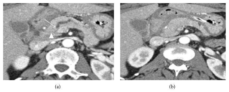
Two-panel contrast-enhanced axial CT scan of the abdomen focused on the pancreatic head during the pancreatic phase. Image (a) demonstrates a markedly dilated pancreatic duct with an abrupt termination in the pancreatic head (marked by an arrow), a classic indirect sign of an obstructing lesion. Despite the ductal obstruction, no discrete hypoattenuating mass is clearly visualized at the point of termination (arrowhead). Image (b), taken 1 cm inferiorly, shows the pancreatic head exhibiting homogeneous enhancement and normal morphology, lacking typical signs of malignancy such as mass effect or hypoattenuation. This visual represents a 'CT-isoattenuating' pancreatic ductal adenocarcinoma, where the tumor's density matches the surrounding parenchyma, making it occult on standard imaging despite its significant size (3.9 cm). This educational case highlights the importance of recognizing secondary imaging signs like pancreatic ductal dilation and abrupt 'cut-off' when a primary mass is not visible.
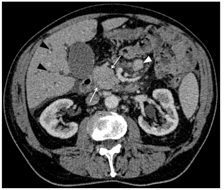
This diagnostic image is an axial contrast-enhanced computed tomography (CT) scan of the abdomen during the portal venous phase. The focal point is a homogeneous, purely solid mass located within the pancreatic head (indicated by white arrows). Associated secondary findings include prominent intrahepatic bile duct dilatation (black arrowheads), suggesting biliary obstruction. An enlarged mesenteric lymph node is also visible (white arrowhead). The image demonstrates signs of peritoneal involvement, notably the presence of ascites within the abdominal cavity. Anatomical landmarks include the liver, gallbladder, both kidneys, and the vertebral body. This case illustrates the radiological presentation of a secondary pancreatic lesion, specifically metastasis (in this clinical context from lung adenocarcinoma), which can mimic primary pancreatic ductal adenocarcinoma but often presents with distinct imaging characteristics such as well-defined margins and varying degrees of ductal dilatation.
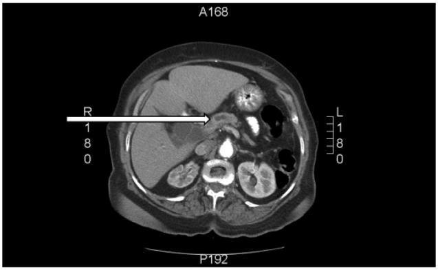
This diagnostic image is an axial contrast-enhanced CT scan of the abdomen at the level of the pancreatic head. The scan demonstrates a significant pathological finding in the pancreatic region: an ill-defined, hypo-attenuating mass located at the head of the pancreas, indicated by a horizontal white arrow. This lesion causes an abrupt ductal cutoff, resulting in secondary dilatation of the proximal biliary tree and the main pancreatic duct (the 'double duct sign'). Surrounding anatomical structures are clearly visible, including the liver in the right upper quadrant, the spleen in the left upper quadrant, and both kidneys in the retroperitoneal space posteriorly. The stomach and bowel loops are seen centrally and anteriorly. This imaging pattern is highly characteristic of pancreatic ductal adenocarcinoma (PDAC) causing obstructive jaundice. The scan is intended for intermediate to advanced medical learners to illustrate the radiological presentation of distal biliary obstruction and pancreatic malignancy.
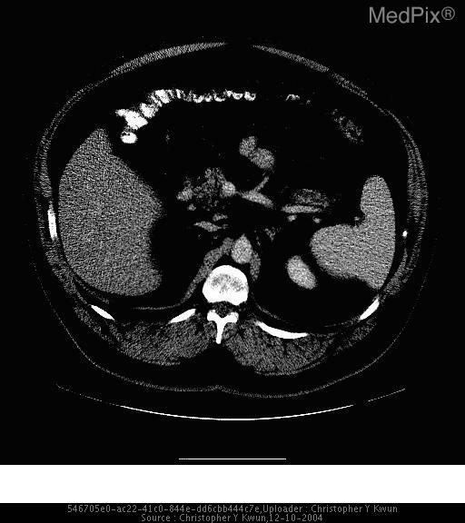
**Imaging Modality:** Contrast-enhanced axial Computed Tomography (CT) scan.
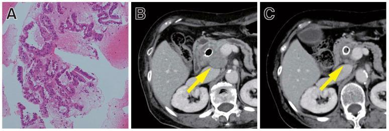
A multi-panel medical image illustrating the clinical response of microsatellite instability-high (MSI-H) pancreatic cancer to immunotherapy. Panel A is a histopathological micrograph (H&E stain) of a pancreatic tissue sample obtained via EUS-FNB, showing irregular, hyperchromatic glandular structures characteristic of adenocarcinoma. Panels B and C are axial contrast-enhanced CT scans of the abdomen at the level of the pancreas. Scan B depicts a baseline state with a large, ill-defined, heterogeneous mass in the head of the pancreas (indicated by a yellow arrow) that was refractory to prior chemotherapy. Scan C shows the same anatomical region 8 weeks into pembrolizumab therapy, demonstrating a significant reduction in the size and density of the pancreatic head mass (indicated by a yellow arrow), representing a partial clinical response. The image serves to demonstrate the utility of biomarker testing (MSI-H) in tailoring immunotherapy for pancreatic ductal adenocarcinoma.
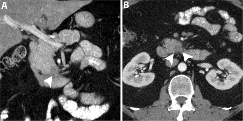
This diagnostic image consists of two panels: (A) a coronal and (B) an axial contrast-enhanced CT scan in the pancreatic phase, focused on the abdominal cavity. The images demonstrate a small, subtle hypoattenuating mass located within the uncinate process of the pancreas, highlighted by white arrowheads. Notably, despite the presence of this focal lesion, there is a distinct absence of secondary signs typically associated with pancreatic ductal adenocarcinoma (PDAC), such as dilatation of the common bile duct (CBD) or the main pancreatic duct (MPD). The overall morphology of the pancreatic head, body, and tail appears otherwise unremarkable with preserved parenchymal enhancement. This case illustrates a diagnostic challenge in early-stage uncinate process PDAC, where the tumor's specific anatomical location prevents biliary or pancreatic ductal obstruction, potentially delaying detection. Such findings are critical for radiologists and oncology specialists to recognize as 'subtle' pancreatic masses during routine or targeted abdominal imaging.
pancreaticoduodenectomy Whipple procedure surgical anatomy illustration
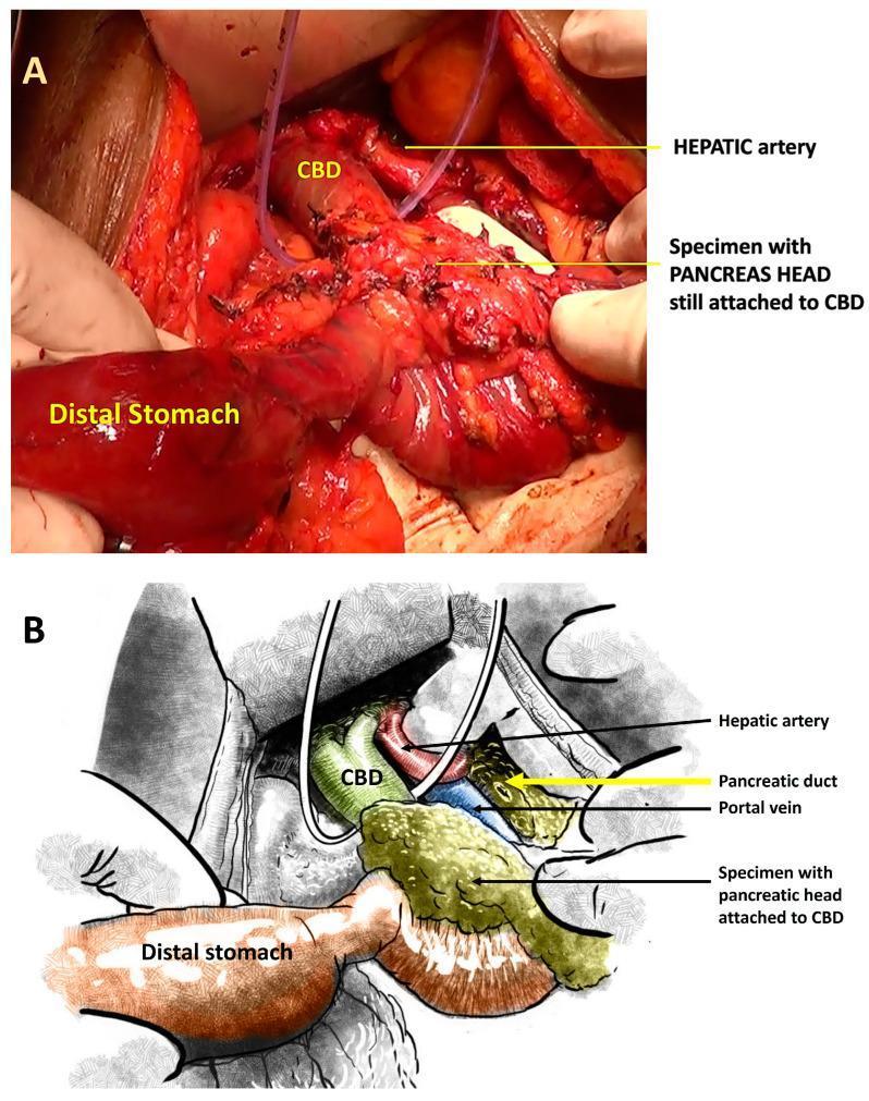
Educational panel illustrating hepatobiliary surgical anatomy during a pancreaticoduodenectomy (Whipple procedure). Image A is an intraoperative clinical photograph displaying a surgical specimen consisting of the pancreatic head remaining attached to the common bile duct (CBD), positioned adjacent to the distal stomach. Visible medical elements include surgical sutures on the CBD and a vascular loop or drain for vessel retraction. Image B is a corresponding schematic illustration that clarify the spatial relationships of the deep vasculature. It depicts the hepatic artery superiorly, the portal vein located posteriorly to the CBD and pancreatic head, and labels the pancreatic duct. This visual aids in understanding the COMBILAST surgical sequence, which emphasizes late division of the bile duct to minimize biliary spillage and surgical site infection (SSI). The content is intended for advanced medical education in surgical oncology and gastrointestinal surgery, focusing on anatomical preservation and operative technique.
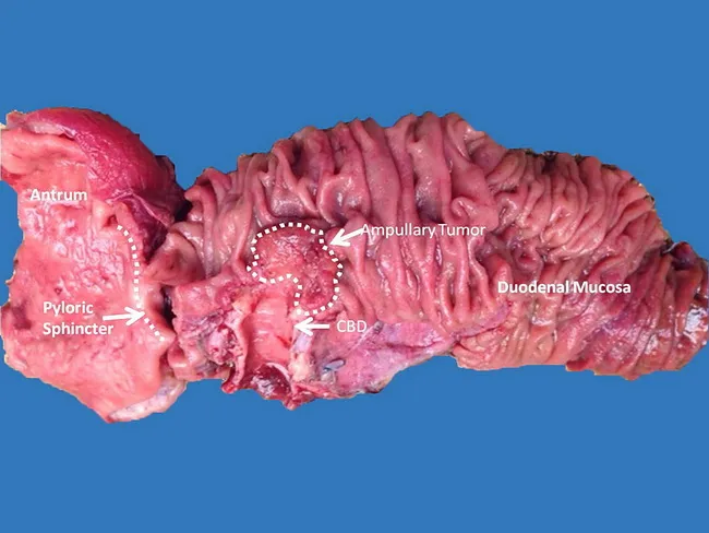
This is a macroscopic photograph of a freshly dissected pancreaticoduodenectomy (Whipple) specimen illustrating an ampullary carcinoma at the ampulla of Vater. The resected tissue includes the duodenum with its mucosa, the common bile duct (CBD), the pancreatic head region, and the surrounding periampullary structures. The labeled structures in the image identify the Antrum and pyloric sphincter on the proximal stomach side, the CBD as a tubular channel, and the ampullary tumor located at the major duodenal papilla. The tumor appears as a nodular, irregular lesion at the ampullary region, with tumor margins that abut the surrounding duodenal mucosa and biliary tract. Although imaging studies such as computed tomography (CT) and endoscopic ultrasound (EUS) are used preoperatively, the lesion itself may be occult on routine imaging and definitive assessment relies on histopathology of cytologic material, endoscopic biopsy, or transduodenal sampling. The Whipple procedure remains the standard surgical management for invasive ampullary carcinoma due to complex regional anatomy and the need for en bloc resection of the pancreatic head, duodenum, bile duct, and often part of the stomach. In noninvasive or limited disease, transduodenal resection may be considered. Image courtesy of Dr. Terri Crook, Dallas, Texas, for educational and diagnostic illustration.
mkdir -p "/tmp/workspace/pancreatic-cancer-case-presentation/src" "/tmp/workspace/pancreatic-cancer-case-presentation/assets" "/tmp/workspace/pancreatic-cancer-case-presentation/output" && python /tmp/skills/shared/scripts/fetch_images.py "https://cdn.orris.care/cdss_images/pmc_clinical_VQA_1ca9176ceb4574fecdfa769212b93d61d44d66ff43fe1ad44bb3c93a6398d531.jpg" "https://cdn.orris.care/cdss_images/pmc_clinical_VQA_0989e0e819d933b464f4e8496dd9e2abc88b7065d5c993b1beb83c5c6b94727e.jpg" > "/tmp/workspace/pancreatic-cancer-case-presentation/assets/images.json"
python -c "import json; d=json.load(open('/tmp/workspace/pancreatic-cancer-case-presentation/assets/images.json')); print([(x.get('url'), bool(x.get('base64')), x.get('error')) for x in d])"
python - <<'PY' import json,base64 from pathlib import Path p=Path('/tmp/workspace/pancreatic-cancer-case-presentation/assets') d=json.loads((p/'images.json').read_text()) for i,x in enumerate(d): raw=base64.b64decode(x['base64'].split(',',1)[1]) (p/f'image_{i+1}.jpg').write_bytes(raw) PY cat > "/tmp/workspace/pancreatic-cancer-case-presentation/src/create_deck.py" <<'PY' from pptx import Presentation from pptx.util import Inches, Pt from pptx.dml.color import RGBColor from pptx.enum.shapes import MSO_SHAPE from pptx.enum.text import PP_ALIGN, MSO_ANCHOR from pptx.enum.dml import MSO_THEME_COLOR from pptx.enum.shapes import MSO_CONNECTOR from pathlib import Path OUT=Path('/tmp/workspace/pancreatic-cancer-case-presentation/output/Pancreatic_Cancer_Case_Presentation_GMC_Ongole.pptx') AS=Path('/tmp/workspace/pancreatic-cancer-case-presentation/assets') prs=Presentation(); prs.slide_width=Inches(13.333); prs.slide_height=Inches(7.5) blank=prs.slide_layouts[6] NAVY=RGBColor(8,31,52); TEAL=RGBColor(20,130,137); GOLD=RGBColor(231,174,53); PALE=RGBColor(239,246,247); INK=RGBColor(26,39,50); GREY=RGBColor(94,109,120); RED=RGBColor(183,55,52); WHITE=RGBColor(255,255,255); LIGHT=RGBColor(221,232,235) def rect(sl,x,y,w,h,color, radius=False, line=None): sh=sl.shapes.add_shape(MSO_SHAPE.ROUNDED_RECTANGLE if radius else MSO_SHAPE.RECTANGLE, Inches(x), Inches(y), Inches(w), Inches(h)) sh.fill.solid(); sh.fill.fore_color.rgb=color; sh.line.color.rgb=line or color if radius: sh.adjustments[0]=0.08 return sh def text(sl,s,x,y,w,h,size=20,color=INK,bold=False,align=None,font='Aptos',val=None): tb=sl.shapes.add_textbox(Inches(x), Inches(y), Inches(w), Inches(h)); tf=tb.text_frame tf.clear(); tf.word_wrap=True; tf.margin_left=0; tf.margin_right=0; tf.margin_top=0; tf.margin_bottom=0 p=tf.paragraphs[0]; p.text=s; p.font.name=font; p.font.size=Pt(size); p.font.bold=bold; p.font.color.rgb=color if align: p.alignment=align if val: tf.vertical_anchor=val return tb def bullet_box(sl,items,x,y,w,h,size=18,accent=TEAL): box=rect(sl,x,y,w,h,WHITE,True,line=LIGHT) top=y+0.25 for i,item in enumerate(items): rect(sl,x+0.22,top+0.12,0.10,0.10,accent,True) text(sl,item,x+0.42,top,w-0.65,0.48,size,INK) top+=0.68 return box def header(sl,title,kicker='CASE PRESENTATION'): rect(sl,0,0,13.333,0.34,NAVY); text(sl,kicker,0.55,0.48,2.2,0.22,9,TEAL,True) text(sl,title,0.55,0.75,12.0,0.52,27,NAVY,True) rect(sl,0.55,1.38,1.35,0.05,GOLD) def footer(sl,n): rect(sl,0,7.22,13.333,0.28,NAVY); text(sl,'Department of General Surgery | Government Medical College, Ongole',0.55,7.275,8.6,0.13,8,WHITE) text(sl,str(n),12.25,7.27,0.45,0.15,9,WHITE,True,PP_ALIGN.RIGHT) def title_slide(): sl=prs.slides.add_slide(blank); rect(sl,0,0,13.333,7.5,NAVY); rect(sl,0,0,0.28,7.5,TEAL) # abstract pancreas-style arcs for x,y,w,h,c in [(9.7,0.6,4.0,4.0,RGBColor(14,61,83)),(10.8,1.6,2.8,2.8,TEAL),(9.2,4.55,3.1,1.1,GOLD)]: sh=sl.shapes.add_shape(MSO_SHAPE.ARC, Inches(x), Inches(y), Inches(w), Inches(h)); sh.line.color.rgb=c; sh.line.width=Pt(6); sh.fill.background() text(sl,'CASE PRESENTATION',0.75,0.95,3.0,0.25,11,RGBColor(124,211,204),True) text(sl,'Pancreatic Cancer',0.75,1.48,8.4,0.75,42,WHITE,True) text(sl,'A resectable pancreatic head adenocarcinoma',0.77,2.38,7.4,0.38,22,RGBColor(218,233,238)) rect(sl,0.76,3.20,1.55,0.06,GOLD) text(sl,'For General Surgery Residents',0.77,3.55,5.0,0.3,17,WHITE,True) text(sl,'Government Medical College, Ongole',0.77,3.93,5.4,0.28,16,RGBColor(191,213,220)) text(sl,'Illustrative teaching case | Clinical decisions require multidisciplinary discussion and institutional protocols.',0.77,6.68,10.8,0.28,11,RGBColor(191,213,220)) def add_case_slide(n): sl=prs.slides.add_slide(blank); header(sl,'Clinical vignette') text(sl,'A focused story that should trigger a pancreatic protocol CT',0.55,1.65,8.6,0.3,16,GREY) rect(sl,0.55,2.20,3.0,3.85,PALE,True); text(sl,'MR',1.05,2.55,1.0,0.55,34,TEAL,True); text(sl,'62-year-old man',1.05,3.20,1.9,0.35,20,NAVY,True) text(sl,'Farmer • ECOG 1\nNo previous surgery',1.05,3.72,1.9,0.7,16,INK) rect(sl,1.05,4.82,1.3,0.35,GOLD,True); text(sl,'DAY 0',1.28,4.91,0.8,0.12,9,NAVY,True,PP_ALIGN.CENTER) items=['Progressive painless jaundice for 3 weeks','Dark urine, pale stools, generalized pruritus','Unintentional weight loss: 6 kg in 2 months','New-onset diabetes mellitus diagnosed 4 months earlier','No fever, biliary colic, or alcohol-related pancreatitis history'] bullet_box(sl,items,3.85,2.20,8.85,3.85,17) text(sl,'Surgical problem representation',0.55,6.35,2.9,0.25,13,TEAL,True) text(sl,'Older adult with obstructive jaundice + weight loss + recent diabetes: pancreatic head / periampullary malignancy until proven otherwise.',0.55,6.67,11.9,0.3,17,NAVY,True) footer(sl,n) def generic_slide(n,title,subtitle,left_title,left_items,right_title,right_items, note=None): sl=prs.slides.add_slide(blank); header(sl,title); text(sl,subtitle,0.55,1.65,12.0,0.3,15,GREY) text(sl,left_title,0.60,2.12,5.7,0.3,17,TEAL,True); text(sl,right_title,6.93,2.12,5.7,0.3,17,TEAL,True) bullet_box(sl,left_items,0.55,2.55,5.95,3.65,16) bullet_box(sl,right_items,6.83,2.55,5.95,3.65,16,GOLD) if note: text(sl,note,0.60,6.52,11.9,0.28,13,NAVY,True) footer(sl,n); return sl def image_slide(n,title,subtitle,img,caption,points): sl=prs.slides.add_slide(blank); header(sl,title); text(sl,subtitle,0.55,1.65,12,0.30,15,GREY) rect(sl,0.55,2.18,6.65,3.80,RGBColor(20,20,20),True) sl.shapes.add_picture(str(img), Inches(0.67), Inches(2.3), width=Inches(6.4), height=Inches(3.55)) text(sl,caption,0.67,6.08,6.3,0.42,11,GREY) bullet_box(sl,points,7.52,2.18,5.23,3.80,16) text(sl,'Imaging point',7.55,6.30,1.3,0.2,12,TEAL,True); text(sl,'Vascular interface and metastatic survey determine the operative pathway.',7.55,6.57,5.0,0.28,13,NAVY,True) footer(sl,n) def flow_slide(n): sl=prs.slides.add_slide(blank); header(sl,'Diagnostic pathway and staging') text(sl,'Do not let relief of jaundice delay oncologic staging.',0.55,1.65,12,0.3,15,GREY) steps=[('1','Confirm obstruction','LFT pattern, US'),('2','Protocol imaging','Pancreas CT, chest CT'),('3','Tissue when needed','EUS-FNB'),('4','MDT decision','Resectability + biology')] x=0.6 for i,(num,ttl,sub) in enumerate(steps): rect(sl,x,2.35,2.72,1.75,PALE,True,line=LIGHT); rect(sl,x+0.18,2.55,0.48,0.48,TEAL if i<3 else GOLD,True); text(sl,num,x+0.18,2.64,0.48,0.15,14,WHITE,True,PP_ALIGN.CENTER) text(sl,ttl,x+0.18,3.18,2.3,0.25,16,NAVY,True); text(sl,sub,x+0.18,3.56,2.3,0.32,13,INK) if i<3: text(sl,'→',x+2.83,3.00,0.45,0.4,27,TEAL,True,PP_ALIGN.CENTER) x+=3.17 text(sl,'Current case',0.60,4.72,1.5,0.25,14,TEAL,True) bullet_box(sl,['Pancreas-protocol CT: 3.1 cm head mass; no arterial contact; patent SMV-PV confluence; no distant metastases','EUS-FNB: adenocarcinoma; baseline CA 19-9 interpreted after bilirubin improves','CT chest: no metastasis; nutrition, performance status, anaesthetic fitness assessed'],0.60,5.05,12.1,1.42,15) footer(sl,n) def resect_slide(n): sl=prs.slides.add_slide(blank); header(sl,'Resectability: the operative language') text(sl,'Classify using high-quality cross-sectional imaging and a pancreas MDT.',0.55,1.65,12,0.3,15,GREY) cols=[('Resectable',TEAL,['No distant metastases','No arterial tumor contact','No SMV/PV contact or limited contact without contour irregularity']),('Borderline resectable',GOLD,['Limited arterial interface may be present','Short-segment SMV/PV involvement potentially reconstructible','Usually neoadjuvant systemic therapy first']),('Locally advanced',RED,['Arterial encasement / unreconstructible venous occlusion','No upfront resection','Systemic therapy, reassess biology and response'])] x=0.55 for t,c,items in cols: rect(sl,x,2.25,4.0,3.8,WHITE,True,line=LIGHT); rect(sl,x,2.25,4.0,0.52,c,True); text(sl,t,x+0.25,2.45,3.4,0.3,18,NAVY,True); bullet_box(sl,items,x+0.18,3.05,3.64,2.55,15,c); x+=4.18 text(sl,'Case classification: anatomically resectable pancreatic head adenocarcinoma.',0.58,6.48,11.5,0.28,16,NAVY,True) footer(sl,n) def surgery_slide(n): sl=prs.slides.add_slide(blank); header(sl,'Operation: pancreaticoduodenectomy') text(sl,'For a fit patient with resectable head/uncinate PDAC, pancreaticoduodenectomy is the potentially curative operation.',0.55,1.65,12,0.35,15,GREY) rect(sl,0.55,2.18,5.85,3.85,RGBColor(245,245,245),True); sl.shapes.add_picture(str(AS/'image_2.jpg'), Inches(0.68), Inches(2.31), width=Inches(5.6), height=Inches(3.58)) bullet_box(sl,['Classic or pylorus-preserving approach according to tumour location and surgical judgement','En-bloc resection: pancreatic head, duodenum, distal CBD, gallbladder ± distal stomach','Reconstruction: pancreaticojejunostomy, hepaticojejunostomy, gastrojejunostomy','Goal: R0 resection with standard lymphadenectomy; avoid routine arterial resection'],6.73,2.18,6.0,3.85,16,GOLD) text(sl,'Key intraoperative checks: liver/peritoneal survey, vascular plane, frozen section only if it changes strategy, meticulous pancreatic anastomosis.',0.60,6.42,11.9,0.32,13,NAVY,True) footer(sl,n) def postop_slide(n): return generic_slide(n,'Postoperative care and complications','A structured pathway detects the problems that matter early.','Routine priorities',['Analgesia and early mobilisation','VTE prophylaxis','Glycaemic monitoring','Enteral nutrition and pancreatic enzyme replacement when indicated','Drain policy guided by intraoperative risk and drain amylase'],'Complications to anticipate',['Postoperative pancreatic fistula (drain amylase-guided)','Delayed gastric emptying','Post-pancreatectomy haemorrhage','Bile leak, intra-abdominal collection, surgical-site infection','Chyle leak and nutritional decline'],'Escalate promptly for haemodynamic instability, falling Hb, sepsis, rising drain output/amylase, or persistent vomiting.') def treatment_slide(n): return generic_slide(n,'Multimodal treatment plan','Systemic therapy is part of curative-intent care, not an afterthought.','Resectable disease',['Proceed to surgery after MDT assessment','Adjuvant chemotherapy once recovered and fit','Modified FOLFIRINOX is a common fit-patient option; alternatives depend on fitness and local protocols','Review final pathology: margins, nodes, grade, response'],'Borderline / locally advanced',['Tissue confirmation before neoadjuvant treatment','Systemic therapy first, commonly modified FOLFIRINOX for fit patients','Restage with CT and clinical/CA 19-9 trajectory','Consider exploration/resection only if biology and anatomy permit'],'Molecular testing: germline testing for all PDAC patients; tumour profiling can identify biomarker-directed systemic options.') def outcome_slide(n): sl=prs.slides.add_slide(blank); header(sl,'Case plan and take-home messages') text(sl,'This case is resectable anatomically. The next decision is not simply “operate or not”, but how to deliver safe multimodal care.',0.55,1.65,12,0.35,15,GREY) rect(sl,0.55,2.23,4.0,3.82,PALE,True); text(sl,'MDT PLAN',0.85,2.57,2.1,0.25,15,TEAL,True); text(sl,'1',0.85,3.15,0.35,0.35,27,NAVY,True); text(sl,'Optimize cholestasis, nutrition, diabetes, fitness',1.35,3.22,2.7,0.55,16,INK); text(sl,'2',0.85,4.16,0.35,0.35,27,NAVY,True); text(sl,'Pancreaticoduodenectomy at HPB-capable centre',1.35,4.23,2.7,0.55,16,INK); text(sl,'3',0.85,5.17,0.35,0.35,27,NAVY,True); text(sl,'Pathology review, recovery, adjuvant systemic therapy',1.35,5.24,2.7,0.55,16,INK) bullet_box(sl,['Painless jaundice plus weight loss warrants urgent malignancy work-up','Pancreas-protocol CT determines staging and vascular anatomy','Resectability is an anatomic category, but treatment is biological and multidisciplinary','Obstructive jaundice is not an automatic indication for preoperative biliary drainage','Curative treatment combines high-quality surgery, perioperative optimisation, and systemic therapy'],4.90,2.23,7.83,3.82,16,GOLD) text(sl,'Questions for the resident: What would change your plan? (cholangitis, poor performance status, borderline vessel interface, distant disease, or BRCA/PALB2 mutation)',0.60,6.45,11.9,0.32,13,NAVY,True) footer(sl,n) def refs_slide(n): sl=prs.slides.add_slide(blank); header(sl,'Selected references') text(sl,'Use current institutional, national and MDT protocols for patient-specific decisions.',0.55,1.65,12,0.3,15,GREY) refs=[ 'Laheru D. Pancreatic Cancer. Goldman-Cecil Medicine, 27th ed. Textbook content used for epidemiology and risk context.', 'Sleisenger and Fordtran’s Gastrointestinal and Liver Disease. Pancreatic Cancer. Textbook content used for disease overview.', 'Current Surgical Therapy, 14th ed. Borderline resectable and locally advanced pancreatic cancer. Textbook content used for systemic-treatment principles.', 'Conroy T, et al. Pancreatic cancer: ESMO Clinical Practice Guideline for diagnosis, treatment and follow-up. Ann Oncol. 2023;34:987-1002.', 'Khan MA, et al. Morbidity and Mortality Following Surgery for Pancreatic Cancer in Low- and Middle-Income Countries: systematic review and meta-analysis. J Surg Oncol. 2025. PMID: 39444276.', 'Images are educational figures retrieved from the indexed clinical image library. CT image: pancreatic head mass with double-duct sign. Operative image: pancreaticoduodenectomy anatomy.' ] y=2.22 for r in refs: rect(sl,0.62,y+0.1,0.12,0.12,TEAL,True); text(sl,r,0.90,y,11.6,0.5,14,INK); y+=0.67 footer(sl,n) # build title_slide(); add_case_slide(2) generic_slide(3,'Initial assessment','Symptoms and signs suggest distal biliary obstruction.','Examination',['Icterus with scratch marks','Palpable, non-tender gallbladder','No ascites or supraclavicular node','Soft abdomen; no palpable mass'],'Initial laboratory panel',['Total bilirubin: 12.4 mg/dL; direct fraction elevated','ALP/GGT markedly elevated; INR and albumin assessed','Hb 10.8 g/dL; creatinine and electrolytes checked','CA 19-9: baseline value only after considering cholestasis'],'Differential: pancreatic head adenocarcinoma, distal cholangiocarcinoma, ampullary carcinoma, benign distal CBD stricture.') flow_slide(4) image_slide(5,'Pancreas-protocol CT','Representative axial CT image: pancreatic head lesion with abrupt ductal obstruction.',AS/'image_1.jpg','Educational image from indexed clinical image library. White arrow indicates the pancreatic head mass; features illustrate the double-duct sign.',['Hypoattenuating pancreatic head lesion','Abrupt pancreatic and common bile duct cutoff: “double-duct sign”','Assess SMA, celiac axis, common hepatic artery, SMV-PV confluence','Survey liver, peritoneum, nodes, lungs for metastases']) resect_slide(6) generic_slide(7,'Tissue diagnosis, markers and drainage','Plan investigations to support the next treatment step.','Tissue and biomarkers',['EUS-guided fine-needle biopsy if neoadjuvant therapy or diagnosis is required before systemic treatment','Histology: pancreatic ductal adenocarcinoma in this case','CA 19-9 supports monitoring but is not diagnostic','Interpret CA 19-9 cautiously in biliary obstruction and Lewis-antigen-negative patients'],'Biliary decompression',['No routine preoperative biliary drainage for every resectable patient','Drain for cholangitis, severe symptomatic obstruction, renal dysfunction, anticipated delay, or neoadjuvant therapy','Endoscopic metal stent is commonly preferred when drainage is indicated','Correct coagulopathy; optimize nutrition and glycaemic control'],'Case: EUS-FNB confirms PDAC. No cholangitis. Surgery is planned after optimisation; stent only if a clinical indication develops.') surgery_slide(8) postop_slide(9) treatment_slide(10) generic_slide(11,'Pathology report: what the surgeon needs','The pathology result directs prognosis, adjuvant discussion and audit.','Minimum data set',['Tumour site and maximum size','Histological type and grade','pT category, lymph-node count and positive nodes','Lymphovascular and perineural invasion','All margin status, particularly SMA/uncinate margin'],'Case pathology (illustrative)',['Moderately differentiated PDAC, 3.2 cm','pT2 pN1 (2/22 nodes), R0 resection','Perineural invasion present','No distant disease identified','Discuss adjuvant modified FOLFIRINOX after recovery and fitness assessment'],'R0 does not mean “low risk”. Prompt recovery, pathology review and timely systemic therapy are central to care.') outcome_slide(12) refs_slide(13) prs.save(OUT) print(OUT) PY python "/tmp/workspace/pancreatic-cancer-case-presentation/src/create_deck.py"
python - <<'PY' from pptx import Presentation from pathlib import Path p=Path('/tmp/workspace/pancreatic-cancer-case-presentation/output/Pancreatic_Cancer_Case_Presentation_GMC_Ongole.pptx') prs=Presentation(p) print('exists', p.exists(), 'bytes', p.stat().st_size, 'slides', len(prs.slides)) for i,s in enumerate(prs.slides,1): texts=[sh.text.strip().replace('\n',' | ') for sh in s.shapes if getattr(sh,'has_text_frame',False) and sh.text.strip()] pictures=sum(1 for sh in s.shapes if sh.shape_type==13) print(i, 'pictures=',pictures, 'title=',texts[1] if len(texts)>1 else texts[0] if texts else '') PY python - <<'PY' from zipfile import ZipFile p='/tmp/workspace/pancreatic-cancer-case-presentation/output/Pancreatic_Cancer_Case_Presentation_GMC_Ongole.pptx' with ZipFile(p) as z: print('pptx zip OK; media:', [n for n in z.namelist() if n.startswith('ppt/media/')]) PY
Pancreatic Cancer Case Presentation GMC Ongole
Presentation · PPTX