
Fungal and parasitic respiratory tract infection
Reading File
Now I'll search the library for relevant sections on fungal and parasitic respiratory infections in parallel.
Finding Sources
Finding Sources
Finding Sources
Finding Sources
Finding Sources
Finding Sources
Reading File
Finding Sources
Finding Sources
Reading File
Reading File
Finding Sources
Reading File
Reading File
Reading File
Reading File
Reading File
Reading File
Finding Sources
Reading File
Reading File
Loading Image
I now have comprehensive information from multiple authoritative textbooks. Let me compile a thorough, well-structured answer.
Fungal and Parasitic Respiratory Tract Infections
PART 1: FUNGAL INFECTIONS
Overview
Fungi are eukaryotic organisms that cause respiratory disease via three main mechanisms: direct tissue invasion (opportunistic infections), hypersensitivity reactions (ABPA), or saprophytic colonization. They are classified as:
- Yeasts (unicellular, e.g., Candida, Cryptococcus)
- Molds - septate hyaline (Aspergillus, Fusarium), aseptate/sparsely septate (Mucor, Rhizopus), or melanized/dematiaceous
- Dimorphic fungi - mold at room temperature, yeast at body temperature (endemic fungi: Histoplasma, Blastomyces, Coccidioides)
The epidemiology of opportunistic mycoses is evolving, driven by expanding immunomodulating therapies, more invasive procedures, climate change, and antiretroviral therapy in HIV patients.
- Murray & Nadel's Textbook of Respiratory Medicine, p. 1301
1. Aspergillosis
Organism: Aspergillus fumigatus (most common), A. flavus, A. niger, A. terreus (intrinsically amphotericin B-resistant)
Key morphology: Septate, narrow (3-6 µm) hyphae with acute angle (45°) branching in tissue specimens (methenamine silver stain).
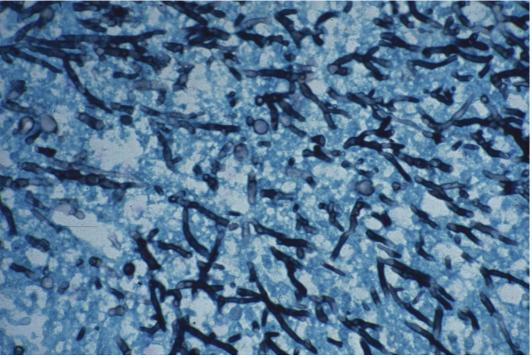
Micrograph showing invasive Aspergillus infection: septate hyphae branching at acute 45° angles (Methenamine silver stain, x450 magnification).
Two major pulmonary forms:
a) Invasive Pulmonary Aspergillosis (IPA)
- Risk factors: Prolonged neutropenia (AML on chemotherapy), hematologic malignancies, HCT/SOT recipients (especially lung transplant), advanced HIV, chronic granulomatous disease, critical illness (COPD, cirrhosis, DM, renal failure), severe influenza
- Pathogenesis: Hyphae invade respiratory mucosa and alveolar capillaries → angioinvasive disease → intravascular thrombosis, hypoxia, coagulative necrosis ("halo sign" on CT)
- Imaging: Halo sign (nodule surrounded by ground-glass opacity), air crescent sign, cavitation
- Diagnosis: CT chest, serum/BAL galactomannan (sensitivity ~70% in hematologic malignancies), beta-D-glucan, BAL culture, tissue biopsy
- Treatment (first-line): Voriconazole (oral/IV) - superior to amphotericin B in landmark trial. Alternative: isavuconazole. Lipid formulations of AmB (ABLC 5 mg/kg/day or liposomal AmB 3-5 mg/kg/day) for refractory/intolerant cases. Echinocandins (caspofungin) as salvage therapy.
b) Chronic Pulmonary Aspergillosis (CPA)
- Occurs in patients with underlying structural lung disease (prior TB, COPD, bullae)
- Subtypes: simple aspergilloma, chronic cavitary, subacute invasive
- Aspergilloma = fungus ball within pre-existing cavity; hemoptysis is the main complication
c) Allergic Bronchopulmonary Aspergillosis (ABPA)
-
Hypersensitivity to Aspergillus antigens in asthmatics/CF patients
-
Features: episodic bronchospasm, fleeting pulmonary infiltrates, central bronchiectasis, elevated IgE (total and specific), eosinophilia, positive skin test
-
Murray & Nadel's Textbook of Respiratory Medicine, pp. 1310-1315
2. Histoplasmosis
Organism: Histoplasma capsulatum (dimorphic fungus)
Endemic areas: Ohio and Mississippi River valleys (USA), Latin America; found in soil enriched by bird/bat droppings; exposure in caves (attack rate 50-100%)
Pulmonary syndromes (Fishman's classification):
| Syndrome | Age | Course | Imaging | Treatment |
|---|---|---|---|---|
| Acute | Any | 1-2 weeks | Diffuse airspace disease, lymphadenopathy | Only if prolonged or severe |
| Subacute | Any | Weeks to months | Focal airspace disease, lymphadenopathy | If symptoms >1 month |
| Chronic | >45, structural lung disease | Months to years | Upper lobe cavitary, thick-walled bullae, emphysema | Yes (itraconazole) |
| Progressive disseminated (PDH) | Immunocompromised | 1-2 weeks | Diffuse airspace opacities | Yes (AmB then itraconazole) |
Mediastinal complications: Mediastinal adenitis (young), mediastinal granuloma, fibrosing mediastinitis (most severe - can obstruct airways/vessels)
Diagnostics (sensitivity varies by syndrome):
- Histoplasma urinary antigen: 83% acute, 92% PDH (best overall test)
- Serology (CF/ID): 95% subacute, 83% chronic
- Culture: 42-74% (gold standard but slow)
- BAL culture: positive ~50% acute; BAL fungal stain: 70% diffuse PDH in AIDS
Treatment:
-
Mild/moderate: Itraconazole
-
Severe/PDH: Liposomal AmB initially, then step-down to itraconazole
-
Monitor with serial urine antigen levels (declines with treatment, rises with relapse)
-
Fishman's Pulmonary Diseases and Disorders, pp. 2360-2363
3. Cryptococcosis
Organism: Cryptococcus neoformans (most common, HIV-associated), C. gattii (immunocompetent)
Source: Pigeon droppings (C. neoformans); inhalation is the portal of entry
Clinical features:
- Portal of entry is the lung, but pulmonary infection is often asymptomatic or minimally symptomatic
- Only <5% present with isolated pneumonia
- Most dangerous manifestation: cryptococcal meningitis (especially CD4 <100 cells/µL)
- Symptoms when present: cough ± hemoptysis, fever, malaise, pleuritic chest pain, dyspnea
- Chest radiograph: diffuse bilateral interstitial opacities (65%), hilar adenopathy (27%), pleural effusion (14%), focal consolidation (13%), nodular opacities (11%), cavitation (11%), or normal CXR
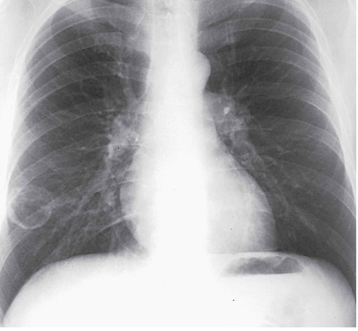
Frontal chest radiograph: HIV patient with cavitary cryptococcal lesion in the right lower lung field.
Diagnosis:
- Cryptococcal antigen (CRAG) test on serum, CSF, urine, BAL - very sensitive and specific
- Negative serum CRAG virtually excludes cryptococcal meningitis
- India ink stain of CSF (polysaccharide capsule)
- Culture of sputum, BAL, or blood
Treatment:
-
Non-CNS mild-moderate (immunocompetent): Fluconazole
-
Severe/CNS disease (HIV/immunocompromised): Liposomal AmB + flucytosine (induction) → fluconazole (consolidation/maintenance)
-
Murray & Nadel's Textbook of Respiratory Medicine, pp. 1835-1838; Frameworks for Internal Medicine, p. 1945
4. Mucormycosis (Zygomycosis)
Organisms: Mucor, Rhizopus, Cunninghamella (order Mucorales)
Key morphology: Aseptate (or sparsely septate), broad, ribbon-like hyphae with irregular, wide-angle branching (>90°) - distinguishes from Aspergillus
Forms:
- Rhinocerebral (most common, diabetics with DKA)
- Pulmonary (neutropenic patients, hematologic malignancy, prolonged steroid use, iron overload/deferoxamine therapy)
- Cutaneous, GI, disseminated
Pulmonary mucormycosis:
- Presents with fever, cough, chest pain; rapidly progressive consolidation, cavity, "reverse halo sign" (CT)
- Angioinvasive → thrombosis and infarction
- Mortality very high (>50%)
Diagnosis: Tissue biopsy with culture (serum beta-D-glucan and galactomannan are negative - important distinction from Aspergillus)
Treatment:
-
Liposomal AmB (first-line, 5-10 mg/kg/day)
-
Isavuconazole or posaconazole (step-down or salvage)
-
Surgical debridement essential
-
Control underlying predisposing condition (glucose control, reduce immunosuppression)
-
Murray & Nadel's Textbook of Respiratory Medicine, p. 1316
5. Pneumocystis jirovecii Pneumonia (PCP)
Although taxonomically a fungus (formerly thought to be a protozoan), Pneumocystis jirovecii causes a distinct form of opportunistic pneumonia.
Risk factors: CD4 <200 cells/µL (HIV), organ transplant, prolonged corticosteroids, cytotoxic chemotherapy
Clinical features: Subacute onset of progressive dyspnea, dry cough, low-grade fever; hypoxemia disproportionate to clinical examination
Imaging: Bilateral perihilar ground-glass opacities on CT; "bat-wing" pattern on CXR; can be normal in early disease
Diagnosis:
- BAL with immunofluorescence (gold standard, sensitivity >90%)
- Serum beta-D-glucan elevated
- PCR on BAL or sputum
Treatment:
- Trimethoprim-sulfamethoxazole (TMP-SMX) - first-line; high-dose IV for severe disease
- Adjunctive corticosteroids if PaO2 <70 mmHg or A-a gradient >35 mmHg
- Alternatives: Pentamidine, atovaquone, clindamycin + primaquine
- Prophylaxis: TMP-SMX when CD4 <200 cells/µL
6. Antifungal Drug Classes (Summary)
| Class | Agents | Mechanism | Key Uses |
|---|---|---|---|
| Polyenes | Amphotericin B (lipid formulations preferred) | Binds ergosterol → membrane disruption | Mucormycosis, severe IFI |
| Azoles | Fluconazole, Itraconazole, Voriconazole, Posaconazole, Isavuconazole | Inhibit ergosterol synthesis (CYP51) | Aspergillosis (voriconazole 1st line), Candida, endemic fungi |
| Echinocandins | Caspofungin, Micafungin, Anidulafungin | Inhibit beta-1,3-glucan synthase → cell wall disruption | Candida (1st line), salvage in Aspergillus |
- Murray & Nadel's Textbook of Respiratory Medicine, p. 1301
PART 2: PARASITIC RESPIRATORY INFECTIONS
1. Pulmonary Hydatid Disease (Echinococcosis)
Organism: Echinococcus granulosus (cystic) and E. multilocularis (alveolar)
Life cycle and transmission:
- Definitive hosts: carnivores (dogs, foxes)
- Intermediate hosts: sheep, cattle
- Humans = accidental hosts via faeco-oral transmission
- Larvae travel hematogenously → liver first, then lungs (~25%)
Pathology:
- Cyst has 3 layers: pericyst (host-derived fibrous), ectocyst (laminar), endocyst (germinal/inner)
- Daughter cysts may form within maternal cyst
Clinical features:
- Often asymptomatic until cyst enlarges or ruptures
- Symptoms: chest pain, cough, dyspnea
- Rupture → "hydatid vomica" (expectoration of salty cyst fluid + daughter cysts), anaphylaxis, pleural seeding ("white cancer"), bronchopleural fistula
Imaging:
- CT/CXR: round or oval well-defined cyst
- "Water lily sign" (floating membranes after partial collapse)
- Gharbi classification describes ultrasonographic stages
Diagnosis:
- Imaging appearance is characteristic
- Serology confirms
- Do NOT aspirate - risk of anaphylaxis and seeding
Treatment:
-
Albendazole (medical treatment, also perioperative to prevent seeding)
-
Surgical resection (definitive) - avoid spilling cyst contents
-
PAIR (Puncture, Aspiration, Instillation of 95% alcohol, Re-aspiration) for liver cysts
-
E. multilocularis: primarily albendazole (often requires prolonged/lifelong therapy)
-
Rosen's Emergency Medicine, p. 2676; Grainger & Allison's Diagnostic Radiology, p. 1484
2. Pulmonary Paragonimiasis
Organism: Paragonimus westermani (lung fluke)
Endemic: Southeast Asia, West Africa, Central/South America
Transmission: Ingestion of raw/undercooked crabs or crayfish containing metacercariae
Life cycle: Metacercariae → penetrate intestinal wall → peritoneum → diaphragm → pleura → lung parenchyma → form cysts containing adult flukes
Clinical features:
- Chronic cough (can mimic tuberculosis)
- Rusty-brown or blood-tinged sputum (hemoptysis)
- Pleuritic chest pain
- Pleural effusion with high eosinophil count
Imaging: Irregular cystic lesions, "ring shadow," pleural effusion
Diagnosis:
- Sputum/stool microscopy - Paragonimus eggs (operculated)
- Serology (ELISA)
- Peripheral eosinophilia is characteristic
Treatment: Praziquantel (first-line); triclabendazole (alternative)
3. Pulmonary Toxocariasis (Visceral Larva Migrans)
Organism: Toxocara canis or T. cati (dog/cat roundworm larvae)
Transmission: Ingestion of contaminated soil with embryonated eggs
Pulmonary manifestation:
- Transient pulmonary infiltrates + severe eosinophilia ("Löffler's syndrome")
- Wheezing, cough, dyspnea
- Affects mainly children
Diagnosis: Serology (ELISA for Toxocara); biopsy shows eosinophilic granuloma with larval fragments
Treatment: Albendazole or mebendazole; corticosteroids for severe pulmonary disease
4. Pulmonary Involvement in Malaria
- Plasmodium falciparum can cause acute lung injury/ARDS (pulmonary malaria)
- Occurs in ~10% of severe falciparum malaria
- Mechanism: sequestration of parasitized RBCs in pulmonary vasculature, cytokine storm
- Features: rapid progression to respiratory failure
- Treatment: Artesunate IV (preferred over quinine for severe malaria) + respiratory support
5. Pulmonary Strongyloidiasis
Organism: Strongyloides stercoralis
Special feature: Can cause hyperinfection syndrome in immunocompromised (especially corticosteroid use) - filariform larvae invade gut wall en masse, disseminate carrying gut bacteria → gram-negative sepsis + pneumonia
Pulmonary features:
- Löffler-like syndrome during larval migration
- Hyperinfection: bilateral interstitial infiltrates, sometimes respiratory failure
- Larvae may be found in sputum/BAL
Treatment: Ivermectin (first-line); albendazole (alternative)
6. Pulmonary Amoebiasis
Organism: Entamoeba histolytica
Mechanism: Extension of hepatic amoebic abscess through the diaphragm into the right pleural space/lung base
Clinical features:
- Right lower lobe consolidation or pleural effusion
- Chocolate-brown ("anchovy sauce") sputum if hepatobronchial fistula forms
- "Anchovy sauce" pus is pathognomonic
Treatment: Metronidazole (tissue amoebicide) followed by diloxanide furoate (luminal)
7. Ascariasis - Pulmonary Phase
Organism: Ascaris lumbricoides
Pulmonary phase: Larvae migrate through pulmonary capillaries into alveoli → "Löffler's syndrome" (transient eosinophilic pneumonitis, bilateral migratory infiltrates, cough, fever, wheeze)
Treatment: Albendazole or mebendazole (for the intestinal phase); pulmonary phase is typically self-limiting
Key Distinguishing Points Summary
| Feature | Aspergillus | Mucormycosis | Cryptococcus | Histoplasma | Hydatid Cyst | PCP |
|---|---|---|---|---|---|---|
| Hyphae | Septate, 45° acute branching | Aseptate, wide-angle | Encapsulated yeast | Dimorphic yeast in tissue | N/A (helminth) | Cysts/trophozoites |
| Key host | Neutropenic, HCT | DKA, iron overload | CD4 <100 (HIV) | Normal or immunocompromised | Any (endemic area) | CD4 <200 |
| 1st-line Rx | Voriconazole | Liposomal AmB + surgery | Fluconazole (mild) or AmB+5FC (CNS) | Itraconazole or AmB | Albendazole + surgery | TMP-SMX |
| Galactomannan | Positive | Negative | Negative | Negative | N/A | Negative |
| Beta-D-glucan | Positive | Negative | Positive | Positive | N/A | Elevated |
Sources:
- Murray & Nadel's Textbook of Respiratory Medicine, 2-Volume Set
- Fishman's Pulmonary Diseases and Disorders, 2-Volume Set
- Rosen's Emergency Medicine
- Frameworks for Internal Medicine
- Grainger & Allison's Diagnostic Radiology
This is a shared conversation. Sign in to Orris to start your own chat.