
Structure of hemoglobin
Hemoglobin is a tetrameric protein made of four polypeptide chains, each carrying a heme group with an iron atom at its center. The most common adult form (HbA) has two alpha and two beta chains. Each iron atom can bind one oxygen molecule, giving a total capacity of four O2 per hemoglobin molecule. The quaternary structure shifts between a "tense" (deoxy) and "relaxed" (oxy) conformation, which underlies cooperative oxygen binding.
Searching sources...
Reading File
Finding Sources
Finding Sources
Good results. Now I'll read the key textbook sections in parallel - Lippincott Biochemistry (the most structured), Guyton physiology, and Harper's - while also fetching the relevant images.
Reading File
Reading File
Reading File
Now let me read the Lippincott section on hemoglobin quaternary structure and T/R conformation, and also fetch the key images.
Reading File
Loading Image
Loading Image
I now have rich textbook content and excellent images. Let me fetch two more key images (T/R conformational change diagram and the globin chain synthesis chart) to complete the answer.
Loading Image
Loading Image
I now have everything needed for a thorough, well-illustrated answer. Here is the complete response:
Structure of Hemoglobin
Hemoglobin is a tetrameric hemeprotein found exclusively in red blood cells (RBCs). It is the body's principal oxygen carrier, transporting O2 from the lungs to peripheral tissues, and also carries CO2 and H+ in the reverse direction.
1. Overall Architecture
Hemoglobin A (HbA), the predominant adult form, consists of four polypeptide (globin) chains - two alpha (α) and two beta (β) - held together by noncovalent interactions. Each chain cradles one heme prosthetic group. The complete molecule therefore carries four heme groups and four iron atoms, allowing it to bind up to four molecules of O2.
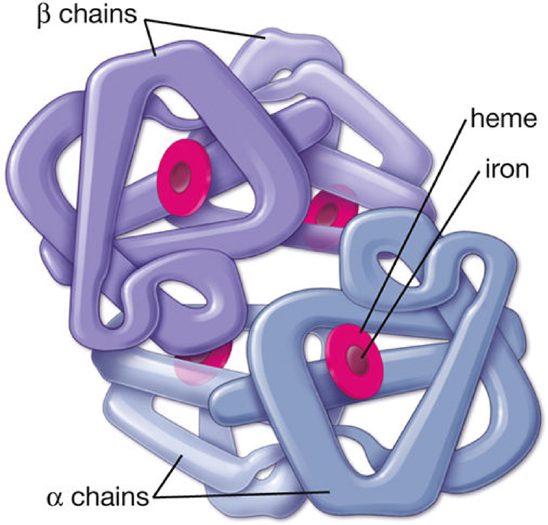
Structural diagram of the hemoglobin molecule - Histology: A Text and Atlas
2. The Heme Group
Each heme is a planar porphyrin ring (protoporphyrin IX) with a ferrous iron (Fe²⁺) coordinated at its center by bonds to four nitrogen atoms of the ring. The iron forms two additional bonds perpendicular to the plane:
- 5th coordination bond: links to the imidazole nitrogen of the proximal histidine (His F8) of the globin chain
- 6th coordination bond: the site that reversibly binds O2
A second histidine, the distal histidine (His E7), does not bond to iron but sits nearby, stabilizing the O2 and preventing irreversible oxidation of Fe²⁺ to Fe³⁺ (methemoglobin).
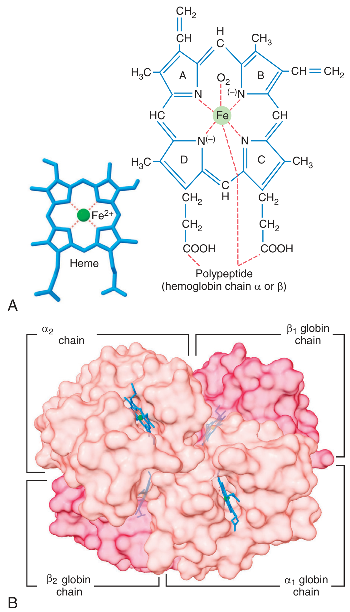
Heme structure and the hemoglobin tetramer surface - Guyton & Hall Textbook of Medical Physiology
3. The Globin Chains
Each globin subunit:
- Is ~80% α-helical, organized into 8 helical segments labeled A through H
- Has a hydrophobic interior (nonpolar residues packed inward)
- Has a central hydrophobic heme-binding pocket that excludes water, protecting the Fe²⁺ from oxidation
- Has polar amino acids on its surface, where they interact with water and other subunits
The α chains contain 141 amino acids and the β chains contain 146 amino acids.
4. Quaternary Structure and the T/R Conformational Switch
The tetramer is best understood as two identical αβ dimers (αβ₁ and αβ₂):
- Within each dimer: the α and β subunits are held together by strong hydrophobic interactions at their interface
- Between the two dimers: the interactions are primarily weaker polar bonds (ionic bonds + hydrogen bonds), which allow relative movement
This inter-dimer mobility is the structural basis for hemoglobin's cooperativity:
| Form | State | Bonds between dimers | O2 affinity | Iron position |
|---|---|---|---|---|
| T (Taut/Tense) | Deoxy | Strong ionic + H-bonds (many) | Low | Pulled out of heme plane |
| R (Relaxed) | Oxy | Fewer polar bonds (some broken) | High | Pulled into heme plane |
When O2 binds to the first subunit (in the T state), it pulls Fe²⁺ into the porphyrin plane. Since Fe²⁺ is covalently linked to the proximal histidine, this movement tugs the attached helix, altering the αβ₁/αβ₂ dimer interface and partially converting the molecule toward the R state. This makes the next O2 binding easier - the molecular basis of cooperative oxygen binding (sigmoidal O2-dissociation curve).
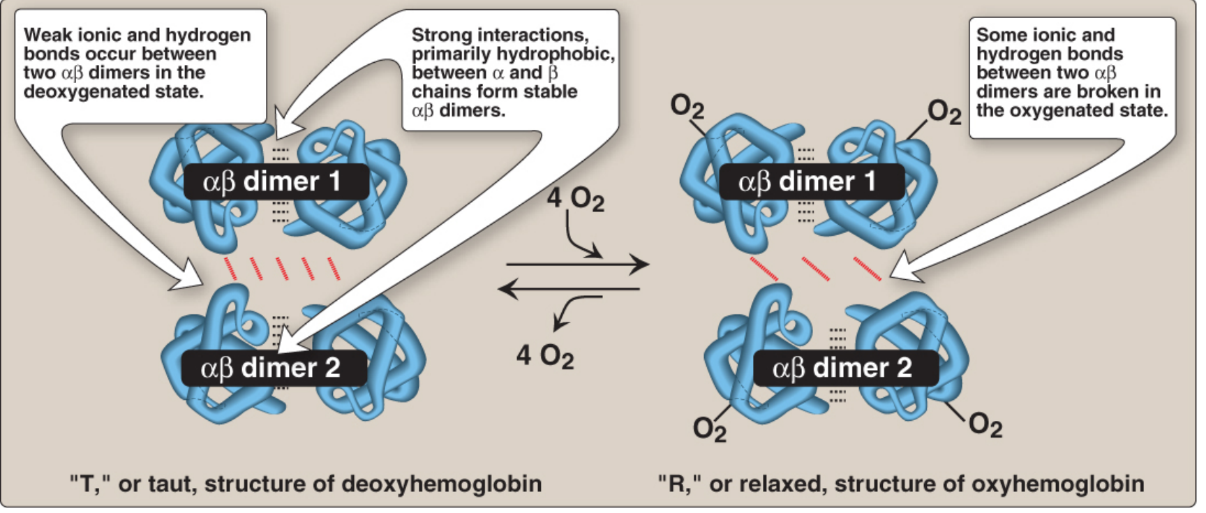
T and R conformational states - Lippincott Illustrated Reviews: Biochemistry, 8th ed.
5. Types of Normal Hemoglobin
Different globin chain combinations produce distinct hemoglobin variants. The type of chain determines O2 affinity and physiological function:
| Type | Chains | Adults (%) | Notes |
|---|---|---|---|
| HbA | α₂β₂ | ~96% | Major adult hemoglobin |
| HbA2 | α₂δ₂ | 1.5-3% | Minor adult variant |
| HbF | α₂γ₂ | <1% adult | Fetal form; higher O2 affinity (less 2,3-BPG binding) |
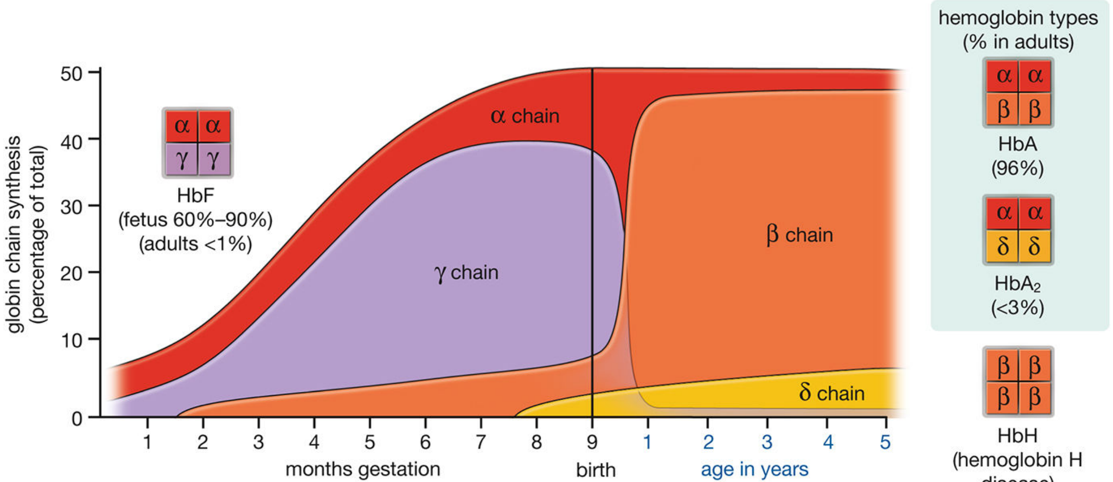
Globin chain synthesis timeline - Histology: A Text and Atlas
HbF has higher O2 affinity than HbA because γ-chains bind 2,3-bisphosphoglycerate (2,3-BPG) less avidly, allowing more efficient O2 uptake from the maternal circulation.
6. Allosteric Effectors (Summary)
The R→T equilibrium (and therefore O2 affinity) is modulated by several allosteric factors:
- 2,3-BPG: binds in the central cavity of the T state, stabilizing it and lowering O2 affinity; important for O2 delivery to tissues
- H+ (Bohr effect): increased [H+] (lower pH) stabilizes T form, promoting O2 release in acidic tissues
- CO2: carbamino groups form on N-terminal amino groups, also stabilizing T form
- Temperature: higher temperature lowers O2 affinity, aiding release during exercise
Sources
- Lippincott Illustrated Reviews: Biochemistry, 8th ed., Ch. 3 (Hemeproteins)
- Guyton & Hall Textbook of Medical Physiology, Ch. 33 (Hemoglobin formation)
- Histology: A Text and Atlas, Ch. 10 (Erythrocytes and hemoglobin types)
This is a shared conversation. Sign in to Orris to start your own chat.