
Write an article for lung health tips for general public Write as a pulmonologist
healthy vs diseased lung comparison diagram
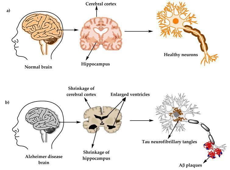
Educational comparison diagram illustrating the macroscopic and microscopic changes in the human brain associated with Alzheimer’s disease (AD). The visual is divided into two sections: (a) Healthy state and (b) Alzheimer’s disease state. At the macroscopic level, cross-sectional brain illustrations highlight a normal cerebral cortex and hippocampus versus a diseased brain showing significant shrinkage of the cerebral cortex, severe shrinkage of the hippocampus, and enlarged ventricles. At the microscopic level, the diagram compares neuronal morphology. Healthy neurons are depicted with intact cell bodies and axons. In contrast, the AD neuron demonstrates classic hallmarks of neurodegeneration: internal accumulation of tau neurofibrillary tangles (NFTs) within the cell body and the external deposition of amyloid-beta (Aβ) plaques around the axon terminals. This illustration serves to teach the pathophysiology of AD, focusing on structural atrophy and the proteinopathy involved in dementia progression. Key educational concepts include neuroanatomy, neurodegenerative pathology, and the amyloid/tau hypothesis.
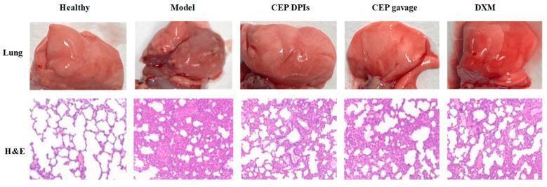
A composite medical comparison image demonstrating lung pathology across five experimental conditions: Healthy, Model (Acute Lung Injury), CEP DPIs, CEP gavage, and DXM (Dexamethasone). The top row contains macroscopic clinical photographs of whole lung tissue. The 'Healthy' lung is light pink and smooth, while the 'Model' lung shows significant dark red discoloration, bruising, and edema. Treatment groups (CEP and DXM) show macroscopic recovery towards a normal pink appearance. The bottom row presents corresponding histopathological sections stained with hematoxylin and eosin (H&E) at 100x magnification. The 'Healthy' section reveals clear, thin-walled alveolar sacs. In contrast, the 'Model' section shows severe alveolar architectural destruction, marked thickening of alveolar septa, and dense inflammatory cell infiltration. The treatment groups, particularly the Cepharanthine dry powder inhaler (CEP DPIs) and DXM, demonstrate significant histological attenuation of injury, characterized by reduced septal thickening and decreased leucocyte infiltration, illustrating the anti-inflammatory efficacy of these interventions in a model of chemical-induced lung injury.
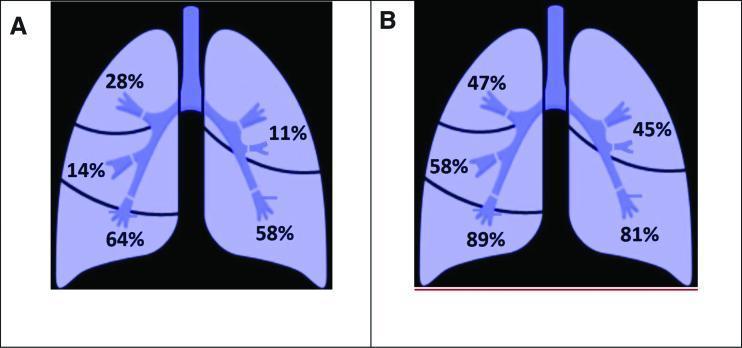
This medical comparison chart consists of two anatomical diagrams (A and B) representing the human lungs in a coronal view. Diagram A illustrates the anatomical distribution of pulmonary artery thrombosis, while Diagram B shows the distribution of inflammatory lung lesions, likely in the context of COVID-19. Both diagrams divide the lungs into five regions: the right superior, middle, and inferior lobes, and the left superior and inferior lobes. In Diagram A (Thrombosis), percentages range from 11% in the left superior lobe to 64% in the right inferior lobe. In Diagram B (Inflammation), percentages are significantly higher, ranging from 45% in the left superior lobe to 89% in the right inferior lobe. Both visualizations demonstrate a distinct predilection for the lower lobes compared to the upper and middle lobes. This anatomical mapping is intended to correlate the location of vascular complications with parenchymal inflammatory changes, highlighting the regional severity of pulmonary pathology.
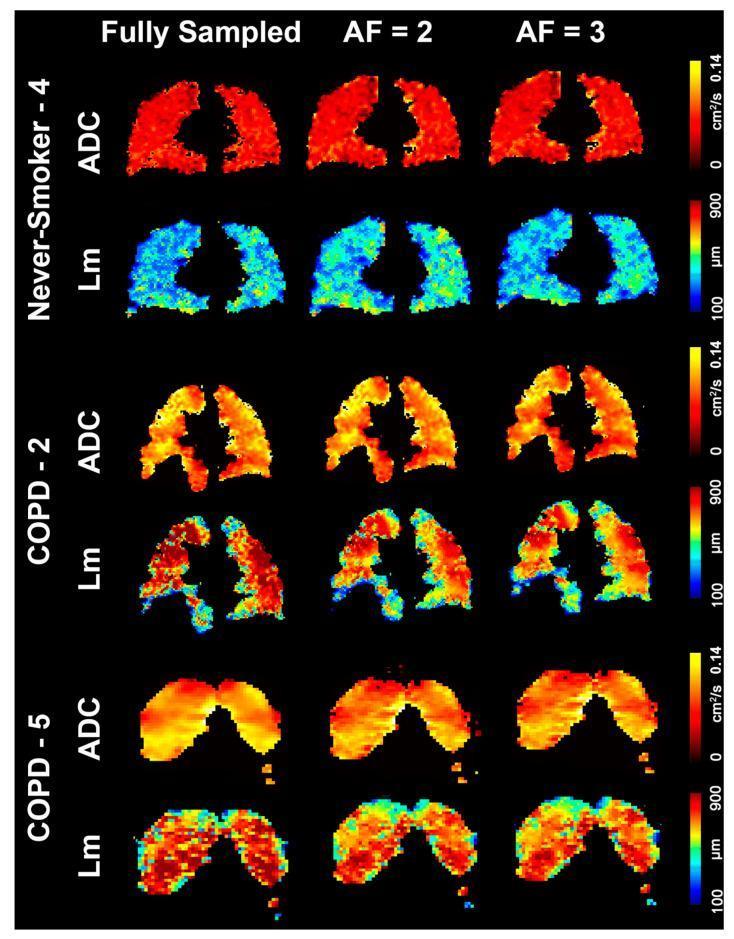
A comparison grid of 129Xe Magnetic Resonance Imaging (MRI) parametic maps showing coronal slices of human lungs. The grid compares three subjects—a healthy never-smoker, a patient with Chronic Obstructive Pulmonary Disease (COPD), and a patient with Alpha-1 Antitrypsin Deficiency (AATD/COPD)—across three data acquisition methods: Fully Sampled, Acceleration Factor (AF) = 2, and AF = 3. Two quantitative parameters are visualized: Apparent Diffusion Coefficient (ADC) and Mean Linear Intercept (Lm). The ADC maps (scale 0 to 0.14 cm²/s) show a progression from uniform low values (red) in the healthy subject to higher, more heterogeneous values (yellow/white) in COPD and AATD, indicating increased gas diffusivity due to alveolar wall destruction (emphysema). The Lm maps (scale 100 to 900 μm) show a shift from predominantly low values (blue/cyan) in the healthy lung to elevated, heterogeneous values (green/red) in the diseased lungs, representing alveolar enlargement. The image demonstrates that undersampled reconstructions (AF 2 and 3) maintain high qualitative and quantitative fidelity compared to fully sampled data in assessing obstructive lung disease.
COPD emphysema smoking lung damage
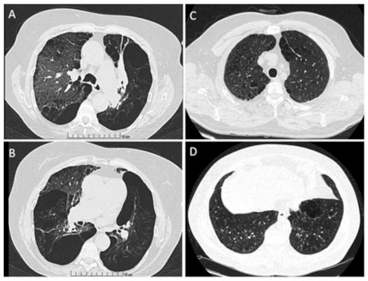
This diagnostic comparison presents four axial High-Resolution Computed Tomography (HRCT) lung windows, categorized by smoking history and anatomical level. Images A and B display the upper and lower lobes of an active smoker with the M/MProcida genotype. These images demonstrate significant structural lung damage, including centrilobular and paraseptal emphysema characterized by areas of low attenuation, architectural distortion, and bronchial wall thickening suggestive of bronchiectasis. Large bullae are particularly visible in the lower lobe periphery (Image B). In contrast, Images C and D display the upper and lower lobes of a never smoker with the M/I genotype. These scans show relatively preserved lung parenchyma with homogenous density and normal pulmonary vascular distribution, though minor areas of ground-glass opacity are present. The comparison highlights the severity of obstructive lung disease manifestations, specifically the progression of emphysematous changes and airway remodeling associated with chronic tobacco exposure in the context of Alpha-1 Antitrypsin Deficiency genetic variants. This material is suitable for medical education regarding radiology of Chronic Obstructive Pulmonary Disease (COPD).
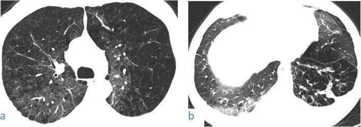
High-resolution axial CT scans of the chest in a 48-year-old male smoker with COVID-19. Image (a) shows the upper lobes featuring marked centrilobular emphysema characterized by multiple lucent spaces centered on secondary pulmonary lobules. This is superimposed with patchy and subpleural ground-glass opacification (GGO), appearing as areas of hazy increased lung density. Image (b) shows the lower lobes, demonstrating peripheral ground-glass opacity in the right lower lobe with associated vascular thickening. The left lower lung shows architectural distortion with visible fibrous stripes and reticular markings. These findings represent a combination of chronic obstructive pulmonary disease (COPD) from smoking history and acute pulmonary manifestations consistent with viral pneumonia (COVID-19), illustrating how pre-existing emphysema can complicate the presentation of infectious lung disease.
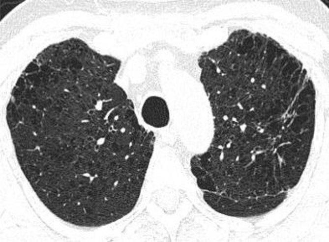
This diagnostic image is an axial non-contrast-enhanced CT scan of the chest at the level of the upper lobes, demonstrating significant chronic obstructive pulmonary disease (COPD) pathology. The lung parenchyma shows extensive centrilobular emphysema, characterized by multiple small, focal areas of low attenuation representing alveolar destruction within the secondary pulmonary lobules. Complementary paraseptal emphysematous changes are visible as subpleural lucencies along the peripheral lung margins. In the apicoposterior segment of the left upper lobe, there is a localized area of increased attenuation consistent with a fibrotic lesion or reticular opacity. The distribution is primarily bilateral and upper-lobe predominant, which is characteristic of smoking-related lung disease. Key educational concepts include the radiographic differentiation between centrilobular and paraseptal emphysema and the identification of concurrent interstitial lung changes in a patient with multi-faceted parenchymal disease.
breathing exercises lung capacity respiratory rehabilitation
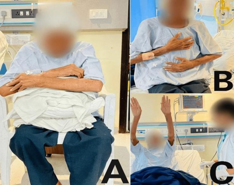
Clinical photograph showing a series of three respiratory physiotherapy interventions (labeled A, B, and C) for a patient in a hospital setting. Panel A depicts the forward-leaning position, with the patient seated and leaning over a supportive pillow to alleviate dyspnea by optimizing accessory muscle use. Panel B shows the patient in a semi-recumbent position performing diaphragmatic breathing exercises, indicated by the hand placement on the chest and abdomen to monitor breath depth and abdominal excursion. Panel C illustrates thoracic expansion exercises, with the patient seated and arms fully abducted and elevated to enhance chest wall mobility and lung volume. The environment includes standard medical equipment such as a wall-mounted cardiac monitor, bedside rails, and a medical professional assisting with the rehabilitation protocol. These interventions are typically used in post-operative or pulmonary rehabilitation contexts to address pleural effusion, atelectasis, and reduced respiratory capacity.
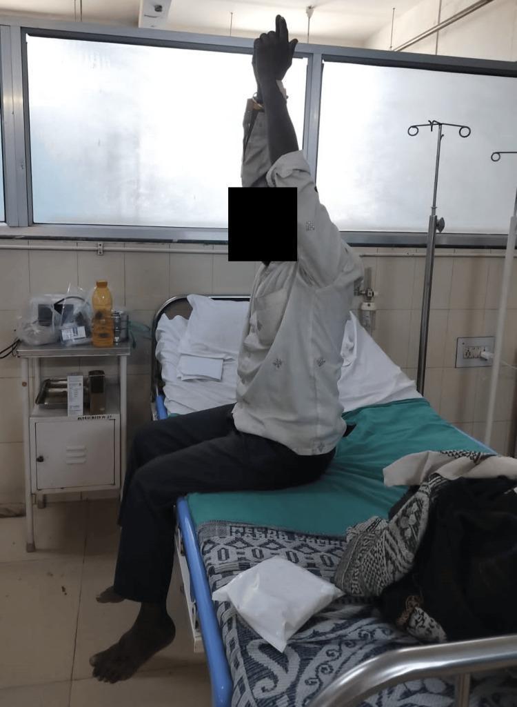
This clinical photograph illustrates a patient performing bedside pulmonary rehabilitation exercises within a hospital setting. The patient is seated upright on the edge of a hospital bed with hips and knees flexed at approximately 90 degrees. One arm is fully extended vertically toward the ceiling with the palm facing inward, demonstrating a thoracic expansion exercise designed to increase lung volume and improve respiratory mechanics. The background features a standard clinical environment, including an IV pole, a bedside table with medical supplies, and a hospital bed with a teal sheet. The photograph serves as an educational visual for physical therapy and cardiorespiratory rehabilitation protocols, specifically for managing extrapulmonary tuberculosis or chronic respiratory conditions. It highlights the integration of active limb movement with breathing techniques to enhance thoracic mobility and reduce dyspnea.
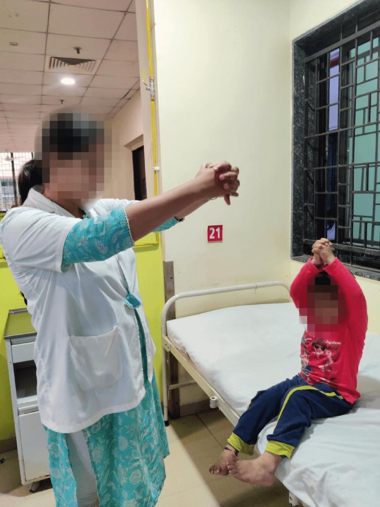
This clinical photograph captures a pediatric patient and a healthcare professional engaged in a respiratory physiotherapy session within a hospital setting. The patient, a young child, is shown in a long-sitting position on a hospital bed with legs extended. Guided by the therapist, the child is performing thoracic expansion exercises, characterized by the raising of both arms overhead with hands clasped to facilitate maximum lung volume and chest wall mobility. The healthcare professional, wearing a white clinical coat, stands adjacent to the bed and demonstrates the movement with their own arms extended and hands clasped. This intervention is typical for post-operative cardiac rehabilitation (following median sternotomy for VSD repair, as indicated by context) to prevent atelectasis and improve pulmonary function. The background reveals a standard clinical environment with a numbered patient station and safety-barred window. This image serves as an educational example of active-assisted breathing techniques and the role of physical therapy in pediatric cardiac recovery.
10 Essential Lung Health Tips: A Pulmonologist's Guide for Everyone
1. Stop Smoking - There Is No Safe Level
- Nicotine replacement therapy (patches, gum, lozenges) combined with counseling achieves quit rates of 25-30%
- Medications like varenicline (Champix/Chantix) and bupropion are effective but work best alongside behavioral support
- On average, people make 8 quit attempts before succeeding - each attempt counts, do not give up
- Even a brief conversation with your doctor about quitting can make a difference
2. Know Your Air - Pollution Is a Silent Lung Threat
- Check daily air quality index (AQI) readings before outdoor exercise - many free apps do this automatically
- On high-pollution days, exercise indoors or reduce the intensity of outdoor workouts
- Avoid exercising near heavy traffic, which concentrates nitrogen dioxide and fine particles
- People with asthma or COPD should carry reliever inhalers on smoggy days
- Gas stoves are a significant source of nitrogen dioxide and can trigger asthma exacerbations
- Wood-burning stoves and fireplaces produce fine particulate matter that penetrates deep into lung tissue
- Kerosene heaters, incense burning, and poorly ventilated cooking all add to indoor pollutant load
- Ensure good ventilation when cooking - open windows or run extractor fans
3. Control Your Asthma Triggers
- House dust mites - the leading trigger worldwide. Encase mattresses and pillows in allergen-proof covers, wash bedding weekly in hot water (>60°C), and reduce indoor humidity below 50%
- Pet dander - proteins from animal skin flakes, saliva, and urine. If rehoming a pet is not possible, keep pets out of bedrooms and use HEPA air filters
- Cockroach allergen - a major trigger in urban environments, particularly affecting children
- Indoor molds - address damp areas immediately; mold remediation is essential for asthma control
4. Exercise - Your Lungs Need Work Too
- Aim for 150 minutes of moderate aerobic activity per week - walking, swimming, cycling
- Diaphragmatic breathing exercises can reduce breathlessness and improve respiratory muscle function - place one hand on your chest and one on your abdomen, and practice breathing so that the abdomen rises, not the chest
- Pursed-lip breathing (breathe in through the nose for 2 counts, breathe out slowly through pursed lips for 4 counts) helps patients with COPD slow respiration, reduce air trapping, and relieve acute breathlessness
- Even patients with moderate lung disease should exercise - physical deconditioning makes breathlessness worse, creating a vicious cycle
5. Get Vaccinated
6. Protect Yourself Occupationally
- Dust (silica, coal, wood, grain) - wear an appropriate respirator (N95 or above), not a paper dust mask
- Asbestos - any building work in structures built before the 1990s may expose you to asbestos fibers; follow proper safety protocols
- Fumes and chemicals - welding fumes, isocyanates (spray painters), formaldehyde (healthcare workers)
- Biological agents - farmers, healthcare workers, and those handling animals
7. Maintain a Healthy Weight and Diet
- Antioxidants (vitamins C and E, beta-carotene) help neutralize oxidative stress caused by pollution and infection
- Omega-3 fatty acids (fatty fish, flaxseed) have anti-inflammatory properties relevant to asthma
- A diet rich in fruits and vegetables is associated with better lung function in epidemiological studies
- Excessive processed food and refined sugar drive systemic inflammation, which worsens airways disease
8. Sleep and Breathing - Do Not Ignore Snoring
- Loud, persistent snoring - especially if it stops and starts
- Witnessed breathing pauses during sleep (your partner may notice this)
- Waking unrefreshed despite adequate sleep time
- Morning headaches (from overnight carbon dioxide retention)
- Excessive daytime sleepiness
- Difficulty concentrating
9. Know the Warning Signs That Need a Doctor
- A cough lasting more than 3 weeks that is new or has changed character
- Coughing up blood (haemoptysis) - always needs investigation, even a small amount
- Progressive breathlessness - if you are becoming less able to do activities you could do a year ago
- Unexplained weight loss with respiratory symptoms
- Recurrent chest infections - more than two per year warrants investigation
- Wheeze in adults who have never had asthma - do not assume it is nothing
10. Avoid What You Can, Manage What You Cannot
| Priority | Action | Impact |
|---|---|---|
| 1 | Never smoke, or quit now | Very high |
| 2 | Avoid secondhand smoke | High |
| 3 | Vaccinate annually | High |
| 4 | Reduce indoor air pollution | Moderate-High |
| 5 | Exercise regularly | Moderate |
| 6 | Use occupational protection | Variable, can be very high |
| 7 | Maintain healthy weight | Moderate |
| 8 | Screen for lung cancer if eligible | Potentially life-saving |
A Final Word from the Clinic
Make In a a5 per sheet
ls -la /tmp/workspace/ 2>/dev/null || mkdir -p /tmp/workspace && echo "Created workspace"
mkdir -p /tmp/workspace/lung-health && python3 << 'PYEOF' from reportlab.lib.pagesizes import A5 from reportlab.lib import colors from reportlab.lib.units import mm from reportlab.platypus import ( SimpleDocTemplate, Paragraph, Spacer, HRFlowable, Table, TableStyle ) from reportlab.lib.styles import getSampleStyleSheet, ParagraphStyle from reportlab.lib.enums import TA_CENTER, TA_LEFT, TA_JUSTIFY from reportlab.platypus import KeepTogether OUTPUT = "/tmp/workspace/lung-health/Lung_Health_Tips_A5.pdf" # A5: 148 x 210 mm doc = SimpleDocTemplate( OUTPUT, pagesize=A5, leftMargin=12*mm, rightMargin=12*mm, topMargin=12*mm, bottomMargin=12*mm, ) W, H = A5 # ── Colour palette ────────────────────────────────────────────── TEAL = colors.HexColor("#1A6B8A") TEAL_LITE = colors.HexColor("#E8F4F8") ORANGE = colors.HexColor("#E8630A") DARK = colors.HexColor("#1C2B36") GREY = colors.HexColor("#5A6A72") WHITE = colors.white # ── Base styles ──────────────────────────────────────────────── base = getSampleStyleSheet() def S(name, **kw): return ParagraphStyle(name, **kw) title_style = S("Title", fontSize=17, leading=21, textColor=TEAL, alignment=TA_CENTER, fontName="Helvetica-Bold", spaceAfter=1*mm) subtitle_style = S("Sub", fontSize=8.5, leading=11, textColor=GREY, alignment=TA_CENTER, fontName="Helvetica-Oblique", spaceAfter=3*mm) section_style = S("Sec", fontSize=8, leading=10, textColor=WHITE, alignment=TA_LEFT, fontName="Helvetica-Bold", spaceBefore=0, spaceAfter=0) body_style = S("Body", fontSize=7.2, leading=10, textColor=DARK, alignment=TA_JUSTIFY, fontName="Helvetica", spaceBefore=1*mm, spaceAfter=1.5*mm) bullet_style = S("Bullet", fontSize=7.2, leading=9.5, textColor=DARK, alignment=TA_LEFT, fontName="Helvetica", leftIndent=8, spaceBefore=0.5*mm, spaceAfter=0) tip_num_style = S("TipNum", fontSize=18, leading=20, textColor=ORANGE, alignment=TA_CENTER, fontName="Helvetica-Bold") footer_style = S("Footer", fontSize=6.5, leading=8, textColor=GREY, alignment=TA_CENTER, fontName="Helvetica-Oblique") warning_style = S("Warn", fontSize=7, leading=9.5, textColor=DARK, alignment=TA_LEFT, fontName="Helvetica-Bold") # ── Content definition ───────────────────────────────────────── tips = [ ( "1", "STOP SMOKING – No Safe Level", "Smoking causes a 20× increased risk of lung cancer and 10× risk of emphysema. " "It is the single most preventable cause of premature death.", [ "Quitting now slows lung function decline — even in COPD", "Use nicotine replacement + counselling for best results (25-30% quit rate)", "Secondhand smoke causes ~50,000 deaths/year — protect your family", "Average 8 attempts before success — every attempt matters", ] ), ( "2", "KNOW YOUR AIR – Pollution Matters", "Particulate matter (PM2.5), ozone, and nitrogen dioxide enter the lungs with every breath, " "causing asthma, accelerated lung decline, and increased mortality.", [ "Check the daily Air Quality Index (AQI) before outdoor exercise", "On high-pollution days, exercise indoors", "Ventilate kitchens — gas stoves produce NO₂ and can trigger asthma", "Test your home for radon — the #2 cause of lung cancer after smoking", ] ), ( "3", "CONTROL ASTHMA TRIGGERS", "Most asthma is preventable with good trigger management. Identify and address your personal triggers.", [ "House dust mites: allergen-proof mattress covers, hot-wash bedding weekly", "Pet dander: keep pets out of bedrooms, use HEPA air filters", "Mould: fix damp areas immediately — mould drives asthma worsening", "Keep indoor humidity below 50%", ] ), ( "4", "EXERCISE YOUR LUNGS", "Regular aerobic exercise trains respiratory muscles and improves breathing efficiency. " "Physical deconditioning makes breathlessness worse — creating a vicious cycle.", [ "Aim for 150 min of moderate aerobic activity per week", "Diaphragmatic breathing: breathe so abdomen rises, not chest", "Pursed-lip breathing: inhale 2 counts, exhale slowly 4 counts", "Even patients with lung disease should stay active", ] ), ( "5", "VACCINATE EVERY YEAR", "Respiratory infections are the leading triggers of COPD and asthma exacerbations. " "Vaccination is one of the simplest lung-protective actions you can take.", [ "Annual flu vaccine — for everyone over 6 months of age", "Pneumococcal vaccine — especially if over 65, a smoker, or diabetic", "COVID-19 boosters — discuss current recommendations with your doctor", ] ), ( "6", "PROTECT YOURSELF AT WORK", "Occupational lung disease — silicosis, asbestosis, occupational asthma — is entirely preventable. " "Many workers only find out too late.", [ "Dust (silica, wood, grain): use N95 respirator, not a paper mask", "Welding fumes and spray paints: use proper respiratory protection", "Any building pre-1990s: asbestos risk — follow safety protocols", "New cough that improves on weekends? Tell your doctor about your job", ] ), ( "7", "MAINTAIN HEALTHY WEIGHT & DIET", "Obesity restricts diaphragm movement, worsens asthma control, and increases sleep apnoea risk. " "Diet quality directly affects lung inflammation.", [ "Antioxidants (vitamins C & E, beta-carotene) reduce oxidative lung stress", "Omega-3 rich foods (oily fish, flaxseed) have anti-inflammatory effects", "Reduce processed food and refined sugar — both drive airway inflammation", "Maintain a healthy BMI to allow full diaphragm excursion", ] ), ( "8", "DON'T IGNORE SNORING", "Obstructive sleep apnoea (OSA) affects ~1 billion people worldwide — most undiagnosed. " "Oxygen drops hundreds of times per night, stressing the heart and brain.", [ "Warning signs: loud snoring, witnessed pauses, morning headaches", "Excessive daytime sleepiness despite adequate sleep time", "Waking unrefreshed — night after night", "CPAP therapy is highly effective. Ask for a sleep study if concerned", ] ), ( "9", "RECOGNISE WARNING SIGNS", "Many lung diseases progress silently for years. By the time COPD causes symptoms, " "50% of lung function may already be lost.", [ "Cough lasting > 3 weeks — always investigate", "Coughing up blood (even small amounts) — see a doctor same day", "Progressive breathlessness — becoming less active year on year", "Recurrent chest infections (>2/year) — needs evaluation", ] ), ( "10", "SCREEN FOR LUNG CANCER", "Lung cancer caught at Stage I has >80% five-year survival. At Stage IV: <10%. " "Screening finds it early — when it is still curable.", [ "Low-dose CT scan recommended for ages 50–80 with heavy smoking history", "20 pack-years (e.g. 1 pack/day × 20 yrs) = screening eligible", "Simple spirometry test takes 15 min — asks your doctor", "Do not wait for symptoms — early disease is often completely silent", ] ), ] story = [] # ── COVER HEADER ────────────────────────────────────────────── story.append(Spacer(1, 2*mm)) story.append(Paragraph("🫁 LUNG HEALTH", title_style)) story.append(Paragraph("10 Essential Tips from a Pulmonologist", subtitle_style)) story.append(HRFlowable(width="100%", thickness=1.5, color=TEAL, spaceAfter=3*mm)) # ── TIPS ────────────────────────────────────────────────────── for num, heading, intro_text, bullets in tips: # Section header bar (simulated with a Table) header_data = [[ Paragraph(f"Tip {num}", S("hn", fontSize=7, fontName="Helvetica-Bold", textColor=WHITE, leading=9)), Paragraph(heading, S("ht", fontSize=7.8, fontName="Helvetica-Bold", textColor=WHITE, leading=9, alignment=TA_LEFT)), ]] header_table = Table(header_data, colWidths=[14*mm, None]) header_table.setStyle(TableStyle([ ("BACKGROUND", (0, 0), (-1, -1), TEAL), ("VALIGN", (0, 0), (-1, -1), "MIDDLE"), ("LEFTPADDING", (0, 0), (-1, -1), 4), ("RIGHTPADDING", (0, 0), (-1, -1), 4), ("TOPPADDING", (0, 0), (-1, -1), 3), ("BOTTOMPADDING",(0, 0), (-1, -1), 3), ("LINEAFTER", (0, 0), (0, 0), 1, WHITE), ])) bullet_paras = [Paragraph(f"• {b}", bullet_style) for b in bullets] block = KeepTogether([ header_table, Paragraph(intro_text, body_style), *bullet_paras, Spacer(1, 2.5*mm), ]) story.append(block) # ── FOOTER ──────────────────────────────────────────────────── story.append(HRFlowable(width="100%", thickness=0.8, color=TEAL_LITE, spaceBefore=2*mm)) story.append(Paragraph( "Sources: Textbook of Family Medicine 9e · Murray & Nadel's Textbook of Respiratory Medicine · " "Fishman's Pulmonary Diseases & Disorders · Harrison's Principles of Internal Medicine 22E (2025)", footer_style)) story.append(Spacer(1, 1*mm)) story.append(Paragraph( "This article is for general public education. Always consult a qualified healthcare professional for personal medical advice.", footer_style)) doc.build(story) print("PDF created:", OUTPUT) PYEOF
Lung Health Tips A5
PDF Document · PDF
- A5 page size (148 × 210 mm) - print-ready, one sheet per side
- All 10 tips formatted with teal header bars, numbered sections, and bullet points
- Intro paragraph + 4 actionable bullets per tip
- Clean professional layout with orange/teal colour scheme suitable for a clinic waiting room or public health handout
- Source citations and disclaimer footer
Write by Dr. Rahul karwa consultant chest and respiratory medicine MD, DNB Add diagrams and photo for general public if possible And make in a pdf a5/ sheet
Generate a PowerPoint presentation with lung health tips