
Tell me about lenticonus
Lenticonus is a rare lens abnormality where the lens surface has a conical or cone-shaped protrusion (anterior or posterior), causing localized myopia and optical distortion. It is strongly associated with Alport syndrome (hereditary nephritis), particularly posterior lenticonus. Anterior lenticonus is rarer and linked to Alport syndrome as well, while posterior lenticonus can also be isolated or sporadic.
Searching sources...
Reading File
Now let me search the medical library and PubMed in parallel for comprehensive information on lenticonus.
Finding Sources
Finding Sources
Searching PubMed
lenticonus Alport syndrome
Good results. Now let me read the relevant textbook sections in parallel.
Reading File
Reading File
Reading File
Reading File
Excellent data. Let me fetch the key textbook image and also get more detail from the Family Medicine text on posterior lenticonus progression/management.
Loading Image
Reading File
Reading File
I now have comprehensive information from multiple textbooks. Here is the full answer:
Lenticonus
Lenticonus is a rare congenital anomaly of lens shape in which the lens surface develops a localized cone-shaped or spherical protrusion. It is classified by location (anterior vs. posterior) and can be unilateral or bilateral.
Anterior Lenticonus
Definition: A bilateral axial projection of the anterior lens surface into the anterior chamber.
Association: Almost all patients with anterior lenticonus have Alport syndrome - a hereditary condition characterized by:
- Progressive sensorineural deafness
- Renal disease (glomerulonephritis leading to kidney failure) due to abnormal glomerular basement membrane
- Mutations in the COL4A5 gene (X-linked, most common), or COL4A3/COL4A4 (autosomal)
- Retinal flecks and posterior polymorphic corneal dystrophy may also occur
Anterior lenticonus is considered nearly pathognomonic for Alport syndrome and is present in a minority of kindreds but is highly diagnostically significant.
Pathophysiology in Alport syndrome: The abnormal type IV collagen (encoded by COL4A3-A5 genes) affects not only the renal and cochlear basement membranes but also the lens capsule, causing it to bulge forward.
Signs:
- "Oil droplet" sign on retroillumination - a round dark reflex in the central lens best seen with a direct ophthalmoscope or retroillumination (see panel C above)
- Localized axial myopia and irregular astigmatism
- Progressive opacification over time
Posterior Lenticonus
Definition: A circumscribed oval or round bulge in the posterior lens capsule and cortex, associated with local thinning or even absence of the posterior capsule.
Epidemiology:
- More common than anterior lenticonus
- The second most common cause of unilateral acquired cataract in children (after traumatic cataract)
- Most cases are unilateral (only about 2/21 patients in one series had bilateral disease), sporadic, and not associated with systemic disease
- May rarely be autosomal dominant
- Can also occur in Alport syndrome and with persistent fetal vasculature (persistent hyperplastic primary vitreous)
Natural history:
- Presents from as early as 3 months to as late as 15 years of age
- The bulge is typically restricted to a 2 x 7 mm axial diameter
- Progressively enlarges with age
- Cortical opacification develops around the protrusion, leading to cataract
- Initially there is a reduced red reflex; cataractous changes develop in the surrounding cortex
- Progression to cataract is variable
Lentiglobus
A closely related but even rarer condition - a generalized hemispherical deformity of the posterior lens, usually unilateral. May be associated with posterior polar opacity.
Signs & Diagnosis
| Feature | Anterior | Posterior |
|---|---|---|
| Laterality | Bilateral | Usually unilateral |
| Association | Alport syndrome (almost always) | Usually isolated/sporadic |
| Protrusion direction | Forward into AC | Backward into vitreous |
| Retroillumination | "Oil droplet" sign | "Oil droplet" sign |
| Cataract risk | Progressive | Common (cortical opacification) |
Key examination finding: The "oil droplet" sign - a dark circular reflex seen centrally on retroillumination or with a direct ophthalmoscope set to a high positive lens. This is considered a pathognomonic feature of Alport syndrome when found in the context of anterior lenticonus.
Management
Refractive: Early correction of induced myopia and astigmatism with glasses or contact lenses to prevent amblyopia (especially in children).
Surgical (when cataract develops):
- Indication for surgery: vision worse than 20/70, or visually significant opacity in a child at risk of amblyopia
- In children under 6 months to 2 years: lensectomy-vitrectomy with 6 mm posterior capsulotomy + anterior vitrectomy (prevents posterior capsule re-opacification, which occurs in >90% of patients under 2 years)
- In children over 2 years: lensectomy with vitrectomy and 4 mm posterior capsulotomy
- Followed by contact lens fitting or intraocular lens (IOL) implantation
- Visual outcomes are generally good with surgery for posterior lenticonus
For anterior lenticonus in Alport syndrome: Refer for nephrology evaluation (hematuria screening, renal biopsy, genetic testing for COL4A mutations) and audiology (sensorineural hearing loss). Lens extraction is performed when vision is significantly affected.
Relation to Alport Syndrome
Anterior lenticonus is among three ocular findings considered almost diagnostic of Alport syndrome (alongside posterior polymorphic corneal dystrophy and retinal flecks). Optical coherence tomography showing temporal retinal thinning is an additional helpful finding. If anterior lenticonus is found, the workup should include:
- Family history (especially male relatives with renal failure on the maternal side, in X-linked cases)
- Urinalysis for hematuria in patient and parents
- Renal function tests
- Audiometry
- Consider renal biopsy and/or genetic testing for COL4A3/A4/A5 mutations
Sources:
- Kanski's Clinical Ophthalmology: A Systematic Approach, 10th ed., pp. 343, 358
- The Wills Eye Manual, p. 536
- National Kidney Foundation Primer on Kidney Diseases, 8th ed., p. 440
- Textbook of Family Medicine, 9th ed., pp. 364-365
A 2022 systematic review (Ramakrishnan et al., J Ophthalmol, PMID 36119140) on ocular manifestations and potential treatments of Alport syndrome provides current evidence-based guidance on monitoring and managing lenticonus and other ocular features in this condition.
Give me images
Let me pull all the lenticonus images from the textbooks.
Finding Sources
Now let me read the image-containing blocks from Kanski's and the Kidney Diseases textbook.
Reading File
Reading File
I have all the image URLs. Let me fetch all of them in parallel now.
Loading Image
Loading Image
Loading Image
Loading Image
Loading Image
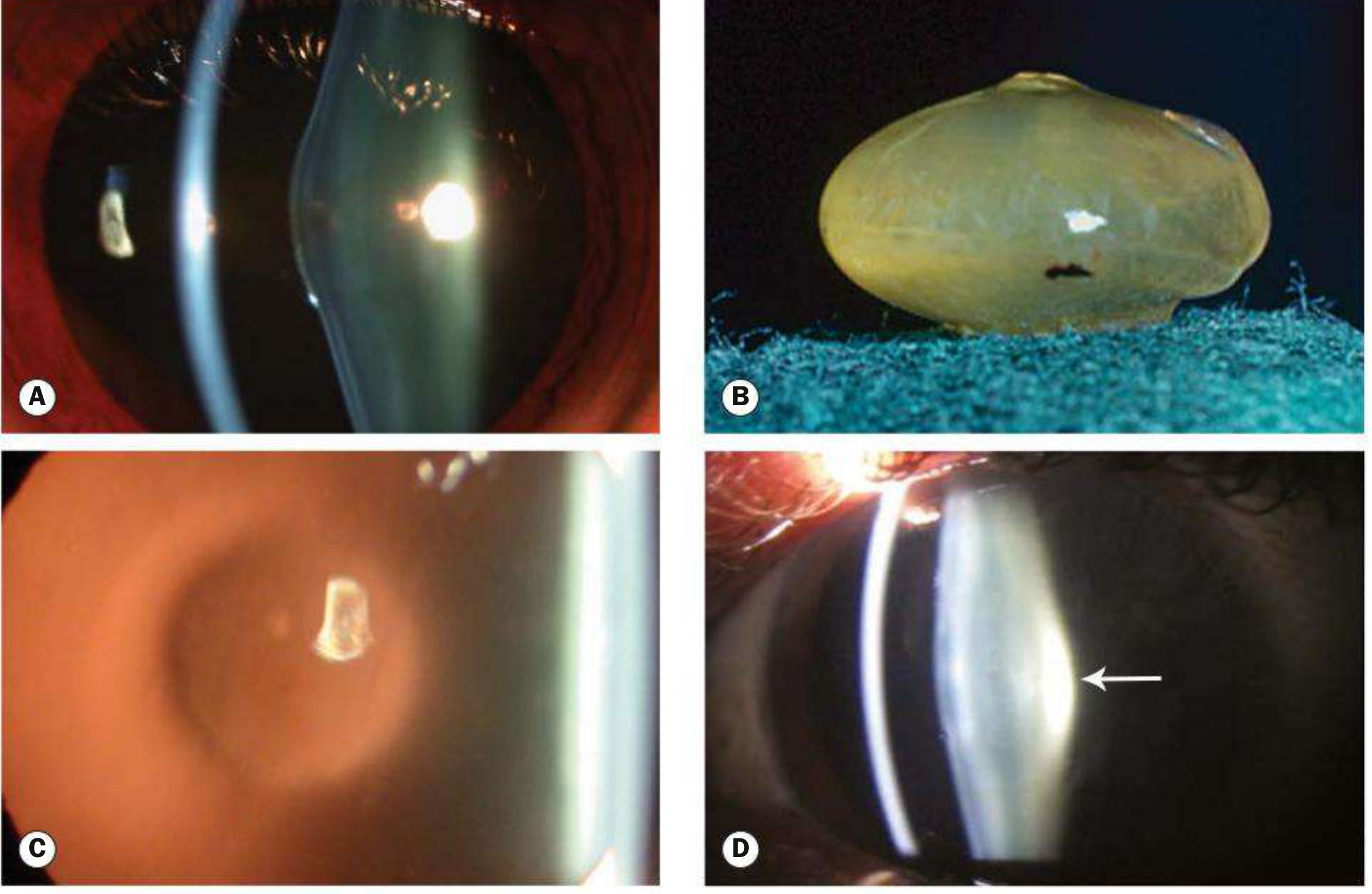
Here are all the textbook images for lenticonus, from Kanski's Clinical Ophthalmology (10th ed.), Fig. 10.31:
Image 1 - Anterior & Posterior Lenticonus (A-D)
(A) Anterior lenticonus in situ - slit-lamp view showing the axial forward protrusion of the anterior lens surface into the anterior chamber.
(B) Extracted lens after intracapsular removal - the conical protrusion of the anterior surface is clearly visible on the excised lens.
(C) "Oil droplet" sign on retroillumination - the pathognomonic appearance in Alport syndrome; the cone-shaped bulge creates a dark central reflex resembling an oil drop against the red fundal reflex.
(D) Posterior lenticonus (arrow) - slit-lamp view showing the posterior bulge of the lens.
Image 2 - Posterior Lenticonus with Capsular Opacification (E)
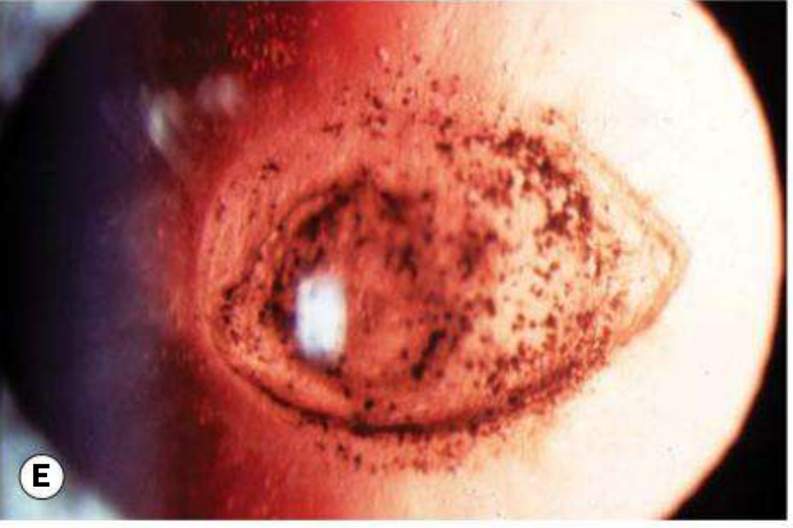
(E) Retroillumination showing opacification of the posterior capsule - a common complication as posterior lenticonus progresses. Dense cortical and posterior subcapsular changes are visible surrounding the cone-shaped protrusion.
Image 3 - Microspherophakia (F) - Related Lens Shape Abnormality
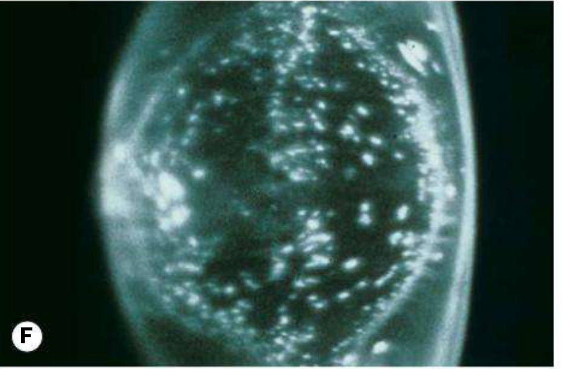
(F) Microspherophakia - a related lens shape anomaly where the lens is small and spherical, shown here with diffuse cortical opacities. Included for comparison as it falls in the differential of congenital lens shape abnormalities.
Image 4 - Microphakia (G)
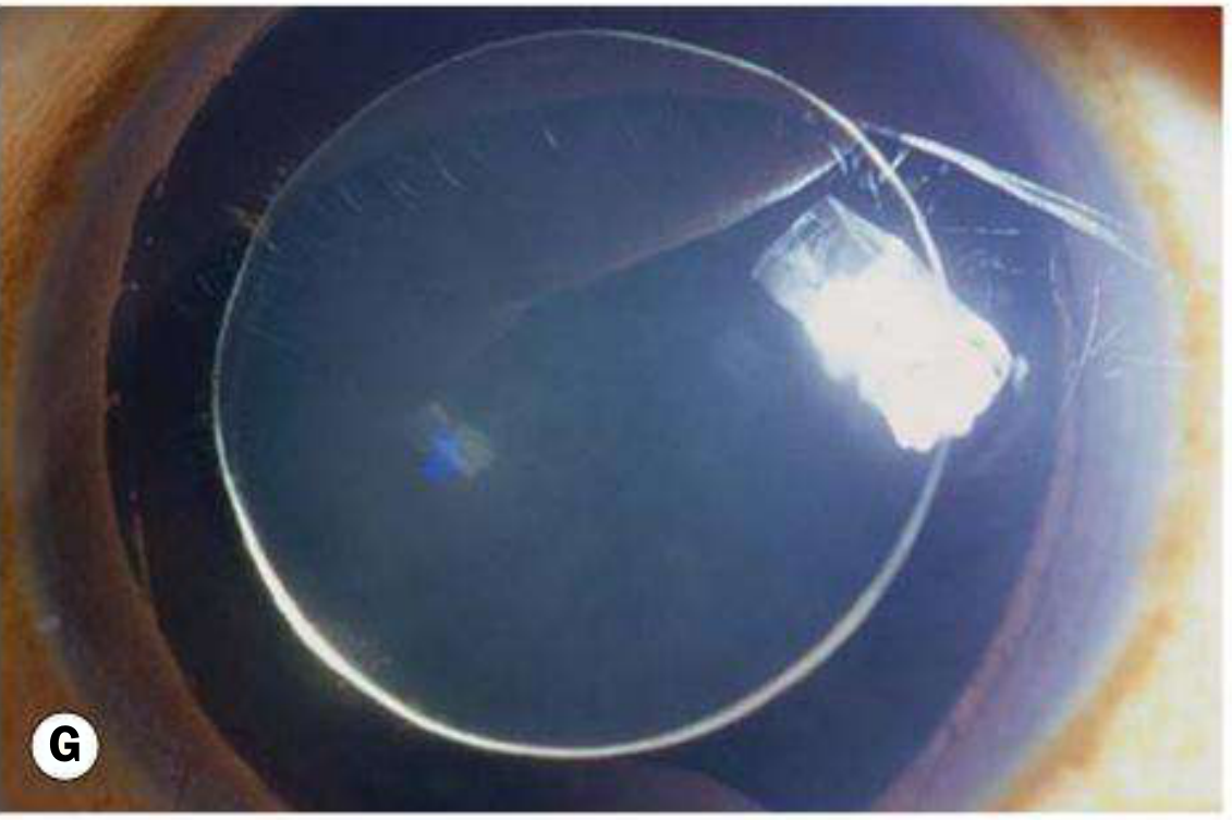
(G) Microphakia - the lens is smaller than normal in diameter. The full circumference of the lens equator is visible within the dilated pupil. Also shown for differential comparison.
Image 5 - Lens Coloboma (H)
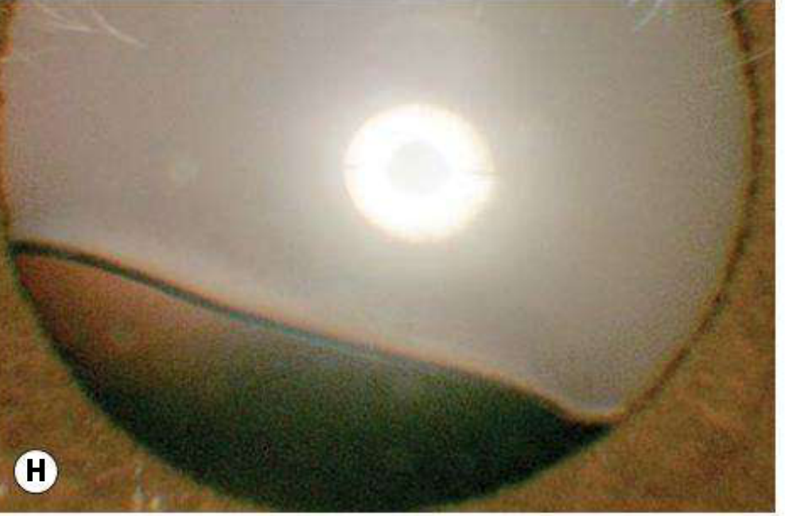
(H) Lens coloboma - a peripheral indentation of the lens due to localized zonular deficiency. Another lens shape abnormality in the differential.
Source: Kanski's Clinical Ophthalmology: A Systematic Approach, 10th ed., Fig. 10.31, p. 358-359
Give me from outside how eye looks
Let me search for external clinical appearance images of lenticonus from the textbooks and web.
Finding Sources
Searching the Web
lenticonus external appearance eye clinical photo slit lamp
Found external clinical images. Let me fetch the most relevant ones from ResearchGate and the textbook (Comprehensive Clinical Nephrology has a slit-lamp image with a cone-shaped arrow annotation).
Loading Image
Loading Image
Loading Image
Finding Sources
ResearchGate images are blocked. Let me read the Comprehensive Clinical Nephrology section which has an annotated slit-lamp image.
Reading File
Loading Image
Found the key annotated slit-lamp image from Comprehensive Clinical Nephrology. Let me fetch it now.
Loading Image
Here are the external clinical appearance images:
Image 1 - Anterior Lenticonus: How the Eye Looks from Outside (Slit-Lamp)
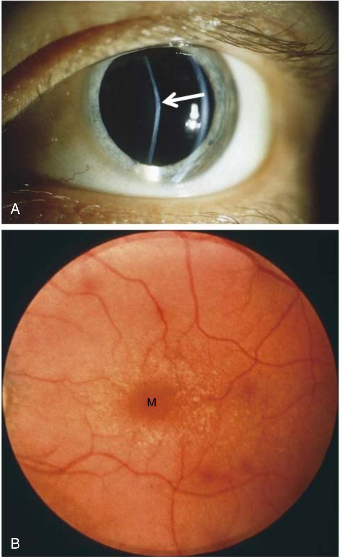
(A) Anterior lenticonus - This is exactly what the eye looks like when examined at the slit lamp. The white arrow points to the apex of the cone-shaped protrusion of the anterior lens surface. Instead of a normal smooth elliptical curve, the lens surface bulges forward into a cone shape - visible here as a kink or step in the reflection of the slit beam off the lens. The rest of the eye (cornea, iris, sclera) looks completely normal from the outside - lenticonus is invisible to the naked eye and requires a slit lamp to detect.
(B) Perimacular flecks - The same patient's fundus, showing white flecks surrounding the macula (M) - the retinal manifestation of Alport syndrome.
Source: Comprehensive Clinical Nephrology, 7th ed., Fig. 48.6
What you see without equipment vs. at the slit lamp
| View | What is visible |
|---|---|
| Naked eye / gross inspection | Eye looks completely normal. No visible external abnormality. |
| Direct ophthalmoscope | "Oil droplet" dark reflex centrally in the red reflex - this is the key bedside clue |
| Slit lamp (optical section) | The slit beam bends/kinks at the apex of the cone - clearly shows the abnormal convex protrusion of the lens surface (as seen in image A above, arrow) |
| Retroillumination | Classic "oil droplet" sign - dark central shadow against the red fundal reflex |
The key clinical point: lenticonus has no external visible abnormality to the naked eye. The eye looks completely normal on casual inspection. It is found only on slit-lamp examination (where the conical lens surface distorts the light beam) or with retroillumination/ophthalmoscopy (where the oil droplet sign appears).
This is a shared conversation. Sign in to Orris to start your own chat.