
60 yr old man progressive develop loss of proprisepction nad vibration..... Tertiary syphilis
tabes dorsalis spinal cord pathology posterior column degeneration syphilis
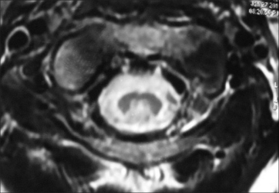
**Imaging Modality:** Magnetic Resonance Imaging (MRI), Axial T2-weighted sequence. **Anatomical Region:** Cervical spinal cord. **Observed Pathology:** Symmetrical intramedullary T2 hyperintensities localized within the posterior columns (dorsal columns) of the spinal cord. **Characteristic Visual Features:** The image demonstrates a distinct "inverted V" or "rabbit ear" pattern of high signal intensity involving the fasciculus gracilis and fasciculus cuneatus. The gray matter H-shape is relatively spared, and the peripheral white matter remains largely isointense. There is no evidence of significant cord expansion or compressive myelopathy at this level. **Clinical Context & Differential:** This radiological sign is highly characteristic of Subacute Combined Degeneration (SCD) of the spinal cord, typically associated with Vitamin B12 deficiency. Other diagnostic considerations for posterior column T2 hyperintensities include copper deficiency myelopathy, Friedreich's ataxia, or infectious etiologies such as tabes dorsalis. **Indexing Keywords:** Axial T2 MRI, Cervical Spinal Cord, Posterior Column Hyperintensity, Inverted V Sign, Subacute Combined Degeneration, Demyelination, Intramedullary Signal Abnormality.
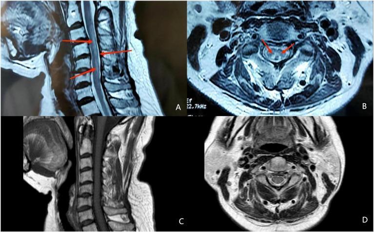
This composite diagnostic image features four MRI panels (A-D) demonstrating subacute combined degeneration of the cervical spinal cord. Panel A is a sagittal T2-weighted MRI showing a longitudinally extensive hyperintense signal within the posterior columns (indicated by red arrows). Panel B displays an axial view at the cervical level, revealing bilateral hyperintensities in the posterior columns that form the characteristic 'inverted V' or 'rabbit ears' sign (red arrows). This pattern is a classic radiological marker for vitamin B12 deficiency and posterior column myelopathy. Panels C (sagittal) and D (axial) represent post-treatment follow-up imaging, demonstrating the complete resolution of the previously seen abnormal hyperintensities. The images serve as an educational comparison of acute pathology versus radiological recovery after therapeutic intervention. These findings are relevant for neurology and radiology students studying metabolic myelopathies and differential diagnoses of spinal cord lesions.
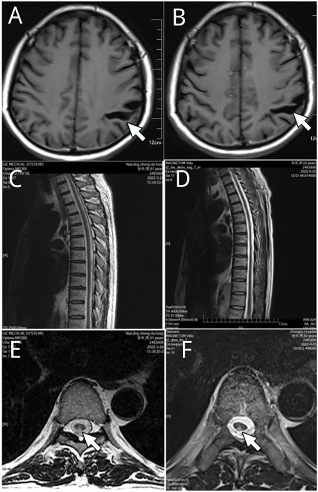
This composite diagnostic image presents a longitudinal Magnetic Resonance Imaging (MRI) comparison of the brain and thoracic spine over a six-month interval (March 2022 and September 2022). Panels A and B are axial T1-weighted cranial scans showing focal cortical atrophy characterized by the widening of the left parietal sulcus (white arrows). Panels C and D provide sagittal T2-weighted views of the thoracic spine, while panels E and F display axial T2-weighted cross-sections. The spinal imaging reveals a hyperintense signal within the posterior columns (dorsal columns) of the spinal cord at the T3-T6 level (white arrows), a finding classically associated with conditions such as neurosyphilis (tabes dorsalis) or subacute combined degeneration. The comparison demonstrates temporal stability of these neurological findings, with no significant progression or resolution between the two time points. This visual material serves as an educational example of identifying specific neuroanatomical pathologies—namely parietal atrophy and dorsal column myelopathy—using multi-planar MRI sequences in the context of chronic infectious or metabolic central nervous system diseases.
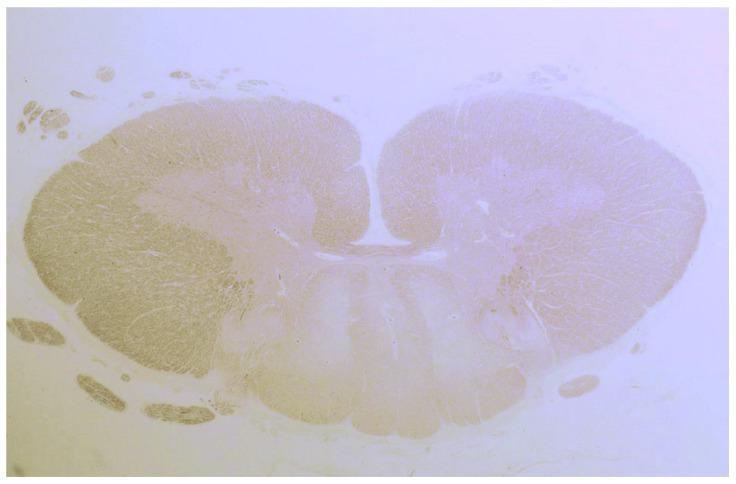
This light microscopy image presents a transverse histological section of a human spinal cord, stained to highlight neural tissue architecture. The section demonstrates the characteristic butterfly-shaped central gray matter with identifiable dorsal and ventral horns surrounding a small central canal. The primary clinical finding is a significant, bilateral pallor within the posterior (dorsal) columns of the white matter. This focal loss of staining intensity represents demyelination and tissue degeneration, contrasting sharply with the more intensely stained lateral and ventral white matter tracts. Such localized degeneration of the fasciculus gracilis and fasciculus cuneatus is histologically characteristic of conditions such as gluten ataxia and sensory ganglionopathy, where dorsal column pathology leads to impaired proprioception and sensory ataxia. Small, darkly stained nerve root bundles are also visible peripheral to the main spinal cord body. The image serves as a high-level educational example of secondary neurodegeneration and systemic-related myelopathy.
Tabes Dorsalis - Tertiary Neurosyphilis
What Is Happening
Pathophysiology
- Chronic syphilitic meningitis leads to subpial inflammation
- T. pallidum damages the dorsal nerve roots at their entry into the spinal cord
- Secondary Wallerian-type degeneration of the fasciculus gracilis and fasciculus cuneatus (posterior columns) follows
- These columns carry proprioception, vibration, fine touch, and 2-point discrimination - all dorsal column modalities
- Spinothalamic (pain/temperature) and motor tracts are largely spared early on
Classic Clinical Features
| Feature | Detail |
|---|---|
| Loss of proprioception | Key symptom - positive Romberg sign |
| Loss of vibration sense | Tuning fork test impaired |
| Sensory (tabetic) ataxia | Wide-based, "slapping" gait - stamping feet to feel ground |
| Lancinating (lightning) pains | Brief, repetitive, shooting pains in legs; also back, thorax, abdomen |
| Areflexia | Absent knee and ankle jerks (dorsal root damage disrupts reflex arc) |
| Argyll Robertson pupils | Bilateral; accommodate but do NOT react to light ("prostitute's pupil") |
| Bladder dysfunction | Atonic bladder, overflow incontinence |
| Visceral crises | Episodic severe abdominal, rectal, or laryngeal pain and vomiting |
| Charcot (neuropathic) joints | Painless joint destruction, classically knees |
The Argyll Robertson Pupil - Key Exam Point
- Light reflex: ABSENT (direct and consensual)
- Near/accommodation reflex: INTACT ("accommodates but does not react")
- Small, irregular, unequal pupils
- Caused by damage in the pretectal region of the midbrain
- Classically described as: "like a prostitute - accommodates but does not react"
Dorsal Column Syndrome vs. Other Posterior Column Disorders
| Feature | Tabes Dorsalis | B12 Deficiency (SCD) |
|---|---|---|
| Columns affected | Posterior only | Posterior AND lateral |
| Motor signs | Absent (early) | Spasticity + UMN signs |
| Cause | T. pallidum | Vitamin B12 deficiency |
| Pain | Lancinating | Paresthesias |
| Reflexes | Absent (LMN) | Initially absent, then hyperreflexia |
| Pupils | Argyll Robertson | Normal |
Investigations
- Serology: VDRL/RPR (screening) + FTA-ABS or TPHA (confirmatory) - almost always positive
- Lumbar puncture - CSF (mandatory in tertiary syphilis):
- Elevated protein
- Mononuclear pleocytosis
- Elevated IgG index (>0.7 = local CNS synthesis)
- CSF VDRL (specific but not sensitive)
- MRI spine: May show posterior column T2 hyperintensity (similar "inverted-V" sign to B12 deficiency, but history/serology distinguishes)
- HIV testing - co-infection is common
Treatment
- Aqueous crystalline penicillin G IV: 18-24 million units/day (3-4 million units IV every 4 hours) for 10-14 days - treatment of choice
- Alternative: Procaine penicillin G 2.4 million units IM daily + probenecid 500 mg orally 4x/day for 10-14 days
- Penicillin-allergic: desensitization is preferred; ceftriaxone 2g IV daily for 10-14 days is an alternative
- Post-treatment: CSF should be re-examined every 6 months until cell count normalizes
- Note: neurological damage (lost reflexes, impaired proprioception) may not fully reverse even with treatment - "tabes dorsalis may burn out even without treatment" but antibiotics prevent further progression
Summary Mnemonic for Tabes Dorsalis
- Posterior column degeneration (proprioception + vibration loss)
- Argyll Robertson pupils
- Sensory ataxia + Stomping/Slapping gait
- Tendon reflexes absent
- Acute lancinating (lightning) pains
Sources: Harrison's Principles of Internal Medicine 22E, p.3612; Bradley & Daroff's Neurology in Clinical Practice; Dermatology 2-Volume Set 5e (Neurosyphilis section); Neuroanatomy through Clinical Cases 3rd Ed.