
Note on masticatory mucosa
masticatory mucosa gingiva hard palate histology keratinized epithelium
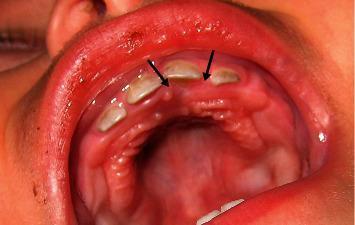
This clinical photograph displays a 1-month postoperative follow-up of a pediatric patient's maxillary arch and hard palate following the excisional biopsy of a pyogenic granuloma. The intraoral view shows the primary deciduous incisors and canines. Two black arrows indicate the previous surgical site on the palatal gingiva, adjacent to the upper left first deciduous incisor. The tissue at the site of the diode laser excision demonstrates complete mucosal healing with no signs of tumor recurrence. The gingiva appears healthy, pink, and well-keratinized, with a smooth texture that matches the surrounding palatal rugae and mucosa. There is no evidence of inflammation, edema, or residual scarring. The primary dentition is properly spaced, and the surrounding oral structures appear within normal physiological limits for an infant. This image serves as a reference for successful soft tissue healing and recurrence monitoring after surgical intervention for benign oral lesions in pediatric patients.
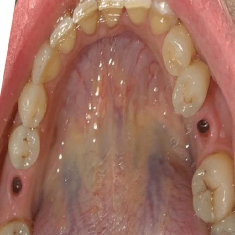
This intraoral clinical photograph provides an occlusal view of the maxillary arch, focusing on the soft tissue healing following dental implant surgery. The image demonstrates the emergence profile of the gingiva in the second surgical stage. Two circular, dark-colored healing abutments are visible protruding from the alveolar ridge at edentulous sites, surrounded by healthy, pink, well-contoured keratinized mucosa. The hard palate shows typical anatomical features, including rugae and a midline raphe, with normal vascular variations and a moist surface. The adjacent natural dentition shows light yellowish coloration with evidence of minor incisal wear and fair oral hygiene, characterized by slight marginal gingival erythema indicative of mild gingivitis. This image is used to illustrate the peri-implant soft tissue management and the establishment of an appropriate gingival emergence profile required before the final prosthetic restoration with zirconia crowns.
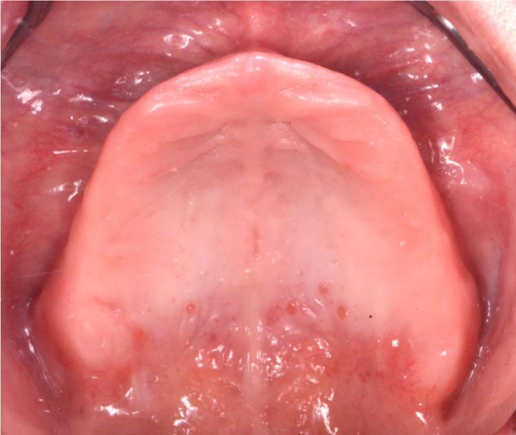
This clinical intraoral photograph provides an occlusal view of an edentulous maxilla, demonstrating significant alveolar ridge resorption consistent with Class III edentulism. The image shows the maxillary alveolar ridge and hard palate. The alveolar mucosa is generally healthy and pink, though the keratinized gingiva covering the crest of the ridge appears thin. In the anterior region of the ridge, several discrete, small erythematous spots are visible, which may indicate mild irritation or chronic atrophic candidiasis (denture stomatitis), given the history of long-term denture wear. Anatomical structures such as the median palatine raphe and palatal rugae are visible but appear somewhat flattened due to tissue atrophy. The surrounding vestibular mucosa appears moist and adequately vascularized. This image is used in prosthodontic education to illustrate the anatomical challenges of severe bone atrophy and the evaluation of soft tissue health prior to fabricating new complete dentures.
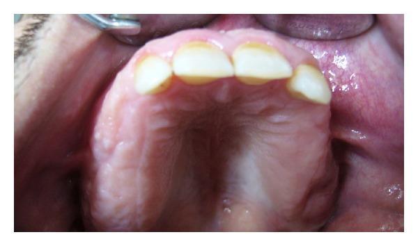
This intraoral clinical photograph provides a 1-year postoperative palatal view of the maxillary arch in a patient previously treated for Florid Cemento-Osseous Dysplasia (FCOD). The image shows the hard palate and the remaining anterior maxillary teeth. The palatal mucosa and gingiva appear healthy, pink, and well-keratinized, with no signs of active inflammation, ulceration, or infection. Notable findings include significant expansion and irregularity of the alveolar ridge, consistent with the underlying fibro-osseous disease process. The visible anterior teeth exhibit yellowish discoloration, incisal wear, and slight malalignment. The posterior areas of the arch appear partially edentulous following surgical recontouring and extractions to facilitate prosthetic rehabilitation. The clinical presentation highlights the stabilized state of the maxillary arch one year after surgical intervention for FCOD, emphasizing the healed soft tissue over the recontoured cortical bone.
Masticatory Mucosa
Classification of Oral Mucosa
| Type | Location | Epithelium | Colour |
|---|---|---|---|
| Masticatory | Gingiva, hard palate | Keratinized / parakeratinized stratified squamous | Pink |
| Lining | Lips, cheeks, floor of mouth, soft palate, alveolar processes, ventral tongue | Non-keratinized stratified squamous | Red |
| Specialized (Gustatory) | Dorsum of tongue (anterior 2/3) | Mixed | Pink-red |
The vermillion border of the lip shares features of both lining and masticatory mucosa and is sometimes regarded as a fourth category.
Definition and Location
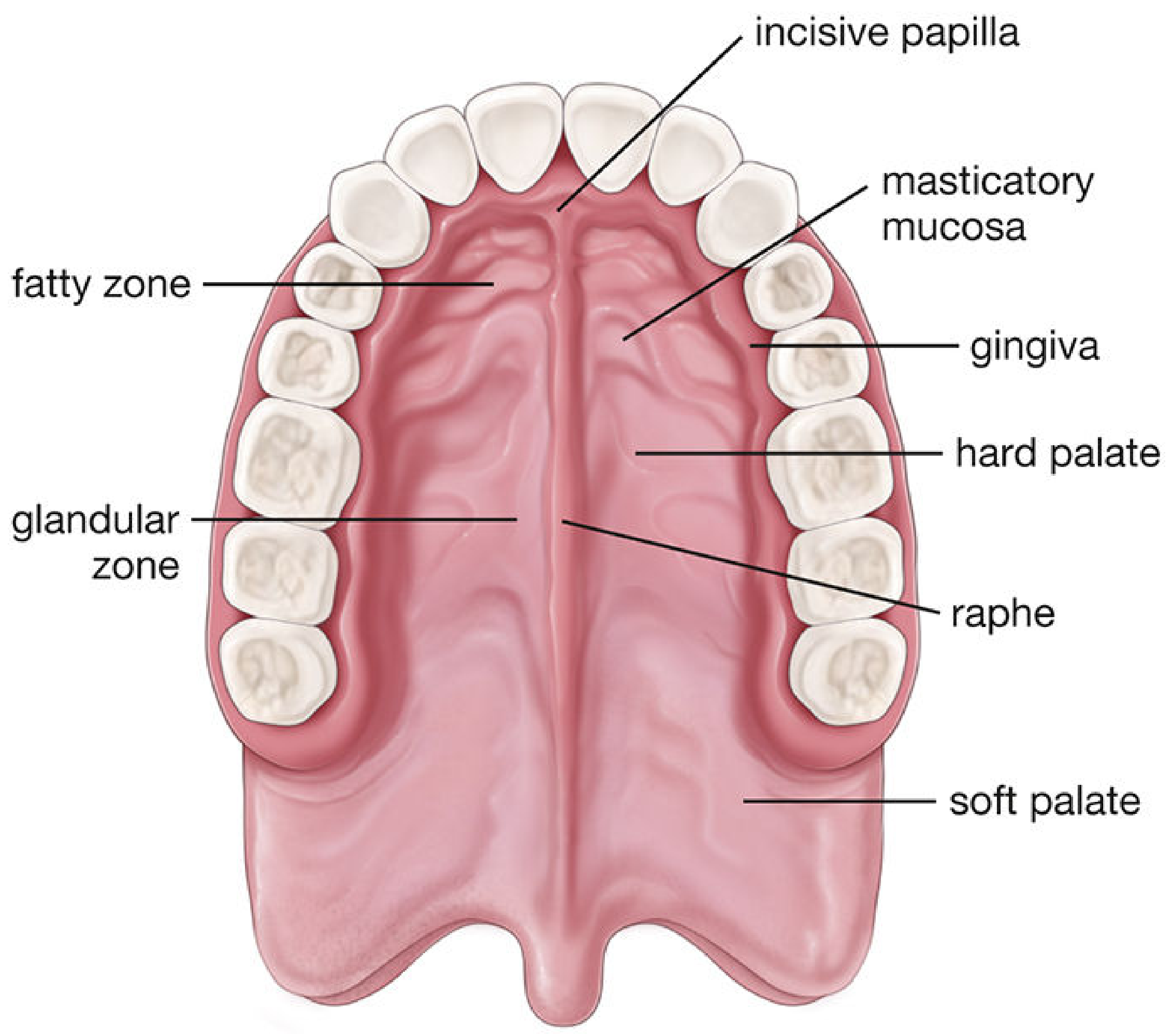
- Gingiva (gums) - surrounds the necks of the teeth and is firmly bound to the alveolar bone
- Hard palate - roof of the mouth containing bone, bisected by the median palatine raphe
Histological Structure
1. Epithelium
- Type: Stratified squamous epithelium that is keratinized in most areas, and parakeratinized in some regions
- Layers in keratinized areas (resembling skin but lacking stratum lucidum):
- Stratum basale
- Stratum spinosum
- Stratum granulosum (with keratohyalin granules)
- Stratum corneum (anucleate, intensely eosinophilic)
- Parakeratinized epithelium: Superficial cells retain their pyknotic (condensed) nuclei and show fewer keratohyalin granules, with less intense eosin staining compared to fully keratinized cells. Cells are still shed (exfoliated) after this stage.
- Cell types present: Keratinocytes, Langerhans cells, melanocytes, and Merkel cells
Key distinction: Fully keratinized cells lose nuclei and stain intensely eosinophilic; parakeratinized cells retain pyknotic nuclei and stain less intensely.
2. Lamina Propria
- Papillary layer (superficial): Loose connective tissue with blood vessels and nerves; some nerve endings penetrate the epithelium as bare axon endings (sensory receptors); some end as Meissner corpuscles
- Reticular layer (deep): Dense connective tissue
- Connective tissue papillae are numerous and deep - this anchors the epithelium firmly, resisting frictional and shearing forces during mastication
- In the gingiva and palatine raphe, the reticular layer blends directly with periosteum - hence no submucosa is present
3. Submucosa
- Absent at the gingiva and midline palatine raphe (mucoperiosteum forms instead)
- Present over most of the hard palate in two zones:
- Fatty zone (anterior): submucosa contains adipose tissue
- Glandular zone (posterior): submucosa contains mucous salivary glands, continuous with those of the soft palate; also contains the greater palatine nerves and vessels
- Thick collagenous bands extend from the mucosa to the underlying bone in submucosal regions
Mucoperiosteum
Functional Adaptations
| Feature | Functional Role |
|---|---|
| Keratinized/parakeratinized epithelium | Resists abrasion, pressure, and friction during chewing |
| Numerous deep connective tissue papillae | Anchors epithelium; prevents shearing; relative immobility |
| Firm attachment to bone (mucoperiosteum) | Prevents displacement under occlusal forces |
| Dense fibrous lamina propria | Structural support and rigidity |
| Pink colour | Reflects moderate vascularity under thick keratinized layer (compare with red lining mucosa) |
Comparison: Masticatory vs. Lining Mucosa
| Feature | Masticatory Mucosa | Lining Mucosa |
|---|---|---|
| Sites | Gingiva, hard palate | Lips, cheeks, floor of mouth, soft palate, ventral tongue, alveolar processes |
| Epithelium | Keratinized / parakeratinized | Non-keratinized (thicker) |
| Epithelial layers | 4 (incl. stratum granulosum, corneum) | 3 (basale, spinosum, superficiale) |
| Connective tissue papillae | Numerous, deep | Few, shallow (allows movement) |
| Submucosa | Absent (gingiva/raphe) or present (rest of palate) | Present (contains fat, minor salivary glands) |
| Mobility | Immobile (bound to bone) | Mobile (overlies muscle) |
| Colour | Pink | Red |
Clinical Relevance
- Recurrent herpes simplex (intraoral): Affects only the keratinized/masticatory mucosa (gingiva, hard palate) - a distinguishing feature from aphthous ulcers, which affect non-keratinized lining mucosa
- Aphthous stomatitis (Behcet disease): Typically spares masticatory mucosa (gingiva, hard palate, dorsum of tongue)
- Gingival surgery/grafts: The firm, keratinized nature makes gingival tissue a preferred donor site for free gingival grafts
- Drug delivery: The lining (not masticatory) mucosa is preferred for transmucosal drug absorption due to its thinner, non-keratinized, highly vascularized epithelium
- Histology: A Text and Atlas with Correlated Cell and Molecular Biology (Ross & Pawlina) - p. 1413-1417
- Junqueira's Basic Histology: Text and Atlas, 17e - p. 736
- Scott-Brown's Otorhinolaryngology Head & Neck Surgery - p. 680