I now have all the material needed. Let me update todos and compose the comprehensive viva answer.---
🧠 HYDROCEPHALUS & ICP MONITORING — Comprehensive Neurosurgery Viva Guide
Sources: Adams & Victor's Principles of Neurology 12e, Grainger & Allison's Diagnostic Radiology, Bradley & Daroff's Neurology in Clinical Practice, Neuroanatomy through Clinical Cases 3e, Goldman-Cecil Medicine, Harriet Lane Handbook 23e
PART 1: HYDROCEPHALUS
1. DEFINITION & OVERVIEW
Hydrocephalus = enlargement of the cerebral ventricles resulting from an imbalance between CSF production and absorption (or obstruction to flow), causing accumulation of CSF under increased pressure.
⚠️ Viva Point: Not all ventriculomegaly = hydrocephalus. Hydrocephalus ex vacuo (atrophic enlargement) must be distinguished — it does NOT benefit from intervention.
2. CLASSIFICATION
A. By Communication of Ventricular System (Dandy & Blackfan, 1914)
| Type | Synonyms | Site of Obstruction | Key Feature |
|---|
| Non-communicating (Obstructive) | Intraventricular obstructive HCP | Within the ventricular system (foramen of Monro, aqueduct, 4th ventricle outlets) | Dye injected into lateral ventricle does NOT reach lumbar subarachnoid space |
| Communicating | Extraventricular obstructive HCP | Basal cisterns, arachnoid villi, convexity subarachnoid spaces | Dye reaches lumbar space; obstruction outside ventricles |
| Hydrocephalus ex vacuo | Compensatory | None | Ventricular enlargement secondary to brain atrophy — NOT true hydrocephalus |
| Normal Pressure Hydrocephalus (NPH) | Hakim-Adams syndrome | Impaired CSF reabsorption (communicating type) | Normal ICP on LP; classic triad |
| Overproduction type | — | Choroid plexus papilloma | Very rare; excess CSF production |
B. By Pathophysiology
- Obstructive (non-communicating): aqueductal stenosis, posterior fossa tumour, colloid cyst of 3rd ventricle
- Communicating: post-meningitis, post-SAH, post-haemorrhagic (neonates), leptomeningeal metastases
- Over-productive: choroid plexus papilloma
C. By Age Group
- Congenital/Infantile (before suture fusion, <2–3 years)
- Childhood (sutures fusing)
- Adult/NPH (idiopathic, elderly)
D. By Onset
- Acute vs. chronic vs. arrested/compensated
3. CSF PHYSIOLOGY (Essential for Viva)
- Production: 0.3–0.35 mL/min (≈ 500 mL/day); mainly by choroid plexus of lateral ventricles
- Total volume: 150 mL (adults); turnover 3–4×/day
- Absorption: Primarily arachnoid granulations → dural venous sinuses; also spinal cord, brain parenchyma
- Flow path: Lateral ventricles → Foramen of Monro → 3rd ventricle → Aqueduct of Sylvius → 4th ventricle → Foramina of Luschka & Magendie → Basal cisterns → Convexity subarachnoid space → Arachnoid granulations
- Monro-Kellie Doctrine: Brain (80–90%) + Blood + CSF = constant volume. Expansion of one = compression of others.
4. ALL TYPES OF HYDROCEPHALUS — IN DETAIL
TYPE 1: CONGENITAL / INFANTILE HYDROCEPHALUS
Demographics
- Age: Neonate to 3 years (before suture fusion)
- M = F (congenital causes); M > F (aqueductal stenosis — X-linked form)
Causes
| Cause | Notes |
|---|
| Aqueductal stenosis | Most common congenital cause; X-linked form (L1CAM mutation) |
| Chiari II malformation | Associated with myelomeningocele; 4th ventricle foramina below foramen magnum |
| Dandy-Walker malformation | Hypoplasia/absence of cerebellar vermis, enlarged 4th ventricle, occipital bossing → dolichocephalic head |
| Post-haemorrhagic | Commonest in premature neonates (IVH → fibrin → obstruction) |
| Post-infective | Post-meningitis: E. coli, Strep. agalactiae, Listeria (neonatal); Neisseria, Strep. pneumoniae (older) |
| Vein of Galen malformation | AV shunting → raised venous pressure → raised ICP |
| Choroid plexus papilloma | Overproduction + obstruction |
Clinical Presentation
- Macrocephaly — head circumference crossing centiles (>97th); most reliable early sign
- Tense, bulging anterior fontanelle (even in upright position)
- Sutural diastasis (widened sutures on palpation)
- Frontal bossing; scalp vein distension; calvarial thinning
- "Setting sun sign" — downward deviation of eyes, sclerae visible above iris; due to pressure on midbrain tectum (Parinaud's phenomenon)
- Lateral rectus palsy (CN VI) — false localizing sign
- Leg spasticity (stretching of corticospinal tracts around dilated ventricles)
- Fretfulness, poor feeding, vomiting
- Advanced: flexed posture, feeble movements, optic atrophy
⚠️ Viva Trap: No papilledema in infants — the fontanelle acts as a safety valve.
TYPE 2: CHILDHOOD HYDROCEPHALUS (Sutures fusing, >2 years)
Causes
- Posterior fossa tumours (most common): Medulloblastoma (#1 in children), Ependymoma, Cerebellar astrocytoma
- Aqueductal stenosis (acquired: gliosis, ependymitis, tumour)
- Pineal region tumours → compress aqueduct
Clinical Presentation (sutures rigid/fusing)
- Headache — worse in the morning (due to lying flat → increased ICP), vomiting (projectile)
- Papilledema (present — skull cannot expand)
- CN VI palsy (false localizing)
- Leg spasticity, ataxia
- Altered conscious level
- Parinaud's syndrome (loss of upward gaze) — from tectal compression
- Cushing's triad (hypertension + bradycardia + irregular respirations) — terminal, indicates herniation
TYPE 3: NORMAL PRESSURE HYDROCEPHALUS (NPH)
Named after: Adams, Fisher & Hakim (1965)
Demographics
- Age: >60 years (idiopathic NPH); can be younger in secondary NPH
- Prevalence: ~0.2% in 70–79 years; ~5.9% in >80 years
Classic Hakim-Adams Triad (order of appearance):
- Gait disturbance (earliest & most prominent) — "magnetic gait," short shuffling steps, widened base, difficulty on stairs, cannot lift feet; mimics Parkinsonism but without rigidity/tremor
- Dementia — "frontal" type: apathy, dullness, slowed thinking, memory impairment; may mimic Alzheimer's
- Urinary incontinence — urgency initially → frank incontinence; bowel incontinence late; "frontal incontinence" (patient indifferent)
Mnemonic: "Wet, Wobbly, and Wacky"
Causes:
- Idiopathic (most common)
- Secondary: SAH, meningitis, TBI, craniotomy, Paget's disease of skull
CSF Pressure in NPH:
- Normal (70–200 mm H₂O on LP) by definition — but may be intermittently elevated (Lundberg B waves may be present on continuous monitoring)
TYPE 4: HYDROCEPHALUS EX VACUO
- Ventricular enlargement secondary to brain parenchymal loss (atrophy, infarct, surgery)
- NOT true hydrocephalus — NO increased ICP
- Does NOT require treatment
- Key differentiator: prominent sulci and cortical atrophy accompanying ventricular enlargement
TYPE 5: ARRESTED/COMPENSATED HYDROCEPHALUS
- Longstanding hydrocephalus where a new equilibrium has been reached
- Ventricles may be large but ICP is normal or near-normal
- May decompensate with minor insults
TYPE 6: EXTERNAL HYDROCEPHALUS (Benign External Hydrocephalus of Infancy)
- Enlarged subarachnoid spaces over hemispheres + mild ventricular enlargement
- Usually self-limiting
- Must exclude subdural hygroma/hematoma, arachnoid cyst
5. INVESTIGATIONS
A. NEUROIMAGING
Non-Contrast CT Head (First Line)
Indications: Acute presentation, suspected raised ICP, macrocephaly, post-trauma, sudden-onset headache
Positive findings in hydrocephalus:
| Finding | Significance |
|---|
| Dilated temporal horns (>2 mm) | Earliest and most reliable sign of hydrocephalic dilatation |
| Frontal horn rounding/ballooning | Periventricular transependymal edema (hypodense halo) |
| Dilatation of 3rd ventricular recesses (supraoptic, infundibular) | Obstructive hydrocephalus |
| Periventricular lucency | Transependymal CSF seepage — indicates active, acute hydrocephalus |
| Effaced sulci/basal cisterns | High ICP |
| Evans Index > 0.3 | Max width of frontal horns / Max inner skull diameter |
| Bicaudate index (min caudate width/skull width) | >95th percentile for age = abnormal |
| Copper-beaten/beaten silver skull | Chronic raised ICP (children) |
| Erosion of dorsum sellae | Chronic raised ICP |
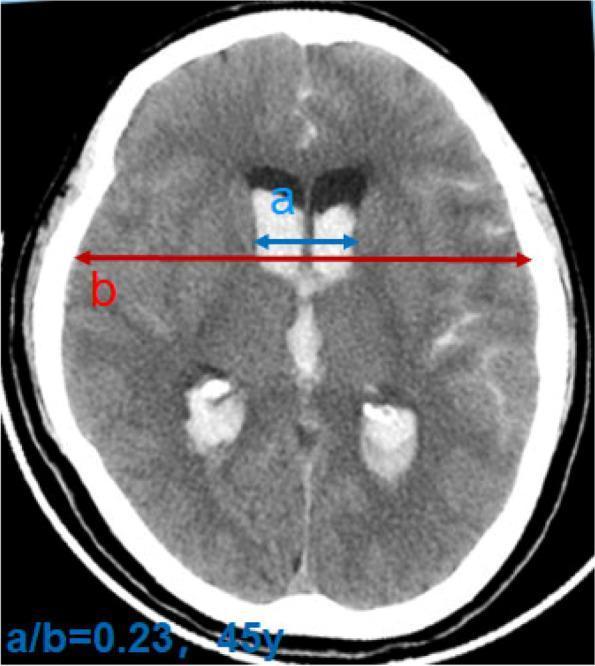
Axial non-contrast CT: Acute obstructive hydrocephalus with IVH, dilated lateral ventricles, bicaudate index = 0.23 (>normal 0.17 for age 45)
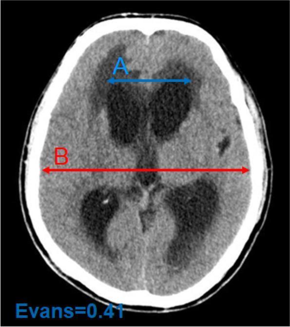
Evans Index = 0.41 (>0.3). Periventricular hypodensity indicating transependymal edema.
MRI Brain (Preferred for Detailed Assessment)
Indications: Suspected cause (tumour, aqueductal stenosis), NPH workup, post-shunt assessment, ETV patency check
Positive findings:
- FLAIR: Periventricular hyperintensity (transependymal edema)
- T2: Dilated ventricles, aqueductal flow void (absent = stenosis, present = patent)
- Sagittal T1: Aqueduct morphology, floor of 3rd ventricle, site of obstruction
- Callosal angle (coronal view): Angle between roof of lateral ventricles at posterior commissure
- NPH: <90° (typically 50–80°)
- Atrophy: >100°
- DESH sign in NPH: Disproportionately Enlarged Subarachnoid-space Hydrocephalus — tight sulci at high convexity + Sylvian fissure enlargement
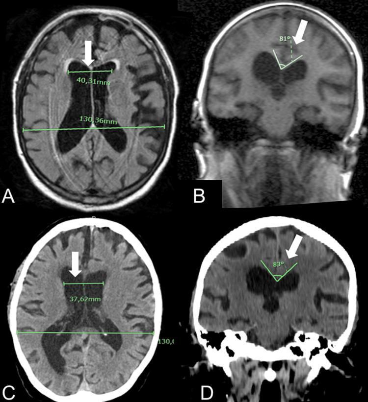
NPH: Evans Index (A, C) and callosal angle 81–83° (B, D) on MRI and CT respectively
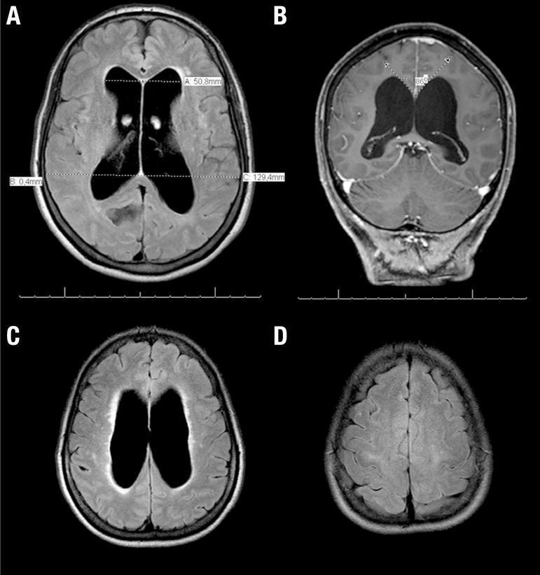
NPH: FLAIR showing Evans Index 0.39, callosal angle 86°, periventricular edema, DESH pattern
ETV patency on MRI: Sagittal T2/FIESTA — large hypointense flow void at stoma in floor of 3rd ventricle → confirms patent ETV.
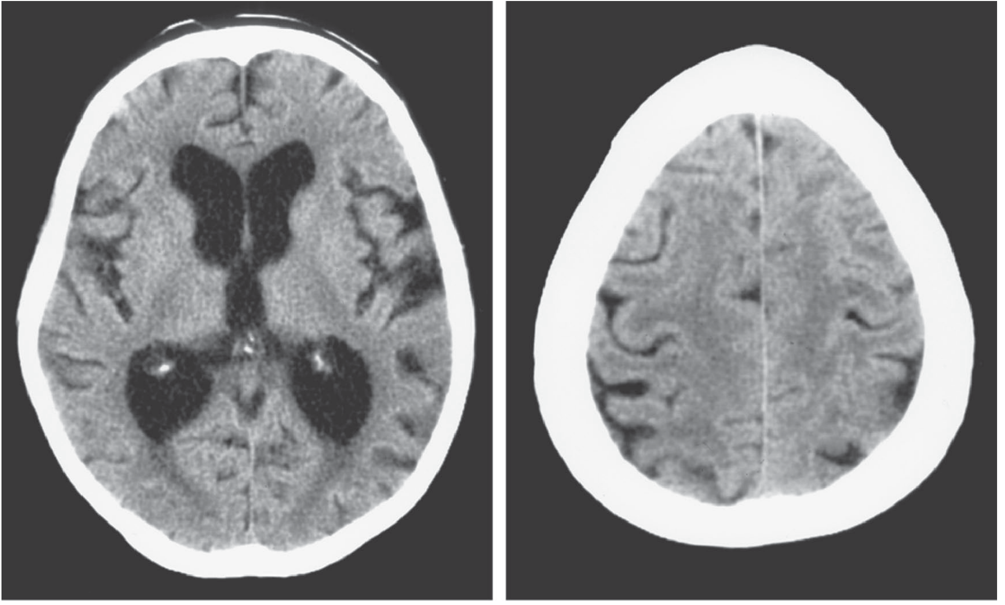
CT of NPH (Adams & Victor, Fig. 29-4):
NPH: Enlarged frontal horns (frontal horn span >39 mm) disproportionate to cortical atrophy (right). This ventricular-sulcal disproportion is the hallmark.
Skull X-ray
- Beaten silver / copper-beaten appearance (chronic ICP — children)
- Sutural diastasis
- Shunt series: assess tubing integrity, disconnection, calcification
B. LUMBAR PUNCTURE
Indications: Suspected communicating HCP, NPH diagnosis, post-infective HCP, opening pressure measurement
NPH High-volume LP tap test:
- Remove 20–50 mL CSF
- Assess gait before and 1–4 hours after (formal timed 10-metre walk)
- Improvement in gait = positive tap test → high predictive value for shunt response
- Negative tap test does NOT exclude NPH or shunt benefit
NPH lumbar drain trial: 50 mL/day for 3 days (inpatient) — more definitive than single LP
CSF findings:
- NPH: Opening pressure usually 150–200 mm H₂O (can be lower); normal composition
- Post-meningitis: elevated protein, pleocytosis
- SAH: xanthochromia
C. PATHOLOGICAL INVESTIGATIONS
| Investigation | Indication | Finding |
|---|
| CSF analysis | All causes, especially infective | Cell count, protein, glucose, culture, xanthochromia |
| Genetic testing (L1CAM) | X-linked aqueductal stenosis (males) | Mutation in L1CAM gene on Xq28 |
| Brain biopsy | Rarely — suspected malignancy/metastasis | Tumour type |
| Ophthalmology/fundoscopy | All symptomatic HCP | Papilledema grading (Frisen scale) |
D. SCORES & INDICES
| Score/Index | What it measures | Threshold |
|---|
| Evans Index | Max frontal horn width / Max inner skull diameter | >0.3 = ventriculomegaly |
| Callosal Angle | Angle between lateral ventricle walls at posterior commissure (coronal MRI) | <90° suggests NPH; <40° = severe |
| Bicaudate Index | Min caudate head distance / skull width | Age-adjusted; >95th percentile = abnormal |
| Hunt-Hess Scale | SAH severity (relevant when HCP follows SAH) | Grade I–V; Grade III–V → EVD placement |
| Fisher Scale (Modified) | CT blood burden in SAH → vasospasm risk | Grade 1–4 |
| GCS | All acute HCP | <8 → intubate, ICP monitor |
| ETV Success Score (ETVSS) | Predicts ETV success | Score 0–90; >80 = good prognosis |
6. MANAGEMENT APPROACH
Step 1: Resuscitation & Emergency Management
Acute HCP with raised ICP:
- Elevate HOB 30°
- Normocapnia (PaCO₂ 35–40 mmHg; hyperventilate briefly to 30–35 if herniation imminent)
- Osmotherapy: Mannitol 0.5–1 g/kg IV over 20 min (draws water out of brain)
- Hypertonic saline (3% NaCl) 1–2 mL/kg — alternative if hypotension present
- Avoid hypotension; maintain CPP >60–70 mmHg
- Dexamethasone 10 mg IV loading dose then 4 mg every 6h — if oedema around tumour (NOT for TBI or ischaemic stroke)
- Levetiracetam 2000 mg IV loading → 1000 mg BD — seizure prophylaxis (especially post-SAH)
- Emergency EVD if acute obstructive HCP causing herniation
Step 2: Medical Management (temporizing)
| Drug | MOA | Dose | Special Points |
|---|
| Acetazolamide | Carbonic anhydrase inhibitor → reduces CSF production by choroid plexus (by 50%) | 25 mg/kg/day in 3 divided doses (neonates); 250–500 mg TDS (adults) | Used temporarily in post-haemorrhagic HCP of prematurity; also used in IIH. Metabolic acidosis, renal stones, hypokalaemia. Sulfonamide allergy caution. |
| Furosemide | Loop diuretic → reduces CSF production (secondary mechanism); reduces choroid plexus Na⁺-K⁺-ATPase activity | 1 mg/kg/day PO (children) | Used with acetazolamide; monitor electrolytes |
| Mannitol | Osmotic diuretic → ↑ plasma osmolality → draws water from brain into blood | 0.5–1 g/kg IV over 20–30 min | Max effect at 30–60 min; duration 4–6h; do NOT use if serum osmolarity >320 mOsm; risk of rebound oedema |
| Hypertonic saline (3%) | Osmotherapy → ↓ ICP by osmotic dehydration | 1–2 mL/kg bolus; maintain Na 145–155 | Preferred over mannitol in hypotension/shock; monitor serum Na |
| Dexamethasone | Glucocorticoid → ↓ BBB permeability → ↓ vasogenic oedema around tumours | 4–10 mg IV/8h | Do NOT use in TBI or ICH; excellent for tumour-related HCP/oedema |
Step 3: Surgical Management
SURGERY OPTION 1: EXTERNAL VENTRICULAR DRAIN (EVD)
When to use:
- Acute/obstructive hydrocephalus (emergency decompression)
- SAH (Hunt-Hess grade III–V), IVH with HCP
- Monitoring ICP while awaiting definitive surgery
- Post-haemorrhagic HCP in neonates (bridging)
- Suspected CSF infection (diagnostic + therapeutic)
Procedure:
- Burr hole at Kocher's point (mid-pupillary line, 1 cm anterior to coronal suture; = 11 cm from nasion, 3 cm from midline on right side)
- Catheter passed ~6–7 cm perpendicular to skull → lateral ventricle (aimed at medial canthus of ipsilateral eye in coronal plane + just anterior to ear in sagittal plane)
- External drainage bag set at 15 cm H₂O above foramen of Monro (level of tragus)
Complications: Haemorrhage (2%), infection/ventriculitis (3–8%), malposition, obstruction
Duration: As short as possible; infection risk increases after 5–7 days; antibiotic-impregnated catheters reduce ventriculitis rates
SURGERY OPTION 2: VENTRICULOPERITONEAL (VP) SHUNT
Most common definitive surgery for HCP
When to use:
- Communicating hydrocephalus of all types
- NPH (VP shunt is first line)
- Post-obstructive HCP where cause cannot be removed
- Communicating post-haemorrhagic/post-infective HCP
- Failed or ineligible for ETV
Contraindications to VP shunt:
- Active infection
- Peritoneal pathology (previous surgery, ascites, obesity — consider VA shunt)
- Significant coagulopathy
Procedure:
- Burr hole at Kocher's point (right frontal preferred)
- Ventricular catheter placed in frontal horn of right lateral ventricle
- Tunnelling under skin: neck → chest → abdomen (subcostal)
- Distal catheter placed in peritoneal cavity (leave 15–20 cm extra for growth in children)
- Valve (differential pressure/programmable) interposed between ventricular and peritoneal catheters
Valve Types:
- Fixed pressure: Low (1–4 cm H₂O), medium (4–8), high (8–12) — chosen based on clinical need
- Programmable/adjustable (e.g. Codman Hakim, Medtronic Strata): Allows non-invasive adjustment post-op via magnet; preferred in NPH to prevent overdrainage
- Anti-siphon device (ASD): Prevents overdrainage in upright position
Complications:
| Complication | Notes |
|---|
| Obstruction (most common) | Proximal (ventricular end, choroid plexus) > distal; presents with recurrent HCP symptoms |
| Infection | 1–5%; Staphylococcus epidermidis (most common), S. aureus; requires shunt removal + EVD + IV antibiotics |
| Slit ventricle syndrome | Chronic overdrainage → ventricles collapse → intermittent obstruction; headache paradoxically worsens upright; treat with valve upgrade |
| Overdrainage | Subdural haematoma/hygroma; especially in NPH elderly |
| Peritoneal complications | Pseudocyst, ascites, visceral perforation, hernia at entry site |
| Shunt fracture | Visible on shunt series X-ray (chest–abdomen); calcification at fracture site |
| Isolated 4th ventricle | In Chiari II post-shunt; 4th ventricle isolates and enlarges; needs separate shunt |
SURGERY OPTION 3: ENDOSCOPIC THIRD VENTRICULOSTOMY (ETV)
When to use (PREFERRED over VP shunt for):
- Non-communicating (obstructive) hydrocephalus with intact CSF absorption
- Aqueductal stenosis (best indication — ETV Success Score >80)
- Posterior fossa tumour with aqueduct obstruction (after tumour removal)
- Post-infective HCP in older children (>1 year)
- ETV Success Score (ETVSS) guides patient selection: age, aetiology, prior shunt → score 0–90
NOT suitable for:
- Communicating hydrocephalus (absorptive failure) — ETV won't help
- Infants <6 months (high failure rate; consider ETV+CPC)
- Very small 3rd ventricle, unfavourable anatomy
Procedure:
- Right frontal burr hole near Kocher's point
- Endoscope passed through right frontal lobe → right lateral ventricle → foramen of Monro → 3rd ventricle
- Floor of 3rd ventricle perforated at the tuber cinereum (anterior to mammillary bodies, posterior to infundibulum)
- Stoma dilated with Fogarty balloon catheter
- Liliequist's membrane may need fenestration to allow CSF into interpeduncular/prepontine cistern
- CSF flows: 3rd ventricle → stoma → interpeduncular cistern → basal cisterns → subarachnoid space → absorbed
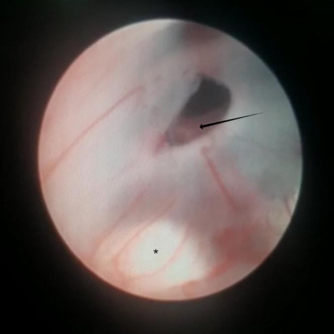
ETV: Black arrow = basilar artery in prepontine cistern; asterisk = mammillary body; dark opening = stoma in floor of 3rd ventricle
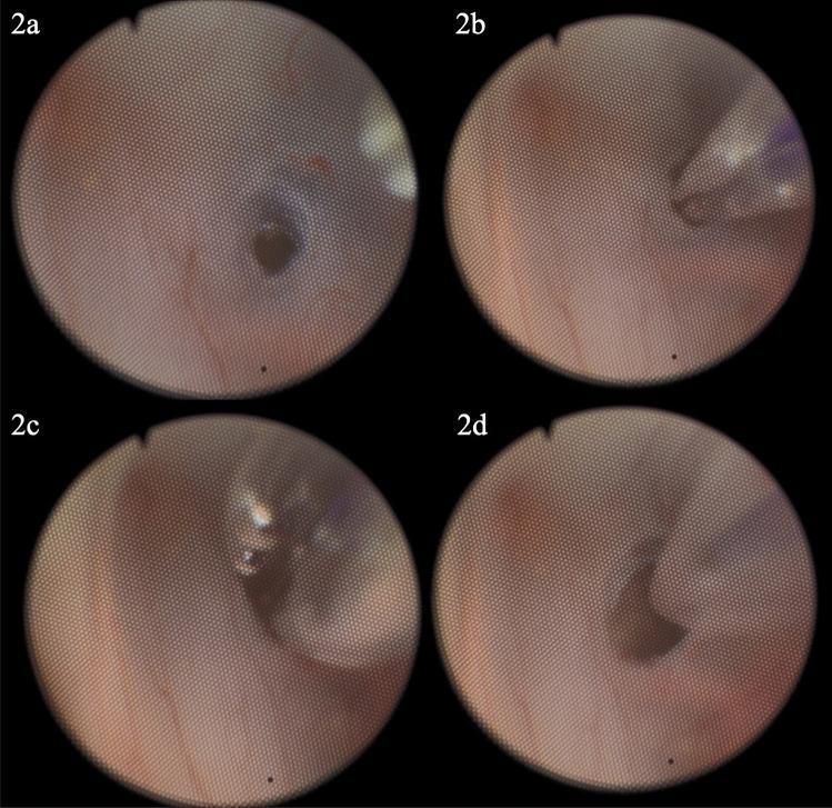
ETV sequence: (a) initial stoma; (b) Fogarty catheter through Liliequist's membrane; (c) balloon inflated; (d) final dilated stoma
Complications:
- Haemorrhage (basilar artery injury — catastrophic)
- CSF leak
- Stoma closure/failure (early failure in <6 months; late failure years later)
- Memory deficit (fornix injury)
- Diabetes insipidus (hypothalamic injury — rare)
ETV + Choroid Plexus Cauterization (ETV+CPC):
- Used in infants <2 years, especially in sub-Saharan Africa settings
- Cauterizes choroid plexus to reduce CSF production
- Success rate >70% in myelomeningocele-related HCP
SURGERY OPTION 4: VENTRICULOATRIAL (VA) SHUNT
- Distal catheter placed in right atrium (via internal jugular vein)
- Used when peritoneal cavity unavailable/unsuitable
- Additional complications: bacteraemia, shunt nephritis (immune complex deposition), pulmonary embolism, arrhythmia, superior vena cava obstruction
- Catheter must be repositioned as child grows
SURGERY OPTION 5: TUMOUR RESECTION (ADDRESSING CAUSE)
- Posterior fossa tumour → surgical excision → may resolve HCP
- Colloid cyst of 3rd ventricle → endoscopic/microsurgical excision
- Pineal tumour → biopsy + radiosurgery or open resection
- Often requires EVD pre-operatively, with reassessment post-resection
PART 2: ICP MONITORING
7. PHYSIOLOGY OF ICP
Normal ICP (supine): < 15 mmHg (measured at foramen of Monro = level of tragus)
Treatment threshold: >20–25 mmHg sustained
CPP = MAP − ICP
- Target CPP: 60–70 mmHg (TBI guidelines)
- Normal CPP: 70–90 mmHg
Monro-Kellie Doctrine: Brain + Blood + CSF = constant. Compensation: CSF shifts to spinal space + venous blood displaced → once exhausted, ICP rises exponentially with minimal volume addition (pressure-volume curve becomes steep at "elbow").
8. ICP WAVEFORMS
Normal ICP Waveform has 3 components per cardiac cycle:
| Peak | Name | Mechanism | Normal Feature |
|---|
| P1 | Percussion wave | Arterial pulsation transmitted from choroid plexus | Tallest peak normally |
| P2 | Tidal wave | Brain tissue compliance | Shorter than P1 normally |
| P3 | Dicrotic wave | Closure of aortic valve | Smallest peak |
⚠️ Pathological sign: When P2 > P1 → reduced intracranial compliance (exhausted compensatory reserve)
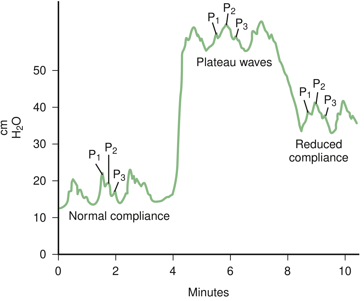
From Bradley & Daroff's Neurology: ICP tracings showing normal compliance (P1>P2>P3) → plateau waves → reduced compliance (P2>P1). Plateau waves = Lundberg A waves; ICP 40–100 mmHg for 5–20 min.
9. LUNDBERG ICP WAVES
| Wave | Description | Clinical Significance |
|---|
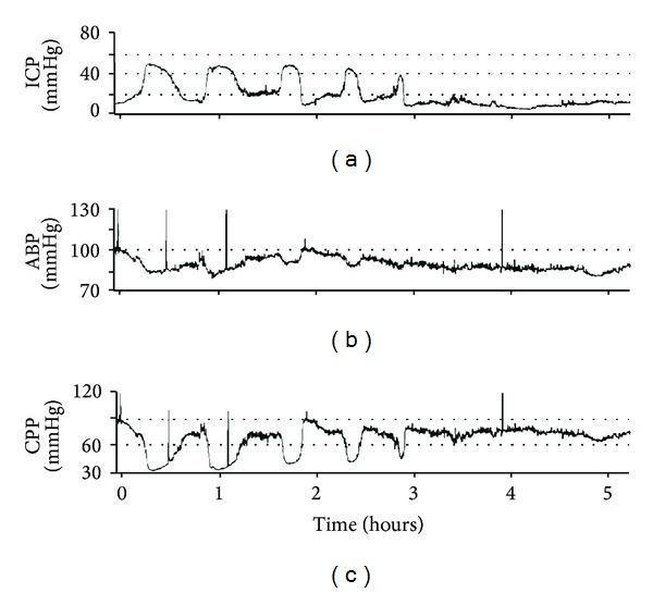
| A waves (Plateau waves) | ICP rises to 40–100 mmHg for 5–20 minutes, then falls suddenly | Most dangerous; indicate severely reduced intracranial compliance; associated with vasodilation and impending herniation |
| B waves | Rhythmic oscillations every 0.5–2 min, amplitude 20–50 mmHg | Associated with sleep, Cheyne-Stokes breathing; may be seen in NPH; less pathological than A waves |
| C waves | Small oscillations (4–8/min, ~20 mmHg) | Corresponds to Traube-Hering-Mayer vasomotor waves; physiological significance uncertain |
Plateau waves: ICP spikes to >50 mmHg → CPP drops to ~40 mmHg → risk of ischaemia
10. ICP MONITORING DEVICES
Indications for ICP Monitoring:
- Severe TBI (GCS ≤8) with:
- Abnormal CT (haemorrhage, contusion, oedema, herniation)
- Normal CT + age >40 years + SBP <90 mmHg + abnormal posturing (any 2 of 3)
- SAH Hunt-Hess Grade III–V
- Acute bacterial meningitis (selected cases)
- Hydrocephalus requiring monitoring during EVD
- Post-operative monitoring after major neurosurgery
- Hepatic encephalopathy with raised ICP
Types of Monitors:
| Device | Site | Gold Standard | Advantages | Disadvantages |
|---|
| Intraventricular Catheter (EVD) | Lateral ventricle | ✅ YES | Calibratable, therapeutic drainage possible, accurate | Difficult to insert with small/shifted ventricles; infection 3–8%; bleeding; cannot monitor when open to drain |
| Intraparenchymal (Camino, Codman) | Brain parenchyma | No (close 2nd) | Easy insertion, safe, continuous reading, low drift, low infection | Cannot drain CSF; cannot recalibrate after insertion; ±2–5 mmHg drift; expensive |
| Subdural bolt | Subdural space | No | Easy insertion | Less accurate, dampened readings, infection, dislodgement |
| Epidural | Epidural space | No | Least invasive | Least accurate; dura acts as barrier; rarely used |
Viva Point: Intraventricular = most accurate = gold standard. Intraparenchymal = most commonly used in practice (easier, safer). The trade-off: can't drain CSF with intraparenchymal.
Antibiotic Prophylaxis for EVD:
- Cefazolin 2 g IV every 8 hours from insertion until 24–48 hours after removal
- OR antibiotic-impregnated catheters (rifampicin/clindamycin or minocycline/rifampicin)
- DO NOT routinely change catheter every 5 days (no benefit; increased risk with multiple insertions)
11. NON-INVASIVE ICP ESTIMATION
(Not yet precise enough for clinical decision-making but promising)
| Method | Principle | Status |
|---|
| Optic nerve sheath diameter (ONSD) — ultrasound or MRI | ONSD >5 mm (U/S) → raised ICP | Screening tool; excellent for bedside |
| Transcranial Doppler (TCD) | Pulsatility index from MCA waveform | Indirect estimate; operator-dependent |
| Tympanic membrane displacement | Changes in perilymph pressure | Experimental |
| Ophthalmoscopy | Papilledema presence/grade | Qualitative only |
| CT/MRI morphological signs | Effaced cisterns, midline shift | Valuable but not continuous |
12. ICP MANAGEMENT ALGORITHM
Raised ICP >20–25 mmHg sustained >5 min
        ↓
TIER 1 (Basics — must do first):
  • HOB 30°
  • Head neutral (avoid venous obstruction)
  • Normothermia
  • Normoglycaemia
  • Avoid hypotension (MAP >80)
  • Normovolaemia
  • Adequate sedation & analgesia (propofol/midazolam + morphine)
        ↓
TIER 2:
  • Drain CSF via EVD (if present)
  • Mannitol 0.5–1 g/kg or Hypertonic saline 3% 1–2 mL/kg
  • Brief hyperventilation (PCO₂ 30–35) — bridge only
        ↓
TIER 3 (Refractory raised ICP):
  • Barbiturate coma (thiopental/pentobarbital)
  • Decompressive craniectomy
  • Hypothermia (controversial)
13. CEREBROVASCULAR PRESSURE REACTIVITY INDEX (PRx)
- Moving correlation between mean ICP and slow MAP fluctuations
- PRx < 0 (negative) → intact autoregulation → arterioles constrict when MAP rises → ICP falls
- PRx > 0 (positive) → impaired autoregulation → ICP passively follows MAP
- PRx guides optimal CPP ("sweet spot" where PRx is most negative)
14. FOLLOW-UP
Post-VP Shunt:
| Time | Modality | What to Check |
|---|
| 24–48 hrs post-op | CT Head | Catheter position, immediate complications, baseline ventricular size |
| 3 months | CT Head | Ventricular size vs. baseline |
| Every 6–12 months × 2 years | CT Head | Size stability, clinical correlation |
| If symptomatic | Urgent CT + Shunt series X-ray | Shunt malfunction (compare with baseline!) |
| Annual (paediatric) | CT Head + Neurodevelopmental assessment | Growth, cognitive milestones |
Viva Point: Always compare with baseline imaging. Absolute ventricular size less important than CHANGE from baseline.
Post-ETV:
| Time | Modality | Purpose |
|---|
| 24–48 hrs | CT Head | Rule out haemorrhage |
| 3 months | MRI (sagittal T2/CISS) | Confirm stoma patency (flow void) |
| 6 months | MRI | Ventricular size reduction |
| Annually | Clinical + MRI/CT | ETV failure (can occur years later) |
NPH (post-shunt):
- Follow-up at 1, 3, 6, 12 months post-shunt
- Assess gait (timed 10m walk), cognition (MMSE/MoCA), continence
- CT at 3 months to assess ventricular decompression
- Programmable valve adjustment guided by clinical response and CT
Congenital HCP (paediatric):
- More frequent follow-up: every 3 months in first 2 years, then 6-monthly
- Monitor: head circumference, fontanelle, developmental milestones
- CT or MRI (prefer MRI for repeat studies to avoid radiation)
- Ultrasound through fontanelle in neonates — no radiation, bedside
15. QUICK VIVA RECAP
| Question | Answer |
|---|
| Gold standard ICP monitor | Intraventricular catheter (EVD) |
| Target ICP | <20 mmHg; treat if >20–25 mmHg |
| Target CPP | 60–70 mmHg (TBI) |
| Normal ICP (adult, supine) | 5–15 mmHg |
| Evans Index threshold | >0.3 = ventriculomegaly |
| Callosal angle NPH | <90° |
| NPH triad order | Gait (first) → Dementia → Incontinence |
| ETV site of fenestration | Tuber cinereum (floor of 3rd ventricle, anterior to mammillary bodies) |
| Lundberg A waves | 40–100 mmHg for 5–20 min; impending herniation |
| Most common congenital HCP cause | Aqueductal stenosis |
| Most dangerous ETV complication | Basilar artery injury |
| Drug that reduces CSF production | Acetazolamide (CA inhibitor); adjunct: furosemide |
| Kocher's point (burr hole site) | 1 cm anterior to coronal suture, mid-pupillary line (right side) |
| EVD set height | 15 cm H₂O above foramen of Monro (level of tragus) |
| P2 > P1 on ICP waveform means | Reduced intracranial compliance |
References: Adams & Victor's Principles of Neurology 12e (pp. 633–638) | Bradley & Daroff's Neurology in Clinical Practice (pp. 1113–1116) | Grainger & Allison's Diagnostic Radiology (pp. 2027–2029) | Neuroanatomy through Clinical Cases 3e (pp. 183–184) | Goldman-Cecil Medicine | Harriet Lane Handbook 23eThis is your complete neurosurgery viva answer on Hydrocephalus and ICP Monitoring. Here's a summary of what's covered:
What was covered:
-
All types of HCP — Non-communicating, Communicating, NPH (Hakim-Adams), Ex vacuo, External (benign infantile), Arrested/Compensated, and Overproduction type — each with age/demographics, causes, and clinical features
-
Clinical presentations by age group — Infant (setting sun sign, macrocephaly, fontanelle bulge) vs. Child (papilledema, morning headache) vs. Adult/NPH ("Wet, Wobbly, Wacky")
-
Investigations — CT (Evans Index, bicaudate index, periventricular lucency), MRI (callosal angle, DESH, flow voids for ETV), LP tap test for NPH, CSF analysis
-
Management — Stepwise: emergency medical (mannitol, dexamethasone, acetazolamide with full MOA and doses) → surgical (EVD at Kocher's point, VP shunt with valve types and complications including slit ventricle, ETV with exact technique and Fogarty balloon, VA shunt)
-
ICP Monitoring — Monro-Kellie, normal values, P1/P2/P3 waveforms, Lundberg A/B/C waves, all device types with pros/cons, PRx index
-
Scores — Evans Index, Callosal Angle, ETVSS, Hunt-Hess, Modified Fisher, GCS thresholds
-
Follow-up protocols — by modality and frequency for each surgical approach