
Tell me about pyogenic liver abscess. Yep.
Reading File
Now let me search the library for pyogenic liver abscess content in parallel.
Finding Sources
Finding Sources
Searching PubMed
"pyogenic liver abscess"[MeSH Terms]
Good. Now let me read the richest sources in parallel - Fischer's Mastery of Surgery, Yamada's Gastroenterology, and Sleisenger & Fordtran.
Reading File
Reading File
Reading File
Good content so far. Let me get the clinical features, diagnosis, and management sections.
Reading File
Reading File
Let me get the treatment/management sections and the Maingot's content on treatment, plus the Sabiston content.
Reading File
Reading File
Excellent - I have rich content. Let me get a bit more on drainage/surgery management and complications/prognosis.
Reading File
Reading File
Let me get the treatment management section from Fischer's and the complications/prognosis from Maingot's.
Reading File
Reading File
I now have comprehensive content. Let me also quickly fetch the CT image of pyogenic liver abscess (double target sign) and the ultrasound image from Maingot's to include visually.
Loading Image
Loading Image
I now have comprehensive textbook content and imaging. Here is a thorough, well-organized answer:
Pyogenic Liver Abscess (PLA)
Definition and Epidemiology
A pyogenic liver abscess is a collection of pus and inflammatory tissue within the hepatic parenchyma caused by bacterial infection. It is the most common type of visceral abscess. Incidence varies significantly by geography - in Taiwan, where PLA is endemic, rates rose from 10.8 to 15.5 cases per 100,000 person-years between 2000-2011, while in Europe and Canada the rate is 1.1-3.7 cases per 100,000. A Minnesota study documented a rise from 2.1 to 3.6 per 100,000 over 35 years. Men and individuals over 65 are disproportionately affected. Morbidity is substantial and mortality ranges from 6% to 10%.
- Fischer's Mastery of Surgery, 8th ed.
- Maingot's Abdominal Operations
Routes of Infection / Pathogenesis
PLA develops through several pathways:
| Route | Mechanism | Examples |
|---|---|---|
| Biliary (most common today) | Direct spread from infected bile ducts | Cholangitis, choledocholithiasis, malignant/benign strictures, instrumentation |
| Portal (pylephlebitis) | Suppurative portal vein thrombophlebitis | Appendicitis, diverticulitis, colorectal malignancy, IBD |
| Hematogenous | Arterial seeding | Endocarditis, severe sepsis, dental/periodontal disease |
| Direct extension | Spread from adjacent structures | Cholecystitis, perinephric abscess |
| Traumatic/Iatrogenic | Necrotic tissue superinfection | Post-TACE, post-ablation, liver surgery, foreign body ingestion (fish/chicken bones) |
| Cryptogenic | No identifiable source | ~35-40% of cases |
Historically, pylephlebitis from untreated appendicitis was the leading cause. Today, biliary disease and instrumentation dominate. PPI use increases risk by reducing gastric acidity and allowing pathogen survival.
- Fischer's Mastery of Surgery; Yamada's Textbook of Gastroenterology, 7th ed.; Sleisenger & Fordtran's GI and Liver Disease
Risk Factors
- Diabetes mellitus
- Malnutrition, immunosuppression
- Prior hepatobiliary/pancreatic disease
- Liver transplant
- Cirrhosis
- History of biliary-intestinal anastomosis or sphincterotomy
- Regular PPI use
- IBD, colorectal malignancy
- Oral flora / periodontal disease (especially in alcoholics - cryptogenic cases)
One population-based study noted a fourfold higher risk of GI malignancy in patients presenting with PLA - screening for occult malignancy is warranted.
Microbiology
Most PLA are polymicrobial. Key organisms:
- E. coli and Klebsiella pneumoniae - most frequently isolated
- Klebsiella pneumoniae (hypervirulent/mucoid) - a distinct syndrome, predominantly in Asia (accounts for ~80% of cases in Taiwan), community-acquired, associated with diabetes, and carries a high risk of metastatic complications (endophthalmitis, meningitis, necrotizing fasciitis)
- Streptococcus milleri group (S. anginosus, S. constellatus, S. intermedius) - normal GI flora, important cause
- Bacteroides fragilis, Fusobacterium necrophorum - most common anaerobes
- Staphylococci - post-trauma/instrumentation or hematogenous; prompt evaluation for infective endocarditis
- Proteus, Pseudomonas spp.
- Rare: Yersinia, Pasteurella, Haemophilus, Listeria, Actinomyces, Clostridium
Anaerobic organisms are likely underrepresented in culture due to suboptimal isolation methods.
- Sleisenger & Fordtran's; Fischer's Mastery of Surgery
Clinical Features
The classic pre-antibiotic triad was spiking fever + RUQ pain + shock. Today, the presentation is typically more indolent, especially in older adults:
- Fever (most consistent finding; can be low-grade)
- Right upper quadrant or epigastric pain - may be pleuritic; may radiate to the right shoulder if the diaphragm is irritated
- Malaise, anorexia, weight loss, nausea
- Symptoms may be present 1 month or more before diagnosis
- Jaundice - uncommon; if present, suggests biliary obstruction
- Normochromic normocytic anemia, weight loss on exam
- Multiple abscesses (often biliary source) tend to present more acutely with sepsis/shock; solitary abscesses are more indolent
Without treatment, mortality approaches 100%. Rupture into the peritoneum is a surgical emergency.
Imaging
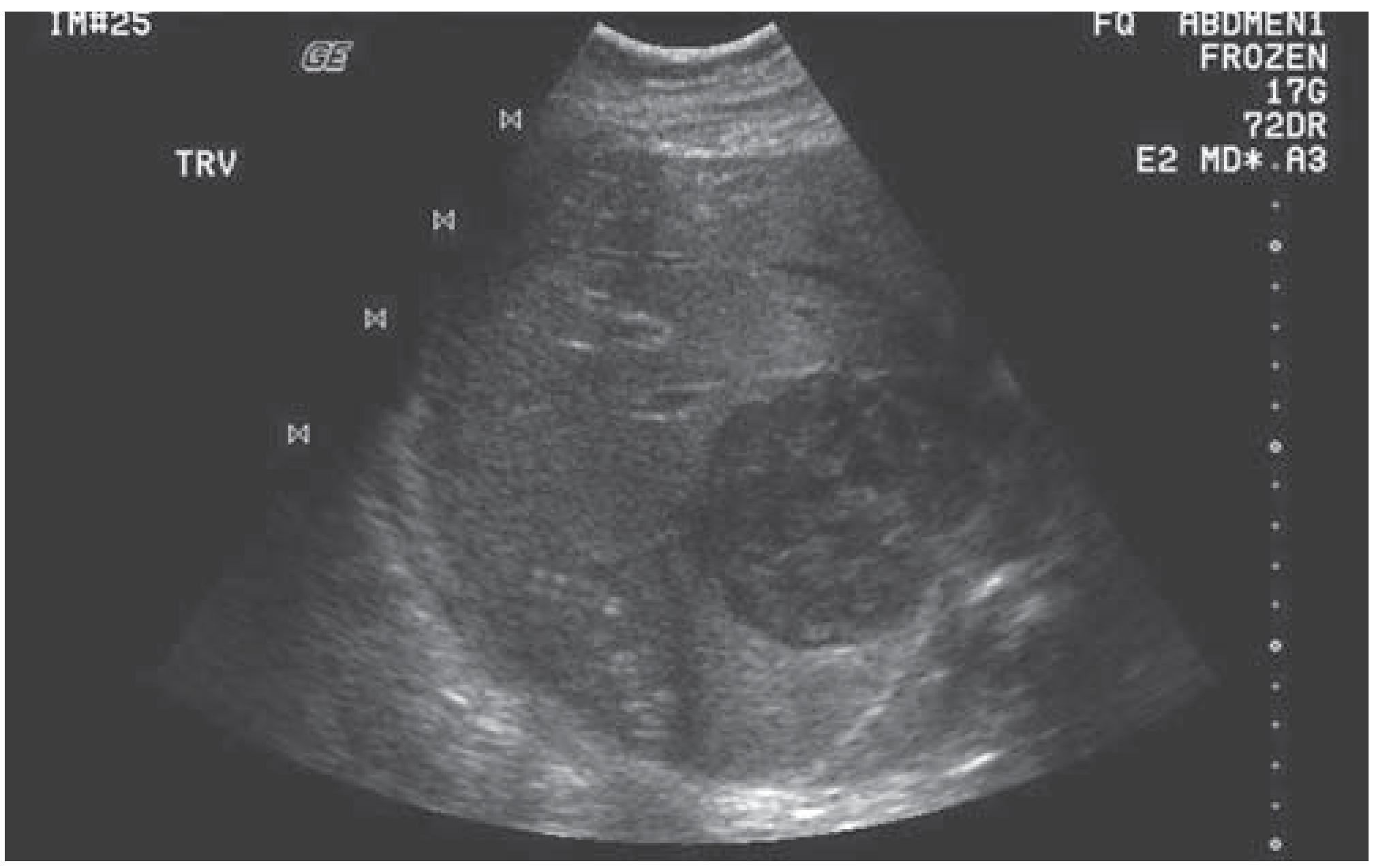
Ultrasound (US)
First-line, widely accessible. Appearance varies with stage:
- Early: mixed hypo/hyperechoic solid-appearing lesion
- Later: complex cystic mass with anechoic spaces and irregular wall, internal septations
- Gas-forming organisms: "dirty shadowing"
- No intracavitary Doppler flow (flow in periphery/septations only)
CT (contrast-enhanced)
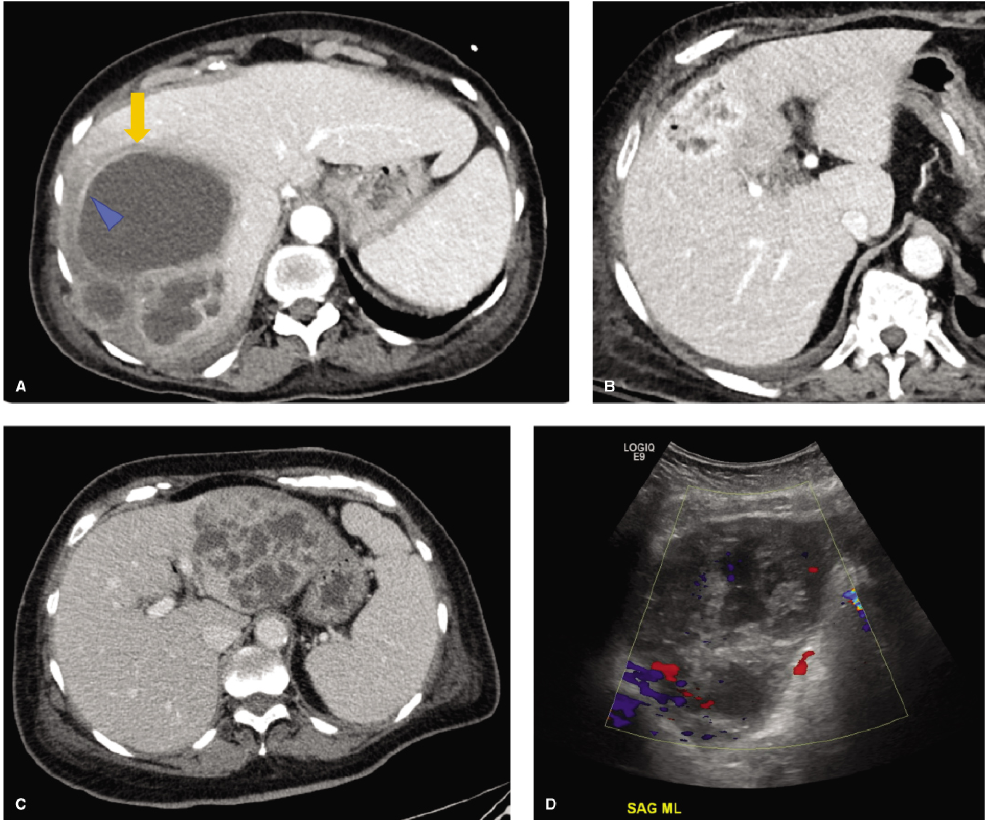
Most useful for characterization and planning:
- Hypoattenuating mass with enhancing rim (most common appearance)
- "Double target sign" - central hypoattenuating pus cavity + enhancing inner rim + outer hypoattenuating rim of parenchymal edema
- "Cluster sign" - multiple small rim-enhancing lesions beginning to coalesce
- "Turquoise sign" - arborizing septa in K. pneumoniae abscess
- Gas within the abscess = gas-forming organism
MRI
- T1: hypointense center
- T2: hyperintense with perilesional edema
- DWI: restricted diffusion (bright DWI, dark ADC) - helpful to distinguish abscess from necrotic tumor
- Superior soft tissue contrast, but not always needed if US/CT is diagnostic
Laboratory Findings
- Elevated alkaline phosphatase (common, often disproportionate)
- Elevated bilirubin, transaminases
- Leukocytosis
- Blood cultures positive in ~50% (up to 95% in K. pneumoniae PLA)
- Anemia
- Hypoalbuminemia in severe/chronic cases
Treatment
Treatment involves three pillars: antibiotics + drainage + source control.
1. Antibiotics
- Draw blood cultures and aspirate abscess before starting antibiotics when possible
- Start broad-spectrum IV antibiotics empirically, covering gram-negatives, gram-positives, and anaerobes
- Classic regimens: aminoglycoside + clindamycin + ampicillin or vancomycin; fluoroquinolones can replace aminoglycosides; metronidazole can replace clindamycin
- Single-agent options: piperacillin-tazobactam, imipenem-cilastatin, or ticarcillin-clavulanate
- Note: K. pneumoniae is intrinsically resistant to ampicillin; first-generation cephalosporins lack adequate anaerobic coverage
- Duration: traditionally 4-6 weeks, though many studies support success with 2 weeks of IV therapy followed by oral step-down based on clinical/culture response
- Empiric anaerobic coverage should always be included in older patients and those with malignancy
2. Percutaneous Drainage
Now the first-line invasive intervention for most PLA:
- Performed under US or CT guidance
- Either needle aspiration (single/repeat) or catheter drainage
- Catheter placement preferred for large, complex, or multiloculated abscesses
- Success rates are high; the method replaced open surgery as standard of care since the 1980s
Contraindications / relative indications to avoid percutaneous drainage:
- Coagulopathy
- Multiple small abscesses (microabscesses)
- Intervening vascular structures (e.g., portal vessels blocking safe access)
- Ascites
- Need for transpleural drainage
- Abscesses requiring surgical source control
3. Surgical Drainage
Reserved for:
- Failure of percutaneous drainage/antibiotics
- Need to treat the underlying intra-abdominal source surgically
- Multiple macroscopic abscesses not amenable to percutaneous approach
- Patient on steroids
- Concomitant ascites
Historically done extraperitoneally via 12th-rib resection; now transperitoneal approach allows simultaneous source control, full liver assessment, and biliary exploration.
- Maingot's Abdominal Operations; Fischer's Mastery of Surgery
Complications
Up to 40% of patients develop complications:
- Sepsis - most common
- Pleural effusion / empyema
- Rupture into peritoneum (peritonitis, high mortality)
- Subphrenic abscess (controlled leak)
- Hemobilia
- Portal vein or hepatic vein thrombosis - can cause portal hypertension or Budd-Chiari syndrome
- Bacteremia with metastatic seeding
K. pneumoniae-specific complications:
-
Bacteremia in ~95% of cases
-
Endophthalmitis (6-61%) - can occur after drainage; often results in permanent visual loss
-
Meningitis, brain abscess, CNS complications
-
Necrotizing fasciitis
-
Septic pulmonary emboli
-
DIC, acute renal failure
-
Maingot's Abdominal Operations; Yamada's Gastroenterology
Prognosis / Poor Prognostic Factors
Historical mortality ranged from 11% to 88%; with modern management it is 6-10% overall, and 6-17% for K. pneumoniae PLA. Poor outcomes are associated with:
| Clinical | Laboratory |
|---|---|
| Age >70 years | WBC >20,000/mm³ |
| Diabetes mellitus | Increasing bilirubin |
| Associated malignancy | Increasing SGOT/AST |
| Biliary etiology | Albumin <2 g/dL |
| Multiple abscesses | |
| Septicemia | |
| Polymicrobial bacteremia | |
| Significant complication |
For K. pneumoniae PLA, abscess >5 cm, intrahepatic gas, APACHE III >40, delayed drainage, and thrombocytopenia are additional poor prognosticators. The main concern with K. pneumoniae PLA today is not mortality per se, but catastrophic disability from irreversible ocular and neurological complications - the K1 genotype is particularly virulent.
Key Distinctions from Amebic Abscess
| Feature | Pyogenic | Amebic |
|---|---|---|
| Cause | Mixed bacteria | Entamoeba histolytica |
| Typical patient | Elderly, comorbidities | Traveler from endemic area (Mexico, S. Asia, Africa); male predominance |
| Microbiology | Polymicrobial | Single organism |
| Serology | Not helpful | Diagnostic (IFA/ELISA) |
| Number | Often multiple | Usually solitary (right lobe) |
| Contents | Pus | "Anchovy paste" fluid |
| Treatment | Abx + drainage | Metronidazole; drainage less often needed |
Sources: Fischer's Mastery of Surgery 8th ed.; Yamada's Textbook of Gastroenterology 7th ed.; Sleisenger & Fordtran's GI and Liver Disease; Maingot's Abdominal Operations; Goldman-Cecil Medicine
This is a shared conversation. Sign in to Orris to start your own chat.