


Identify the two pictures here
Image 1 - Chest X-ray (AP View)
- Left hemithorax: There is a large homogeneous opacity in the left mid-to-lower lung zone, obscuring the left hemidiaphragm and left heart border (positive silhouette sign). The left costophrenic angle is completely obliterated.
- Right hemithorax: Relatively clear with normal bronchovascular markings; right costophrenic angle is sharp.
- Mediastinum/Trachea: Trachea appears central.
Image 2 - Practical Examination Spot (Taste Bud Models)
- Yellow outer layer - surrounding stratified squamous epithelium / tongue papilla
- Orange/red inner section - the barrel-shaped taste bud structure
- Central dark element - gustatory (sensory) cells or nerve fibers leading to the taste pore
Isn't image 2 is cleft palate
cleft palate anatomical model
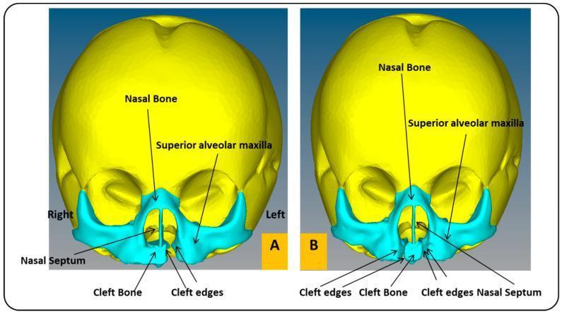
Frontal view of two Finite Element Analysis (FEA) skull models representing a one-month-old infant with craniofacial anomalies. Model A illustrates a Unilateral Cleft Lip and Palate (UCLP), showing a singular discontinuity in the superior alveolar maxilla and cleft bone on the left side, with a noticeable deviation of the nasal septum away from the cleft. Model B illustrates a Bilateral Cleft Lip and Palate (BCLP), characterized by two symmetrical clefts flanking a central prolabium/premaxillary segment (labeled as cleft bone). In the BCLP model, the nasal septum is centrally positioned but isolated by the bilateral voids. Both models use color-coding to differentiate anatomical regions: yellow for the neurocranium and teal for the midface/maxillary structures. Labeled landmarks include the nasal bone, superior alveolar maxilla, nasal septum, cleft bone, and cleft edges. These models are designed for biomechanical simulation of external pressure treatments to correct maxillary alignment in pediatric patients.
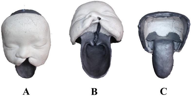
This clinical educational image displays three views (A, B, C) of a plaster cast model derived from a 6-month-old infant with a congenital complete unilateral cleft lip and palate. View A (Anterior) shows the external facial structure, highlighting a significant cleft on the left side that disrupts the upper lip and nasal base. View B (Inferior/Basal) provides a comprehensive look at the internal defect, demonstrating a complete cleft through the alveolar ridge (jaw), hard palate, and soft palate, showing direct communication between the oral and nasal cavities. View C (Posterior) illustrates the dorsal aspect and the pharyngeal extension of the model. The model utilizes white plaster for the midface and forehead, while darker materials define the internal oral and pharyngeal structures. This specimen serves as an anatomical foundation for surgical simulators, specifically designed to teach undergraduate medical and dentistry students about orofacial cleft anatomy and surgical planning for repair procedures.
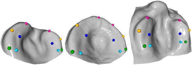
Anatomical diagram and 3D mesh model comparison of different cleft lip and palate (CLP) types, used in orthodontic and surgical planning. The image displays three distinct computer-aided design (CAD) templates. Left: Unilateral cleft lip and palate (UCLP) showing an asymmetrical alveolar ridge with a unilateral indentation. Center: Isolated cleft palate showing a more symmetrical, rounded arch with a central depression. Right: Bilateral cleft lip and palate (BCLP) demonstrating a significant bilateral cleft through the alveolar ridge and a prominent premaxilla. Each mesh is annotated with a standardized set of ten colored landmarks (green, cyan, blue, yellow, and pink spheres) strategically placed to map the surface morphology. The landmarks are concentrated along the alveolar ridges and cleft margins to guide the automated registration of presurgical orthopedic plates. These templates represent the average palatal morphology for each cleft type, serving as educational models for clinical classification and surgical workflow automation in pediatric maxillofacial care.
taste bud histology model practical exam
- A central gap/cleft running through the middle
- A yellow outer arc (could represent the palatal shelves or alveolar ridge)
- An orange/red inner portion (could represent soft tissue or the cleft margins)
- A dark central vertical line - which could represent the cleft itself (the gap in the palate)

I think the answer is one of them in photo tell me which one
- The list is titled "Models" with 9 items
- Model 8 is "Fetal Circulation"
- Model 9 is "Cleft palate"
- The display board in Image 2 is labeled "SPOT 8"
Not not basis on number
- An arch/horseshoe shape
- Yellow outer layer (like two palatal shelves)
- Orange inner tissue
- A central dark gap/cleft running through the middle
- Multiple identical models showing the same structure
- Branchial Arches
- Development of Notochord
- Development of Primitive Streak
- Development of Face
- Rotation of Gut
- TOF (Tetralogy of Fallot)
- Descent of Testis
- Fetal Circulation
- Cleft Palate
Can you explain cleft palate model as shown in image 2
cleft palate model embryology palatine shelves fusion
cleft palate cross section palatine shelves unfused embryology diagram
What the Model in Image 2 Shows
Breaking Down the Colors in the Model
| Color in Model | What it Represents |
|---|---|
| Yellow outer arch | The two lateral palatine shelves (extensions of the maxilla) |
| Orange/red inner tissue | The oral mucosa or soft tissue lining of the palate |
| Dark central gap | The cleft - the midline failure of fusion |
Embryological Explanation
- Around week 6-7, two lateral palatine processes (shelves) grow horizontally from the maxillary prominences
- By week 9, they elevate above the tongue, grow toward each other, and fuse in the midline
- They also fuse with the primary palate anteriorly and the nasal septum superiorly
- The lateral palatine processes fail to fuse in the midline
- This leaves a gap (cleft) communicating between the oral and nasal cavities
- Can involve the soft palate only, the hard palate, or both
Types (as shown in the textbook diagram above)
- B - Cleft uvula (mildest)
- C/D - Unilateral or bilateral cleft of the secondary (posterior) palate
- E/F - Complete unilateral/bilateral cleft of lip + anterior palate
- G/H - Complete bilateral cleft of lip + entire palate (most severe)
Clinical Significance
-
Occurs in ~1 in 700-1000 births
-
Caused by failure of mesenchymal proliferation and fusion of palatal shelves
-
Risk factors: teratogens (alcohol, phenytoin, steroids), genetic factors, folic acid deficiency
-
Leads to feeding difficulties, speech problems, recurrent ear infections (Eustachian tube dysfunction)
-
Treatment: surgical repair (palatoplasty) at around 9-18 months of age
-
The Developing Human: Clinically Oriented Embryology, 10th Ed., p. 537-545