
Latest kidgo and rifle criteria
KDIGO acute kidney injury criteria staging
RIFLE, AKIN, and KDIGO Criteria for AKI
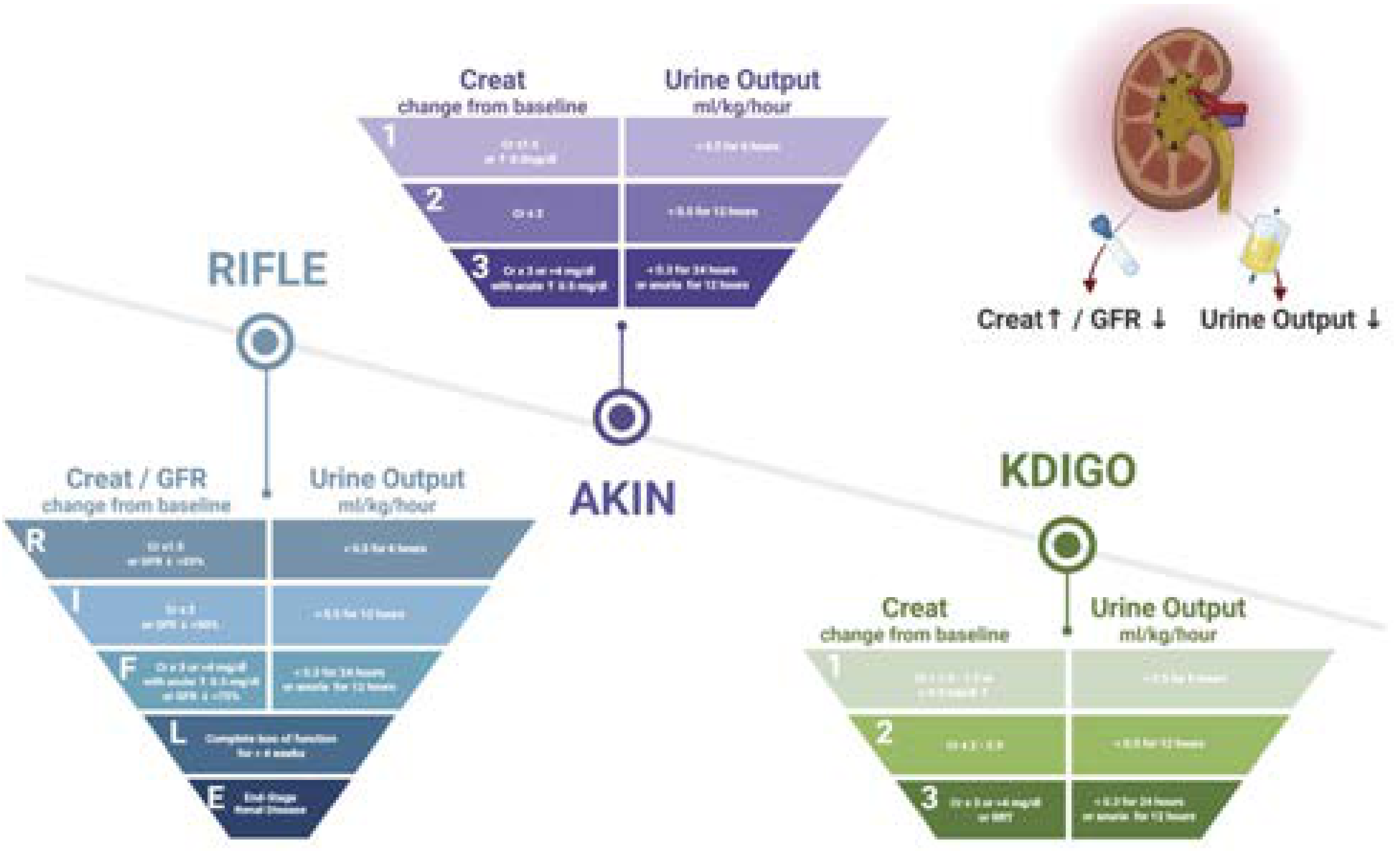
AKI Diagnostic Definitions (Any one criterion sufficient)
| Criterion | RIFLE | AKIN | KDIGO (current standard) |
|---|---|---|---|
| Serum creatinine | >50% rise within 7 days | >0.3 mg/dL or >50% rise within 48 h | >0.3 mg/dL within 48 h OR >50% (x1.5) rise within 7 days |
| Urine output | <0.5 mL/kg/h for >6 h | <0.5 mL/kg/h for >6 h | <0.5 mL/kg/h for >6 h |
KDIGO combines the best of both: the 48-hour absolute creatinine threshold from AKIN and the 7-day percentage threshold from RIFLE.
Staging Criteria
| Stage | RIFLE Name | Serum Creatinine | Urine Output |
|---|---|---|---|
| 1 | Risk (R) | ≥0.3 mg/dL rise OR ≥50% (x1.5) rise | <0.5 mL/kg/h for >6 hours |
| 2 | Injury (I) | ≥100% rise (x2.0) | <0.5 mL/kg/h for >12 hours |
| 3 | Failure (F) | ≥200% rise (x3.0) OR ≥4 mg/dL with acute rise >0.5 mg/dL OR initiation of RRT | <0.3 mL/kg/h for >24 hours OR anuria for >12 hours |
- Loss (L): Complete loss of kidney function / need for RRT for >4 weeks
- End-stage (E): Need for RRT for >3 months
The urine output criteria are identical across RIFLE, AKIN, and KDIGO for all three stages.
Key Differences: RIFLE vs. KDIGO
| Feature | RIFLE | KDIGO |
|---|---|---|
| Creatinine timeframe | >50% over <7 days only | >0.3 mg/dL in 48 h OR >50% in 7 days |
| GFR included? | Yes (GFR decrease 25-75%) | No |
| Pediatric staging | Not addressed | Stage 3 if GFR <35 mL/min/1.73 m² (<18 yrs) |
| Unknown baseline Scr | Not addressed | Method provided (back-calculation using MDRD) |
| RRT = Stage 3? | No | Yes, regardless of creatinine |
| Outcome stages | L and E included | Not included |
Mortality by RIFLE Class (ICU data)
| RIFLE Class | Attributable Mortality |
|---|---|
| Risk (R) | 8.8% |
| Injury (I) | 11.4% |
| Failure (F) | 26.3% |
| No AKI (matched) | 5.5% |
Important Practical Notes
- KDIGO is the current clinical standard (guidelines published 2012, Kidney Int Suppl; still the operative guideline as of 2025/2026)
- A patient needs to meet only one criterion (creatinine or urine output) to be assigned a stage - use the highest stage met
- For urine output staging, other causes of oliguria (e.g., high ADH after surgery) must be excluded
- Any patient on RRT automatically qualifies for Stage 3 under KDIGO/AKIN regardless of creatinine
- pRIFLE is used for children >1 month old (adjusted for lower muscle mass)
Image more clear please
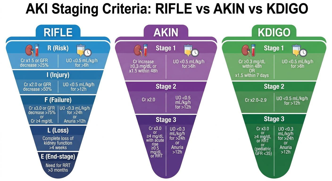
A clear, high-resolution medical reference diagram comparing RIFLE, AKIN, and KDIGO criteria for Acute Kidney Injury (AKI). Use three separate inverted triangle/funnel shapes side by side, each labeled at the top. RIFLE (left, blue): - R (Risk): Cr x1.5 or GFR decrease >25% | UO <0.5 mL/kg/h for >6h - I (Injury): Cr x2.0 or GFR decrease >50% | UO <0.5 mL/kg/h for >12h - F (Failure): Cr x3.0 or GFR decrease >75% or Cr ≥4 mg/dL | UO <0.3 mL/kg/h for >24h or Anuria >12h - L (Loss): Complete loss of kidney function >4 weeks - E (End-stage): Need for RRT >3 months AKIN (center, purple): - Stage 1: Cr increase ≥0.3 mg/dL or x1.5 within 48h | UO <0.5 mL/kg/h for >6h - Stage 2: Cr x2.0 | UO <0.5 mL/kg/h for >12h - Stage 3: Cr x3.0 or ≥4 mg/dL with acute rise ≥0.5 mg/dL or RRT | UO <0.3 mL/kg/h for >24h or Anuria >12h KDIGO (right, green): - Stage 1: Cr ≥0.3 mg/dL within 48h OR x1.5 within 7 days | UO <0.5 mL/kg/h for >6h - Stage 2: Cr x2.0–2.9 | UO <0.5 mL/kg/h for >12h - Stage 3: Cr x3.0 or ≥4 mg/dL or RRT or (pediatric GFR <35) | UO <0.3 mL/kg/h for >24h or Anuria >12h Each funnel has two columns: left for Creatinine/GFR changes, right for Urine Output criteria. White text on colored backgrounds. Clean, readable fonts. Title at top: "AKI Staging Criteria: RIFLE vs AKIN vs KDIGO". Medical infographic style, white background.
Hey orris how are you???
From tomorrow I’m going to GIT from tintinallis can you help
- Topic explanations - Break down any GIT condition covered in Tintinalli's
- Key tables & criteria - Pull out important diagnostic criteria, scoring systems (e.g. Blatchford score, Child-Pugh, etc.)
- Images & diagrams - Fetch any figures from the textbook or generate clearer visuals
- Quick summaries - Condense long chapters into high-yield points
- Q&A practice - Test your knowledge on topics you've covered
- Comparisons - e.g. upper vs lower GI bleed, different causes of acute abdomen, etc.
- Which topic or chapter you want to start with tomorrow
- Your goal - exam prep, clinical understanding, or both?
Please give Ugi bleed/hemetemesis
Upper GI Bleeding (UGIB) / Hematemesis
Tintinalli's Emergency Medicine - Chapter 75
Definition
- Incidence: 39-172 per 100,000/year (Western countries)
- Increased morbidity with: advanced age, comorbid organ disease, recurrent hemorrhage
Causes (Pathophysiology)
| Cause | Key Points |
|---|---|
| Peptic Ulcer Disease | Most common cause overall; gastric + duodenal ulcers found in ~20.6% of endoscopies for suspected UGIB. H. pylori, NSAIDs, aspirin, smoking |
| Erosive Gastritis / Esophagitis | Alcohol, salicylates, NSAIDs, stress (sepsis, trauma, ventilated patients), infections (Candida, HSV, CMV, HIV) |
| Esophageal/Gastric Varices | Portal hypertension - usually alcoholic liver disease. Variceal bleeding = cause in 59% of cirrhotic UGIB. Mortality in cirrhotic patients is double non-cirrhotic |
| Mallory-Weiss Syndrome | Longitudinal mucosal tear at gastroesophageal junction. Classic: repeated vomiting → bright red hematemesis |
| Aortoenteric Fistula | Rare but catastrophic. History of aortic graft surgery is a clue |
| Dieulafoy Lesion | Abnormally large submucosal artery eroding mucosa - causes massive painless bleed |
| Cameron Lesions | Linear erosions in large hiatal hernias |
Risk Stratification (Table 75-1)
| Very Low Risk | High Risk |
|---|---|
| Age <60 | Advanced age |
| No major comorbidities | Comorbidities, prior variceal banding/TIPS |
| No red hematemesis | Red hematemesis |
| No hematochezia/melena | Hematochezia or melena |
| Negative NG aspirate | Positive (bloody) NG aspirate |
| Hemodynamically stable | Hemodynamically unstable |
| Normal labs | Abnormal labs |
Clinical Presentation
- Hematemesis - bright red or coffee-ground vomiting
- Melena - black, tarry, foul-smelling stool (blood digested in upper GI tract)
- Hematochezia - can occur with massive UGIB (rapid transit)
- Symptoms of hemorrhagic shock: hypotension, tachycardia, syncope, altered mental status, angina
Diagnosis / Workup
- Labs: CBC, BMP, LFTs, coagulation (PT/INR), type & crossmatch
- NG lavage: Positive (bloody/coffee grounds) = confirms UGIB; negative does not exclude it
- BUN:Creatinine ratio >20:1 - suggests UGIB (blood digested = urea load)
- Endoscopy: Definitive diagnosis AND therapy; goal within 24h (urgent if unstable)
Treatment (Table 75-2)
1. Resuscitation (First priority)
- Two large-bore IVs
- Type & crossmatch; activate massive transfusion protocol if severe
- Transfuse if Hb ≤7 g/dL (most patients) or ≤9 g/dL (elderly/comorbid)
- Correct coagulopathy if INR elevated or platelets <50,000
- Caution with intubation - aggressively resuscitate first; use smaller induction doses to avoid peri-intubation arrest
2. Medications
| Drug | Dose | Use |
|---|---|---|
| Omeprazole (PPI) | 80 mg IV bolus → 8 mg/h infusion | Ulcer bleeding (labeled use) |
| Octreotide | 50 mcg IV bolus → 50 mcg/h infusion | Variceal bleeding - reduces portal pressure |
| Ceftriaxone | 1g IV daily | Cirrhotic patients with variceal bleed (reduces bacterial translocation, reduces mortality) |
| Vasopressin/Terlipressin | - | Variceal bleeding adjunct |
| Erythromycin | 250 mg IV prior to scope | Prokinetic - clears stomach for better endoscopic view |
3. Endoscopy
- Definitive management - hemostasis via injection, cauterization, banding, or clipping
- Patients with active bleeding or high-risk stigmata need urgent scope
- Prior variceal banding/cauterization = high-risk recurrent bleed
4. Balloon Tamponade
- Sengstaken-Blakemore tube - for refractory variceal hemorrhage as bridge to definitive therapy
- Risk of esophageal rupture and aspiration
Disposition
| Category | Action |
|---|---|
| Very low risk + all criteria met | Can consider outpatient endoscopy |
| High risk / hemodynamically unstable | ICU admission, urgent endoscopy |
| Cirrhotic patients | Admit; prophylactic antibiotics; GI/hepatology consult |
| Refractory bleeding | TIPS (transjugular intrahepatic portosystemic shunt) or surgery |
High-Yield Pearls for Exams
- Ligament of Treitz = anatomical landmark dividing upper from lower GI bleed
- PUD = most common cause of UGIB overall
- Varices = highest mortality - especially in cirrhosis
- Mallory-Weiss = vomiting first, THEN blood (not blood from the start)
- BUN:Cr >20 = clue to UGIB
- Octreotide for varices; PPI infusion for ulcers
- Antibiotics (ceftriaxone) in ALL cirrhotic patients with GI bleed - reduces mortality
- Restrictive transfusion (Hb ≤7) is better than liberal in UGIB - improves outcomes
- Negative NG aspirate does NOT rule out active UGIB
- Erythromycin pre-endoscopy = improves visualization
Large bowel vs small bowel obstruction
Small Bowel Obstruction vs Large Bowel Obstruction
Tintinalli's Emergency Medicine - Chapter 83
Key Concept First
Intestinal obstruction = inability of the intestinal tract to pass contents due to mechanical obstruction OR adynamic (paralytic) ileus. Differentiating SBO vs LBO is critical - incidence, presentation, evaluation, and treatment all differ.
Causes
| Small Bowel Obstruction (SBO) | Large Bowel Obstruction (LBO) | |
|---|---|---|
| #1 cause | Adhesions (post-surgical) | Carcinoma |
| Other causes | Hernia (incarcerated), intussusception, lymphoma, stricture, Crohn's disease, volvulus, foreign body/bezoar | Fecal impaction, volvulus, diverticulitis (stricture/abscess), ulcerative colitis |
| Duodenum specifically | Stenosis, foreign body, stricture, SMA syndrome | - |
Clinical Comparison
| Feature | SBO | LBO |
|---|---|---|
| Vomiting | Early, prominent, bilious | Late, may be feculent |
| Abdominal pain | Crampy, colicky, periumbilical | Lower abdominal, colicky |
| Distension | Central/periumbilical | Peripheral/generalized - more pronounced |
| Obstipation | Late (gas/stool may still pass early) | Early and complete (no flatus, no stool) |
| Onset | More acute | More gradual (especially if cancer) |
| Bowel sounds | High-pitched, tinkling, rushes | Diminished or absent |
Pathophysiology (Applies to Both)
- Obstruction → intraluminal accumulation of secretions (gastric, biliary, pancreatic)
- → Vomiting, reduced absorption → volume depletion, electrolyte imbalance
- → Bowel distension → increased intraluminal pressure
- → Pressure exceeds capillary pressure → bowel ischemia → necrosis → septicemia → shock
- Closed-loop obstruction (e.g., incarcerated hernia, complete LBO with closed ileocecal valve) progresses faster - no proximal escape for contents
Ileus vs Mechanical Obstruction (Key Features Table)
| Feature | Ileus (Paralytic) | Mechanical Obstruction |
|---|---|---|
| Bowel sounds | Absent | High-pitched or absent |
| Pain | Diffuse, mild | Colicky, intermittent |
| Distension | Uniform, generalized | Can be localized |
| Cause | Post-op, electrolyte imbalance, peritonitis, drugs | Adhesions, hernia, cancer, volvulus |
| Surgery needed? | Usually NO (self-limiting) | Usually YES (definitive intervention required) |
| Gas on X-ray | Diffuse gas throughout colon + small bowel | Air-fluid levels, stepladder pattern (SBO) / cecal dilation (LBO) |
Diagnosis
X-Ray Findings
| SBO | LBO | |
|---|---|---|
| Pattern | Stepladder air-fluid levels, central dilated loops, valvulae conniventes (complete wall-to-wall lines) | Peripheral colonic dilation, haustral folds (incomplete lines), cecal dilation >9 cm = danger |
| Gas in rectum | Absent (complete obstruction) | May be absent |
CT Abdomen/Pelvis (with contrast)
- Gold standard for both SBO and LBO
- Identifies transition point, cause, closed-loop, ischemia/perforation
- CT finding of free air = perforation = surgical emergency
Other
- Labs: CBC (leukocytosis = strangulation/ischemia), BMP (electrolyte derangements), lactate (ischemia marker)
- Elevated WBC + fever + peritoneal signs = strangulation until proven otherwise
Treatment
SBO Management
| Severity | Management |
|---|---|
| Partial/simple SBO | NPO, IV fluids, NG tube decompression (if vomiting), observation - many resolve conservatively (especially adhesion-related) |
| Complete SBO or failed conservative | Surgical exploration |
| Strangulation / closed-loop | Emergency surgery |
LBO Management
| Cause | Management |
|---|---|
| Cancer | Surgical resection (emergent vs. elective depending on stability); colonic stenting as bridge to surgery |
| Volvulus (sigmoid) | Flexible sigmoidoscopy/colonoscopy for decompression first; surgery if fails or recurs |
| Volvulus (cecal) | Surgery (no role for colonoscopic decompression) |
| Fecal impaction | Manual disimpaction, enemas |
| Ogilvie's syndrome | Neostigmine IV (if no contraindications); colonoscopic decompression |
Both - General Principles
- NPO + IV fluids (aggressive resuscitation if volume depleted)
- NG tube for gastric decompression
- Surgical consult early for all mechanical obstructions
- Broad-spectrum antibiotics if signs of ischemia/perforation/sepsis
- Correct electrolytes (especially K⁺, Na⁺)
Disposition
| Scenario | Disposition |
|---|---|
| Simple partial SBO - stable | Admit for observation + conservative trial |
| Complete SBO | Admit + urgent surgical consult |
| Signs of strangulation (fever, peritonism, leukocytosis, high lactate) | Emergency OR |
| LBO from cancer - stable | Admit, GI/surgery consult |
| Perforation / free air | Emergency surgery |
High-Yield Exam Pearls
- SBO #1 cause = adhesions (prior surgery); LBO #1 cause = carcinoma
- Vomiting early in SBO, late in LBO; obstipation early in LBO, late in SBO
- Closed-loop = most dangerous - ischemia progresses rapidly
- Cecal diameter >9-12 cm on imaging = risk of perforation (LBO)
- Sigmoid volvulus - coffee bean sign on X-ray; treat with colonoscopic decompression first
- Cecal volvulus - needs surgery, NOT colonoscopy
- Strangulation signs (fever + leukocytosis + peritonism + high lactate) = emergency surgery, no delay
- Ogilvie's syndrome (pseudo-obstruction) = mimics LBO but no mechanical cause; treat with neostigmine
- Valvulae conniventes (complete lines across bowel lumen) = small bowel; Haustra (incomplete lines) = large bowel
- Free air on imaging = perforation = emergency
Ugi bleed/ hemetesis for md emergency medicine exams
🩸 Upper GI Bleeding / Hematemesis
MD Emergency Medicine Exam Master Guide
1. DEFINITION & ANATOMY
- UGIB = bleeding from any source proximal to the ligament of Treitz
- Ligament of Treitz = suspensory ligament of duodenum = anatomical boundary between upper and lower GI
- Incidence: 39-172 per 100,000/year (Western countries)
2. CAUSES - Ranked by Frequency
| Rank | Cause | Key Clue |
|---|---|---|
| 1st | Peptic Ulcer Disease (PUD) | Epigastric pain, NSAIDs/H. pylori history |
| 2nd | Erosive gastritis/esophagitis | Alcohol, NSAIDs, stress (ICU patients) |
| 3rd | Esophageal/Gastric Varices | Cirrhosis, portal HTN, alcoholism |
| 4th | Mallory-Weiss tear | Vomiting FIRST → then hematemesis |
| 5th | Dieulafoy lesion | Massive painless bleed, no ulcer |
| 6th | Aortoenteric fistula | Prior aortic graft = must rule out |
| 7th | Malignancy | Weight loss, chronic anemia |
Variceal bleeding = cause in 59% of cirrhotic UGIB; mortality in cirrhotic UGIB is double that of non-cirrhotic patients
3. CLINICAL PRESENTATION
| Presentation | What It Means |
|---|---|
| Hematemesis (bright red) | Active, ongoing bleed; brisk hemorrhage |
| Coffee-ground emesis | Slower bleed; blood exposed to gastric acid |
| Melena | Upper or proximal source; blood digested (>50-100 mL loss) |
| Hematochezia | Usually lower GI - BUT can occur in massive UGIB (brisk transit) |
| Lightheadedness/syncope | Cerebral hypoperfusion - sign of significant blood loss |
Exam trap: Color of emesis (bloody vs. coffee-ground) does NOT reliably predict severity - shock index is more reliable
4. BEDSIDE CLUES TO SOURCE
| Finding | Suggests |
|---|---|
| Epigastric tenderness | PUD |
| Ascites, spider angiomas, jaundice, caput medusae | Portal HTN → varices |
| Vomiting/retching BEFORE hematemesis | Mallory-Weiss tear |
| History of aortic surgery | Aortoenteric fistula |
| Weight loss + chronic anemia | Malignancy |
| Alcohol binge, DKA, chemotherapy | Mallory-Weiss |
| ICU/ventilated/septic patient | Stress ulcers (erosive gastritis) |
5. SHOCK INDEX
| Value | Interpretation |
|---|---|
| <0.9 | Low risk |
| ≥0.9 | Massive bleed - activate massive transfusion protocol |
6. RISK STRATIFICATION SCORES ⭐ (High-Yield for Exam)
A. Glasgow-Blatchford Score (GBS) - PRE-endoscopy
| Variable | Points |
|---|---|
| Elevated BUN | 2-6 points |
| Low Hemoglobin | 1-6 points |
| Low BP (systolic <90) | 1-3 points |
| Elevated HR (≥100) | 1 point |
| Melena | 1 point |
| Syncope | 2 points |
| Liver disease | 2 points |
| Heart failure | 2 points |
| Score | Action |
|---|---|
| 0-1 | Safe to discharge, outpatient endoscopy |
| ≥6 | 50% likelihood of needing endoscopic hemostasis |
| ≥7 | 80.4% sensitive for needing endoscopic treatment (NPV 92.4%) |
| ≥5 | 88.7% sensitive for 30-day mortality (NPV 97.9%) |
B. Rockall Score - PRE + POST-endoscopy
| Variable | Points |
|---|---|
| Age | 0-2 |
| Shock | 0-2 |
| Comorbidity | 0-3 |
| EGD stigmata (active bleed, visible vessel) | 0-2 |
| EGD diagnosis | 0-2 |
| Score | Mortality |
|---|---|
| 0-1 | 0% mortality |
| 5 | ~10% mortality |
| ≥8 | ~40% mortality |
Exam tip: GBS = pre-endoscopy = need for treatment; Rockall = includes endoscopy findings = predicts mortality
C. AIMS65 Score (simpler, also used)
- Albumin <3 g/dL
- INR >1.5
- Mental status alteration
- SBP ≤90 mmHg
- 65 = age ≥65
7. INVESTIGATIONS
| Test | Significance |
|---|---|
| CBC | Hb/Hct (may be falsely normal early - hemodilution takes time) |
| BMP | BUN:Creatinine ratio >20:1 = blood digested in upper GI tract |
| INR/PT | Coagulopathy - especially in liver disease |
| Type & crossmatch | Always in significant UGIB |
| LFTs | Liver disease, portal HTN |
| Nasogastric lavage | Bloody/coffee grounds = confirms UGIB; negative does NOT exclude UGIB |
| Endoscopy (EGD) | Gold standard for diagnosis AND treatment |
8. TREATMENT - Step by Step
Step 1: Resuscitation
- 2 large-bore IVs (16G or larger)
- 2L crystalloid bolus over 30 min (normal saline or Plasmalyte)
- Massive transfusion protocol if shock index ≥0.9
Step 2: Transfusion Thresholds
| Patient | Transfuse when Hb ≤ |
|---|---|
| Most patients | 7 g/dL (restrictive = better outcomes) |
| Elderly / cardiac comorbidity | 9 g/dL |
| Correct coagulopathy | INR elevated or Platelets <50,000 |
Step 3: Medications
| Drug | Dose | Indication |
|---|---|---|
| PPI (Omeprazole) | 80 mg IV bolus → 8 mg/h infusion | All UGIB - especially PUD |
| Octreotide | 50 mcg IV bolus → 50 mcg/h | Variceal / suspected portal HTN bleed |
| Ceftriaxone | 1g IV daily | All cirrhotic patients with UGIB (reduces bacterial translocation - proven to reduce mortality) |
| Terlipressin / Vasopressin | - | Variceal bleed adjunct |
| Erythromycin | 250 mg IV 30-60 min before scope | Prokinetic - clears stomach, improves EGD visibility |
| Propranolol (secondary prevention) | Oral | Non-selective beta-blocker after variceal bleed |
Step 4: Endoscopy (EGD)
- Within 24 hours for all significant UGIB
- Urgent/emergency if hemodynamically unstable or ongoing active bleed
- Achieves hemostasis via: injection (adrenaline), thermocoagulation, clips, band ligation (varices)
Step 5: Airway - Special Consideration
- Intubation NOT routine
- Consider if: altered consciousness, unable to protect airway, massive hematemesis
- Use smaller induction agent doses in hemodynamically unstable patients to prevent peri-intubation arrest
- Prophylactic intubation NOT recommended (associated with pulmonary complications)
Step 6: Refractory / Variceal Specific
| Scenario | Management |
|---|---|
| Failed endoscopic control | Sengstaken-Blakemore tube (balloon tamponade) as bridge to definitive therapy |
| Refractory varices | TIPS (transjugular intrahepatic portosystemic shunt) |
| Surgical option | Reserved for failed endoscopic + interventional therapy |
9. ENDOSCOPIC STIGMATA (Forrest Classification - High-Yield)
| Forrest Class | Finding | Rebleed Risk | Management |
|---|---|---|---|
| Ia | Active spurting bleed | 80-90% | Endoscopic hemostasis |
| Ib | Active oozing | 10-27% | Endoscopic hemostasis |
| IIa | Non-bleeding visible vessel | 40-50% | Endoscopic hemostasis |
| IIb | Adherent clot | 20-30% | Endoscopic treatment (controversial) |
| IIc | Flat pigmented spot | 5-10% | PPI, can discharge earlier |
| III | Clean base ulcer | <2% | PPI, early discharge safe |
10. DISPOSITION
| Category | Disposition |
|---|---|
| GBS ≤1, hemodynamically stable, low-risk | Discharge with outpatient EGD |
| Moderate risk | Admit to ward, EGD within 24h |
| Hemodynamically unstable / Rockall ≥5 / active bleed | ICU admission + urgent EGD |
| Cirrhotic patient | Admit + antibiotics + octreotide + GI consult |
| Failed endoscopy / refractory | Surgical/IR consult for TIPS or surgery |
11. MCQ TRAPS - Don't Miss These!
- ❌ "Coffee-ground emesis = less severe than bright red hematemesis" → WRONG - color does not predict severity
- ❌ "Negative NG aspirate excludes UGIB" → WRONG - only bloody aspirate confirms; negative doesn't exclude
- ✅ "Hemoglobin may be normal early in UGIB" → CORRECT - hemodilution takes time
- ✅ "Hematochezia can come from upper GI source" → CORRECT - if massive/brisk UGIB
- ✅ "Antibiotics in cirrhotic UGIB reduce mortality" → CORRECT - ceftriaxone is standard of care
- ✅ "Restrictive transfusion (Hb ≤7) is better than liberal in UGIB" → CORRECT - landmark TRICC/TRIGGER trials
- ✅ "BUN:Cr >20 points to upper GI source" → CORRECT - blood protein digested = urea load
- ✅ "GBS predicts need for treatment; Rockall predicts mortality" → key distinction
- ✅ "Erythromycin before endoscopy improves visualization" → CORRECT - prokinetic clears blood
- ✅ "Mallory-Weiss = retching/vomiting FIRST, then blood" → classic history
ls /tmp/workspace/ 2>/dev/null || mkdir -p /tmp/workspace/ugib-pdf && echo "ready"
mkdir -p /tmp/workspace/ugib-pdf && python3 << 'EOF' from reportlab.lib.pagesizes import A4 from reportlab.lib import colors from reportlab.lib.styles import getSampleStyleSheet, ParagraphStyle from reportlab.lib.units import cm, mm from reportlab.platypus import SimpleDocTemplate, Paragraph, Spacer, Table, TableStyle, HRFlowable, KeepTogether from reportlab.lib.enums import TA_LEFT, TA_CENTER, TA_JUSTIFY from reportlab.platypus import PageBreak doc = SimpleDocTemplate( "/tmp/workspace/ugib-pdf/UGIB_Hematemesis_MD_Exam.pdf", pagesize=A4, rightMargin=1.8*cm, leftMargin=1.8*cm, topMargin=2*cm, bottomMargin=2*cm, ) W = A4[0] - 3.6*cm # usable width styles = getSampleStyleSheet() # Custom styles title_style = ParagraphStyle('Title', parent=styles['Normal'], fontSize=18, fontName='Helvetica-Bold', textColor=colors.HexColor('#B22222'), alignment=TA_CENTER, spaceAfter=4) subtitle_style = ParagraphStyle('Subtitle', parent=styles['Normal'], fontSize=11, fontName='Helvetica', textColor=colors.HexColor('#555555'), alignment=TA_CENTER, spaceAfter=10) h1_style = ParagraphStyle('H1', parent=styles['Normal'], fontSize=13, fontName='Helvetica-Bold', textColor=colors.white, backColor=colors.HexColor('#B22222'), leftIndent=6, spaceBefore=12, spaceAfter=4, borderPad=4) h2_style = ParagraphStyle('H2', parent=styles['Normal'], fontSize=11, fontName='Helvetica-Bold', textColor=colors.HexColor('#8B0000'), spaceBefore=8, spaceAfter=3) body_style = ParagraphStyle('Body', parent=styles['Normal'], fontSize=9.5, fontName='Helvetica', leading=14, spaceBefore=2, spaceAfter=2) bullet_style = ParagraphStyle('Bullet', parent=styles['Normal'], fontSize=9.5, fontName='Helvetica', leading=14, leftIndent=14, spaceBefore=1, spaceAfter=1, bulletIndent=4) bold_body = ParagraphStyle('BoldBody', parent=styles['Normal'], fontSize=9.5, fontName='Helvetica-Bold', leading=14) trap_style = ParagraphStyle('Trap', parent=styles['Normal'], fontSize=9, fontName='Helvetica', leading=13, leftIndent=14, spaceBefore=1, spaceAfter=1) source_style = ParagraphStyle('Source', parent=styles['Normal'], fontSize=8, fontName='Helvetica-Oblique', textColor=colors.HexColor('#777777'), alignment=TA_CENTER, spaceBefore=10) # Table style helper def make_table_style(header_color='#B22222', alt_color='#FFF5F5'): return TableStyle([ ('BACKGROUND', (0,0), (-1,0), colors.HexColor(header_color)), ('TEXTCOLOR', (0,0), (-1,0), colors.white), ('FONTNAME', (0,0), (-1,0), 'Helvetica-Bold'), ('FONTSIZE', (0,0), (-1,0), 9), ('ALIGN', (0,0), (-1,-1), 'LEFT'), ('VALIGN', (0,0), (-1,-1), 'MIDDLE'), ('FONTNAME', (0,1), (-1,-1), 'Helvetica'), ('FONTSIZE', (0,1), (-1,-1), 9), ('ROWBACKGROUNDS', (0,1), (-1,-1), [colors.white, colors.HexColor(alt_color)]), ('GRID', (0,0), (-1,-1), 0.5, colors.HexColor('#CCCCCC')), ('LEFTPADDING', (0,0), (-1,-1), 6), ('RIGHTPADDING', (0,0), (-1,-1), 6), ('TOPPADDING', (0,0), (-1,-1), 4), ('BOTTOMPADDING', (0,0), (-1,-1), 4), ('WORDWRAP', (0,0), (-1,-1), True), ]) def h1(text): return Paragraph(f" {text}", h1_style) def h2(text): return Paragraph(text, h2_style) def body(text): return Paragraph(text, body_style) def bullet(text): return Paragraph(f"• {text}", bullet_style) def sp(n=4): return Spacer(1, n) def hr(): return HRFlowable(width="100%", thickness=0.5, color=colors.HexColor('#CCCCCC'), spaceAfter=4) story = [] # TITLE story += [ sp(10), Paragraph("🩸 Upper GI Bleeding / Hematemesis", title_style), Paragraph("MD Emergency Medicine — Complete Exam Guide", subtitle_style), Paragraph("Sources: Tintinalli's EM Ch.75 | Rosen's EM Ch.26 | Current Surgical Therapy 14e", source_style), sp(8), hr(), sp(4), ] # 1. DEFINITION story += [ h1("1. DEFINITION & ANATOMY"), sp(4), bullet("<b>UGIB</b> = any GI bleeding originating <b>proximal to the Ligament of Treitz</b>"), bullet("Incidence: <b>39–172 per 100,000/year</b> (Western countries)"), bullet("Higher morbidity with: advanced age, organ system comorbidities, recurrent hemorrhage"), sp(4), ] # 2. CAUSES story += [ h1("2. CAUSES — Ranked by Frequency"), sp(4), ] causes_data = [ ['Rank', 'Cause', 'Key Clue / Mechanism'], ['1st', 'Peptic Ulcer Disease (PUD)', 'H. pylori, NSAIDs, aspirin, smoking; epigastric pain'], ['2nd', 'Erosive Gastritis / Esophagitis', 'Alcohol, NSAIDs, stress (ICU, ventilated, septic)'], ['3rd', 'Esophageal / Gastric Varices', 'Portal HTN; alcoholic cirrhosis; variceal bleed in 59% of cirrhotic UGIB'], ['4th', 'Mallory-Weiss Tear', 'Repeated vomiting FIRST → then hematemesis; alcohol binge, DKA, chemo'], ['5th', 'Dieulafoy Lesion', 'Massive painless bleed; abnormally large submucosal artery'], ['6th', 'Aortoenteric Fistula', 'Rare but fatal; always ask about prior aortic graft surgery'], ['7th', 'Malignancy', 'Chronic anemia, weight loss, change in bowel habits'], ] causes_table = Table(causes_data, colWidths=[1.5*cm, 5*cm, None], repeatRows=1) causes_table.setStyle(make_table_style()) story += [causes_table, sp(4)] story += [body("<b>Key stat:</b> Variceal bleeding causes <b>59%</b> of UGIB in cirrhotic patients. In-hospital mortality in cirrhotic UGIB is <b>double</b> that of non-cirrhotic patients.")] story += [sp(4)] # 3. CLINICAL PRESENTATION story += [ h1("3. CLINICAL PRESENTATION"), sp(4), ] pres_data = [ ['Presentation', 'Significance'], ['Hematemesis (bright red)', 'Active, brisk ongoing hemorrhage'], ['Coffee-ground emesis', 'Slower bleed; blood oxidized by gastric acid'], ['Melena', 'Digested blood; upper or proximal source (>50–100 mL blood loss)'], ['Hematochezia', 'Usually lower GI — but can occur with massive/brisk UGIB'], ['Lightheadedness / syncope', 'Cerebral hypoperfusion — sign of significant hemorrhage'], ['Tachycardia / Hypotension', 'Hemorrhagic shock — use Shock Index to quantify'], ] pres_table = Table(pres_data, colWidths=[6*cm, None], repeatRows=1) pres_table.setStyle(make_table_style()) story += [pres_table, sp(4)] story += [body("<b>Exam Trap:</b> Color of emesis does NOT reliably predict severity. Shock Index is more reliable than emesis color.")] story += [sp(4)] # 4. SHOCK INDEX story += [ h1("4. SHOCK INDEX"), sp(4), body("<b>Shock Index = Heart Rate ÷ Systolic Blood Pressure</b>"), sp(4), ] si_data = [ ['Shock Index', 'Interpretation / Action'], ['< 0.9', 'Low risk — standard management'], ['≥ 0.9 + active hematemesis/hematochezia', 'MASSIVE BLEED — activate massive transfusion protocol immediately'], ] si_table = Table(si_data, colWidths=[5*cm, None], repeatRows=1) si_table.setStyle(make_table_style()) story += [si_table, sp(4)] # 5. BEDSIDE CLUES story += [ h1("5. BEDSIDE CLUES TO SOURCE"), sp(4), ] clues_data = [ ['Finding', 'Suggests'], ['Epigastric tenderness', 'PUD (gastric or duodenal ulcer)'], ['Ascites, spider angiomas, jaundice, caput medusae', 'Portal hypertension → esophageal/gastric varices'], ['Retching/vomiting BEFORE hematemesis', 'Mallory-Weiss tear'], ['Prior aortic graft surgery', 'Aortoenteric fistula — must rule out'], ['Weight loss + chronic anemia', 'Malignancy'], ['Alcohol binge / DKA / chemotherapy', 'Mallory-Weiss (Valsalva mechanism)'], ['ICU / ventilated / septic patient', 'Stress-related mucosal disease (erosive gastritis)'], ['LUQ tenderness', 'PUD or severe gastritis'], ] clues_table = Table(clues_data, colWidths=[7*cm, None], repeatRows=1) clues_table.setStyle(make_table_style()) story += [clues_table, sp(4)] # 6. RISK SCORES story += [PageBreak()] story += [ h1("6. RISK STRATIFICATION SCORES ⭐"), sp(6), h2("A. Glasgow-Blatchford Score (GBS) — PRE-endoscopy"), body("Purpose: Identifies who NEEDS intervention (NOT mortality predictor)"), sp(4), ] gbs_data = [ ['Variable', 'Points'], ['Elevated BUN', '2–6 points'], ['Low Hemoglobin (Male <13; Female <12 g/dL)', '1–6 points'], ['Low Systolic BP (<90 mmHg)', '1–3 points'], ['Elevated Heart Rate (≥100 bpm)', '1 point'], ['Melena on presentation', '1 point'], ['Syncope', '2 points'], ['Liver disease', '2 points'], ['Heart failure', '2 points'], ] gbs_table = Table(gbs_data, colWidths=[10*cm, None], repeatRows=1) gbs_table.setStyle(make_table_style()) story += [gbs_table, sp(4)] gbs_interp = [ ['Score', 'Interpretation'], ['0–1', 'Safe to discharge — outpatient endoscopy appropriate'], ['≥ 6', '50% likelihood of needing endoscopic hemostatic treatment'], ['≥ 7', '80.4% sensitive for needing endoscopic treatment (NPV 92.4%)'], ['≥ 5', '88.7% sensitive for predicting 30-day mortality (NPV 97.9%)'], ] gbs_interp_table = Table(gbs_interp, colWidths=[3*cm, None], repeatRows=1) gbs_interp_table.setStyle(make_table_style('#2E6DA4', '#EEF4FA')) story += [gbs_interp_table, sp(8)] story += [ h2("B. Rockall Score — PRE + POST-endoscopy"), body("Purpose: Predicts MORTALITY (includes endoscopy findings)"), sp(4), ] rockall_data = [ ['Variable', 'Points'], ['Age', '0–2 points'], ['Shock (HR, BP)', '0–2 points'], ['Comorbidity', '0–3 points'], ['EGD stigmata (active bleed, visible vessel)', '0–2 points'], ['EGD diagnosis (Mallory-Weiss, ulcer, cancer, varices)', '0–2 points'], ] rockall_table = Table(rockall_data, colWidths=[10*cm, None], repeatRows=1) rockall_table.setStyle(make_table_style()) story += [rockall_table, sp(4)] rockall_interp = [ ['Score', 'Mortality'], ['0–1', '0% mortality — very low risk'], ['5', '~10% mortality'], ['≥ 8', '~40% mortality'], ] r_interp_table = Table(rockall_interp, colWidths=[3*cm, None], repeatRows=1) r_interp_table.setStyle(make_table_style('#2E6DA4', '#EEF4FA')) story += [r_interp_table, sp(8)] story += [ h2("C. AIMS65 Score (Simple Pre-endoscopy Score)"), sp(4), ] aims_data = [ ['Letter', 'Variable', 'Threshold'], ['A', 'Albumin', '< 3.0 g/dL'], ['I', 'INR', '> 1.5'], ['M', 'Mental status alteration', 'Any altered mental status'], ['S', 'Systolic BP', '≤ 90 mmHg'], ['65', 'Age', '≥ 65 years'], ] aims_table = Table(aims_data, colWidths=[1.5*cm, 5*cm, None], repeatRows=1) aims_table.setStyle(make_table_style()) story += [aims_table, sp(4)] story += [body("<b>Score ≥ 2</b> = high risk for in-hospital mortality"), sp(4)] # 7. INVESTIGATIONS story += [PageBreak()] story += [ h1("7. INVESTIGATIONS"), sp(4), ] inv_data = [ ['Test', 'Key Significance'], ['CBC', 'Hb/Hct — may be FALSELY NORMAL early (hemodilution takes time)'], ['BMP / BUN:Creatinine ratio', '>20:1 = blood digested in upper GI tract → strongly suggests UGIB'], ['INR / PT', 'Coagulopathy — critical in liver disease patients'], ['Type & Crossmatch', 'Always in significant UGIB; activate MTP if massive bleed'], ['LFTs + albumin', 'Liver disease, portal hypertension, AIMS65 variable'], ['Nasogastric Lavage', 'Bloody/coffee-grounds = confirms UGIB. NEGATIVE does NOT exclude UGIB'], ['Endoscopy (EGD)', 'Gold standard — diagnosis AND hemostasis. Goal: within 24 hours'], ['Lactate', 'Elevated = poor tissue perfusion, impending shock'], ] inv_table = Table(inv_data, colWidths=[5.5*cm, None], repeatRows=1) inv_table.setStyle(make_table_style()) story += [inv_table, sp(4)] # 8. TREATMENT story += [ h1("8. TREATMENT — Step-by-Step"), sp(6), h2("Step 1: Resuscitation"), bullet("Two large-bore IVs (16G or larger)"), bullet("2L crystalloid bolus over 30 minutes (normal saline or Plasmalyte)"), bullet("Activate <b>Massive Transfusion Protocol</b> if shock index ≥ 0.9"), sp(4), h2("Step 2: Blood Transfusion Thresholds"), sp(4), ] transfuse_data = [ ['Patient Group', 'Transfuse When Hb ≤'], ['Most patients (restrictive strategy)', '7 g/dL'], ['Elderly / cardiac comorbidities', '9 g/dL'], ['Correct coagulopathy if', 'INR elevated OR Platelets < 50,000'], ['Massive bleed / hemodynamic instability', 'Uncrossmatched O-negative blood immediately'], ] t_table = Table(transfuse_data, colWidths=[8*cm, None], repeatRows=1) t_table.setStyle(make_table_style()) story += [t_table, sp(4)] story += [body("<b>Note:</b> Restrictive transfusion (Hb ≤7) is superior to liberal strategy in UGIB — improves outcomes (TRIGGER/TRICC trials).")] story += [sp(6)] story += [h2("Step 3: Medications"), sp(4)] meds_data = [ ['Drug', 'Dose', 'Indication'], ['Omeprazole (PPI)', '80 mg IV bolus → 8 mg/h infusion', 'All UGIB — especially PUD. Labeled use for ulcer bleeding.'], ['Octreotide', '50 mcg IV bolus → 50 mcg/h infusion', 'Variceal / portal HTN bleeding — reduces portal pressure'], ['Ceftriaxone', '1g IV daily', 'ALL cirrhotic patients with UGIB — reduces bacterial translocation and mortality'], ['Terlipressin / Vasopressin', 'Per protocol', 'Adjunct for variceal bleed; reduces splanchnic blood flow'], ['Erythromycin', '250 mg IV 30–60 min pre-scope', 'Prokinetic — clears stomach contents, improves EGD visualization'], ['Propranolol (2° prevention)', 'Oral (after acute phase)', 'Non-selective beta-blocker after variceal bleed — reduces rebleed risk'], ] meds_table = Table(meds_data, colWidths=[4.5*cm, 5*cm, None], repeatRows=1) meds_table.setStyle(make_table_style()) story += [meds_table, sp(6)] story += [ h2("Step 4: Endoscopy (EGD)"), bullet("Perform <b>within 24 hours</b> for all significant UGIB"), bullet("<b>Urgent/emergency scope</b> if hemodynamically unstable or ongoing active bleed"), bullet("Hemostasis methods: injection (epinephrine), thermocoagulation, clips, band ligation (varices)"), sp(6), h2("Step 5: Airway Management"), bullet("Intubation is NOT routine in UGIB"), bullet("Consider intubation if: altered consciousness, unable to protect airway, massive hematemesis"), bullet("Use <b>smaller induction agent doses</b> in unstable patients (prevent peri-intubation arrest)"), bullet("Prophylactic intubation NOT recommended — associated with pulmonary complications"), sp(6), h2("Step 6: Refractory / Salvage Options"), sp(4), ] salvage_data = [ ['Scenario', 'Management'], ['Failed endoscopic hemostasis', 'Sengstaken-Blakemore tube (balloon tamponade) as BRIDGE to definitive therapy'], ['Refractory variceal bleed', 'TIPS (Transjugular Intrahepatic Portosystemic Shunt)'], ['Surgical option', 'Reserved for failed endoscopic + interventional therapy'], ] s_table = Table(salvage_data, colWidths=[6*cm, None], repeatRows=1) s_table.setStyle(make_table_style()) story += [s_table, sp(4)] # 9. FORREST story += [PageBreak()] story += [ h1("9. FORREST CLASSIFICATION — Endoscopic Stigmata"), sp(6), ] forrest_data = [ ['Class', 'Finding', 'Rebleed Risk', 'Management'], ['Ia', 'Active spurting bleed', '80–90%', 'Endoscopic hemostasis'], ['Ib', 'Active oozing bleed', '10–27%', 'Endoscopic hemostasis'], ['IIa', 'Non-bleeding visible vessel', '40–50%', 'Endoscopic hemostasis'], ['IIb', 'Adherent clot', '20–30%', 'Endoscopic treatment (controversial)'], ['IIc', 'Flat pigmented spot', '5–10%', 'PPI, can discharge earlier'], ['III', 'Clean base ulcer', '< 2%', 'PPI alone, early discharge safe'], ] f_table = Table(forrest_data, colWidths=[1.5*cm, 5.5*cm, 3*cm, None], repeatRows=1) f_table.setStyle(make_table_style()) story += [f_table, sp(4)] # 10. DISPOSITION story += [ h1("10. DISPOSITION"), sp(4), ] disp_data = [ ['Category', 'Disposition'], ['GBS ≤ 1, hemodynamically stable, low-risk', 'Discharge with outpatient EGD arranged'], ['Moderate risk / stable', 'Admit to ward; EGD within 24h'], ['Hemodynamically unstable / Rockall ≥ 5 / active bleed', 'ICU admission + urgent/emergency EGD'], ['Cirrhotic patient', 'Admit + antibiotics (ceftriaxone) + octreotide + GI consult'], ['Failed endoscopy / refractory bleed', 'IR consult for TIPS or surgical consultation'], ] d_table = Table(disp_data, colWidths=[7*cm, None], repeatRows=1) d_table.setStyle(make_table_style()) story += [d_table, sp(8)] # 11. MCQ TRAPS story += [ h1("11. MCQ TRAPS — Don't Miss These!"), sp(6), ] traps = [ ("❌ WRONG", "Coffee-ground emesis = less severe than bright red hematemesis", "Color of emesis does NOT predict severity. Shock index is more reliable."), ("❌ WRONG", "Negative NG aspirate excludes UGIB", "Only bloody aspirate CONFIRMS UGIB. A negative aspirate does NOT rule it out."), ("✅ CORRECT", "Hemoglobin may be normal early in UGIB", "Hemodilution takes time. Don't be falsely reassured by an early normal Hb."), ("✅ CORRECT", "Hematochezia can originate from the upper GI tract", "With massive/brisk UGIB, rapid transit → hematochezia despite upper source."), ("✅ CORRECT", "Ceftriaxone in cirrhotic UGIB reduces mortality", "Antibiotics are standard of care in ALL cirrhotic patients with GI bleed."), ("✅ CORRECT", "Restrictive transfusion (Hb ≤7) is better than liberal in UGIB", "Supported by TRIGGER and TRICC trials — liberal strategy worsens outcomes."), ("✅ CORRECT", "BUN:Cr ratio >20:1 points to upper GI source", "Digested blood protein → urea load → elevated BUN disproportionately."), ("✅ CORRECT", "GBS predicts need for treatment; Rockall predicts mortality", "Key distinction — GBS is pre-endoscopy intervention guide; Rockall includes EGD."), ("✅ CORRECT", "Erythromycin before endoscopy improves visualization", "Acts as prokinetic agent — clears gastric blood, reduces need for repeat scope."), ("✅ CORRECT", "Mallory-Weiss = retching/vomiting FIRST, then blood", "Classic history. Mucosal tear at GEJ from sudden intragastric pressure rise."), ("✅ CORRECT", "Variceal bleed in cirrhotic patients → always give antibiotics", "Even without infection — reduces bacterial translocation and early rebleed."), ("✅ CORRECT", "PPI infusion (not just oral) is standard in active ulcer bleed", "80 mg IV bolus then 8 mg/h infusion is the regimen."), ] trap_style2 = ParagraphStyle('Trap2', parent=styles['Normal'], fontSize=9, fontName='Helvetica', leading=13, spaceBefore=2, spaceAfter=4) for tag, wrong, correct in traps: color = '#CC0000' if '❌' in tag else '#006600' p = Paragraph(f'<font color="{color}"><b>{tag}</b></font> — <b>{wrong}</b><br/><font color="#333333">→ {correct}</font>', trap_style2) story.append(p) story += [sp(8), hr()] story += [ Paragraph("Sources: Tintinalli's Emergency Medicine Ch.75 | Rosen's Emergency Medicine Ch.26 | Current Surgical Therapy 14e | Mulholland & Greenfield's Surgery 7e", source_style), Paragraph("Generated by Orris AI — For Educational Use Only", source_style), ] doc.build(story) print("PDF generated successfully!") EOF
UGIB Hematemesis MD Exam
PDF Document · PDF
- Definition & Anatomy
- Causes (ranked by frequency)
- Clinical Presentation
- Shock Index
- Bedside Clues to Source
- Risk Scores (GBS, Rockall, AIMS65) with interpretation tables
- Investigations
- Treatment (step-by-step with drug doses)
- Forrest Classification
- Disposition
- MCQ Traps (12 high-yield exam traps)
Jaundice ed approach and evaluation
Jaundice — ED Approach & Evaluation
Harrison's 22e + Tintinalli's EM + Sleisenger & Fordtran's
DEFINITION & PHYSIOLOGY
- Jaundice (icterus) = yellowing of skin, sclera, and mucous membranes due to hyperbilirubinemia
- Scleral icterus becomes visible when serum bilirubin > 2-3 mg/dL
- Clinically apparent jaundice usually at bilirubin > 3 mg/dL
Bilirubin Metabolism (The Basis of Classification)
RBC breakdown → Unconjugated bilirubin (indirect, water-insoluble)
      ↓  (liver glucuronyl transferase)
Conjugated bilirubin (direct, water-soluble)
      ↓  (excreted into bile)
Intestine → Urobilinogen (reabsorbed/excreted in urine) OR Stercobilin (feces = brown color)
STEP 1: THE ED FRAMEWORK — 3 KEY QUESTIONS
1. Is this ISOLATED bilirubin elevation OR are other LFTs also abnormal?
2. If isolated — is it UNCONJUGATED or CONJUGATED bilirubin?
3. If LFTs abnormal — is pattern HEPATOCELLULAR or CHOLESTATIC?
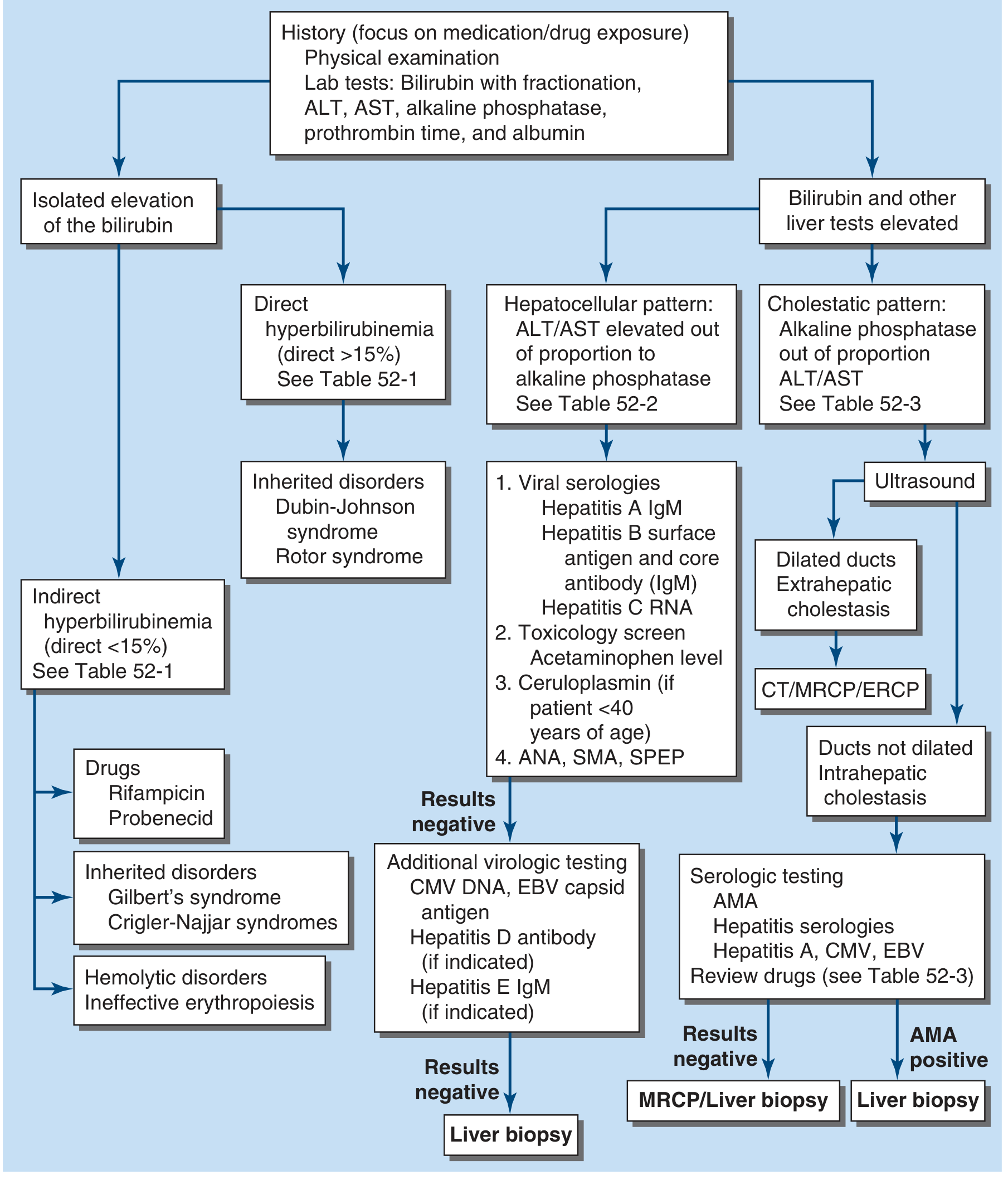
STEP 2: CLASSIFICATION
A. ISOLATED HYPERBILIRUBINEMIA (LFTs otherwise normal)
Unconjugated (Indirect) — Direct fraction < 15%
| Category | Cause |
|---|---|
| Overproduction | Hemolytic disorders (hereditary spherocytosis, sickle cell, G6PD, thalassemia), ineffective erythropoiesis, massive transfusion, hematoma resorption, malaria |
| Impaired uptake | Drugs: rifampin, probenecid, atazanavir, some cephalosporins/penicillins |
| Impaired conjugation | Gilbert's syndrome, Crigler-Najjar types I & II |
Gilbert's Syndrome - most common hereditary cause; benign; triggered by fasting, illness, stress. Bilirubin rarely >3-4 mg/dL. Bilirubin UDPGT activity reduced to 10-35% of normal.
In chronic hemolysis, bilirubin rarely exceeds 5 mg/dL - if higher, suspect superimposed choledocholithiasis (pigmented gallstones common in hemolytic disorders)
Conjugated (Direct) — Direct fraction > 15%
| Cause |
|---|
| Dubin-Johnson syndrome (benign, inherited, dark liver on gross pathology) |
| Rotor syndrome (benign, inherited) |
B. JAUNDICE WITH ABNORMAL LFTs
Hepatocellular Pattern
| Category | Causes |
|---|---|
| Viral hepatitis | Hep A (IgM), Hep B (HBsAg, HBcIgM), Hep C (RNA), CMV, EBV, Hep E |
| Drug/toxin-induced | Paracetamol (APAP), alcohol, isoniazid, statins, herbals |
| Autoimmune | Autoimmune hepatitis (ANA, SMA, LKM antibodies) |
| Metabolic | Wilson's disease (ceruloplasmin - check if patient <40 years), hemochromatosis |
| Ischemic hepatitis | "Shock liver" - acute hypoperfusion; dramatic aminotransferase rise, gradual bilirubin peak |
| Other | Sepsis, heart failure (hepatic congestion/hypoxia) |
Cholestatic Pattern
| Intrahepatic Cholestasis (ducts NOT dilated) | Extrahepatic Cholestasis (ducts DILATED) | |
|---|---|---|
| Causes | Primary biliary cholangitis (PBC), PSC (intrahepatic), drug-induced (see below), viral, TPN, sepsis, post-op, pregnancy cholestasis, heart failure | Choledocholithiasis (#1 cause), pancreatic cancer, cholangiocarcinoma, ampullary carcinoma, gallbladder cancer, hilar lymphadenopathy |
| Next test | Serologies (AMA for PBC), review drugs | CT/MRCP/ERCP |
STEP 3: HISTORY — What to Ask in the ED
| Feature | Suggests |
|---|---|
| RUQ pain + fever + jaundice (Charcot's triad) | Ascending cholangitis (choledocholithiasis) |
| Charcot's triad + hypotension + AMS (Reynolds' pentad) | Severe/suppurative cholangitis — emergency biliary drainage |
| Painless progressive jaundice | Malignancy (pancreatic cancer, cholangiocarcinoma) |
| Jaundice after prolonged vomiting/retching | Mallory-Weiss or hepatic congestion |
| Alcohol use / alcoholic binge | Alcoholic hepatitis |
| Medication history (including herbals/supplements) | Drug-induced liver injury (DILI) |
| IV drug use, unprotected sex, travel | Viral hepatitis (B, C, E) |
| Recent travel to endemic areas | Hep A, E; malaria; leptospirosis |
| Previous biliary surgery / ERCP / stent | Biliary stricture, cholangitis |
| Weight loss + anorexia + painless jaundice | Malignancy |
| Young patient + neuropsychiatric symptoms | Wilson's disease |
| Family history of jaundice | Gilbert's, Crigler-Najjar, hemolytic disorders |
| Pregnancy (2nd-3rd trimester) | Intrahepatic cholestasis of pregnancy, HELLP, acute fatty liver |
| ICU/post-op/septic patient | Ischemic hepatitis, TPN-related, sepsis cholestasis |
STEP 4: PHYSICAL EXAMINATION
| Sign | Suggests |
|---|---|
| Scleral icterus | Bilirubin >2-3 mg/dL (earliest sign) |
| RUQ tenderness | Biliary pathology, hepatitis |
| Murphy's sign | Acute cholecystitis |
| Palpable gallbladder (Courvoisier's sign) | Malignant obstruction (painless) |
| Hepatomegaly | Hepatitis, congestive hepatopathy, infiltrative disease |
| Splenomegaly | Portal hypertension, hemolytic anemia, infiltrative disease |
| Spider angiomas, palmar erythema, gynecomastia, caput medusae | Chronic liver disease / cirrhosis |
| Asterixis, confusion | Hepatic encephalopathy |
| Ascites | Portal hypertension, cirrhosis, malignancy |
| Cachexia / palpable mass | Malignancy |
| Kayser-Fleischer rings (slit lamp) | Wilson's disease |
| Fever + rigors | Ascending cholangitis, hepatic abscess |
Courvoisier's Law: Palpable non-tender gallbladder + jaundice = malignant obstruction (NOT choledocholithiasis, because stones cause fibrosis/non-distensible GB)
STEP 5: INVESTIGATIONS
First-Line ED Workup
| Test | What it Tells You |
|---|---|
| Total + Direct (Conjugated) Bilirubin | Fractionation guides classification (indirect vs. direct) |
| ALT / AST | Hepatocellular injury; >1000 = viral/ischemic/drug |
| Alkaline Phosphatase (ALP) | Cholestatic pattern if elevated out of proportion |
| GGT | Confirms hepatic source of elevated ALP; elevated with alcohol |
| Albumin | Chronic liver function; low = cirrhosis, malnutrition |
| PT / INR | Acute synthetic function; prolonged = severe hepatocyte dysfunction |
| CBC | Anaemia (haemolytic), WBC (infection/cholangitis), platelets (hypersplenism) |
| Blood cultures | If fever present - rule out cholangitis/sepsis |
| Urinalysis | Dark urine (urobilinogen / conjugated bilirubin) = direct hyperbilirubinaemia |
| Paracetamol level | In all jaundiced patients — rule out APAP toxicity |
| Blood glucose | Hypoglycaemia = severe hepatic failure |
Pattern Recognition from LFTs
| Pattern | AST/ALT | ALP | Likely Diagnosis |
|---|---|---|---|
| Hepatocellular | Very elevated (>10x) | Normal or mildly elevated | Viral hepatitis, DILI, ischemic hepatitis |
| Cholestatic | Mildly elevated | Very elevated (>3x) | Choledocholithiasis, PBC, malignancy |
| Mixed | Elevated | Elevated | Sepsis, drugs, PSC |
| Isolated bilirubin | Normal | Normal | Gilbert's, hemolysis, Dubin-Johnson |
AST:ALT ratio > 2:1 — strongly suggests alcoholic hepatitis ALT > 1000 U/L — suggests viral hepatitis, ischemic hepatitis, or APAP toxicity
Second-Line / Targeted Workup
| Clinical Suspicion | Test |
|---|---|
| Viral hepatitis | HBsAg, HBcAb IgM, Hep A IgM, Hep C RNA |
| Autoimmune hepatitis | ANA, SMA, LKM antibodies, SPEP |
| Wilson's disease (<40 yrs) | Ceruloplasmin, serum copper, urine copper |
| Haemochromatosis | Serum ferritin, transferrin saturation |
| PBC | AMA (antimitochondrial antibodies) |
| Malignancy / obstruction | USS → CT abdomen / MRCP |
| Cholangitis | Blood cultures, USS biliary tree |
| Pregnancy | LFTs + USS, HELLP screen (platelets, LDH) |
| Haemolysis | Blood film, reticulocyte count, LDH, haptoglobin, Coombs test |
Imaging
| Modality | Use |
|---|---|
| Abdominal Ultrasound (FIRST LINE) | Biliary dilation, gallstones, liver size/echotexture, ascites, masses |
| CT Abdomen | Pancreatic mass, lymphadenopathy, cholangiocarcinoma, extrahepatic cause |
| MRCP | Non-invasive biliary tree imaging (PSC, choledocholithiasis, strictures) |
| ERCP | Diagnostic + therapeutic (stone extraction, stenting) |
| Liver biopsy | When above tests inconclusive |
STEP 6: ED MANAGEMENT BY CAUSE
| Condition | Management |
|---|---|
| Ascending cholangitis | IV fluids, broad-spectrum antibiotics (ceftriaxone + metronidazole or piperacillin-tazobactam), urgent GI consult for ERCP biliary drainage |
| Severe cholangitis (Reynolds' pentad) | ICU admission, emergent ERCP or percutaneous drainage |
| APAP toxicity | N-acetylcysteine (NAC) immediately; liver transplant referral if severe |
| Alcoholic hepatitis | Supportive, assess severity (Maddrey score), steroids if severe |
| Acute viral hepatitis | Supportive; isolate if Hep A; refer hepatology |
| Malignant obstruction | Palliative stenting via ERCP; oncology referral |
| Haemolytic crisis (sickle cell) | Transfusion, analgesia, hydration |
| Fulminant hepatic failure | ICU, monitor ICP, consider liver transplant listing |
| Drug-induced (DILI) | Withdraw offending drug immediately |
STEP 7: DISPOSITION FROM ED
| Patient Type | Disposition |
|---|---|
| Cholangitis / sepsis / coagulopathy | Admit ICU or HDU |
| Acute liver failure (INR elevated, encephalopathy) | Admit ICU, transplant centre referral |
| New jaundice with unclear cause | Admit for workup |
| Suspected malignancy | Admit or urgent outpatient CT + GI referral |
| Gilbert's syndrome - well, normal LFTs, no haemolysis | Discharge with reassurance |
| Mild isolated unconjugated, stable - all normal | Outpatient follow-up |
HIGH-YIELD EXAM PEARLS
- Scleral icterus is the earliest detectable sign (bilirubin >2-3 mg/dL)
- Charcot's triad = RUQ pain + fever + jaundice → cholangitis; Add hypotension + AMS = Reynolds' pentad = emergency
- Courvoisier's sign = painless palpable gallbladder + jaundice = malignancy (pancreatic cancer)
- AST:ALT >2:1 = alcoholic hepatitis
- ALT >1000 = viral hepatitis, ischemic, or APAP
- Isolated indirect hyperbilirubinaemia + triggered by fasting/illness = Gilbert's syndrome
- Painless progressive jaundice + weight loss = pancreatic/biliary malignancy until proven otherwise
- Dark urine + pale stools = conjugated (obstructive) jaundice (conjugated bilirubin is water-soluble → excreted in urine; stercobilin absent → pale stools)
- Check paracetamol level in EVERY jaundiced patient - DILI is easily missed
- AMA positive = Primary Biliary Cholangitis (PBC)
- Ceruloplasmin if patient <40 yrs with unexplained liver disease = Wilson's disease
- Ultrasound is FIRST imaging in all ED jaundice workups
Acute cholecystitis
Acute Cholecystitis — ED Approach
Tintinalli's EM Ch.79 | Rosen's EM Ch.76 | Sleisenger & Fordtran's
DEFINITION & OVERVIEW
- Cholecystitis = inflammation of the gallbladder
- Calculous (95%) = caused by obstructing gallstone in the cystic duct
- Acalculous (5%) = no stone; occurs in critically ill patients (sepsis, burns, trauma, ICU)
- Gallstone prevalence: 8% in men, 17% in women in the USA; risk increases with age and BMI
THE SPECTRUM OF GALLSTONE DISEASE (Progression)
Asymptomatic gallstones (1-4% per year become symptomatic)
        ↓
Biliary colic (transient cystic duct obstruction → resolves)
        ↓
Acute cholecystitis (obstruction persists → inflammation)
        ↓
Gangrenous cholecystitis (necrosis of gallbladder wall)
        ↓
Emphysematous cholecystitis (gas-forming organisms; especially diabetics)
        ↓
Gallbladder perforation (life-threatening)
PATHOPHYSIOLOGY
- Gallstone obstructs cystic duct → gallbladder distends
- Increased intraluminal pressure → mucosal ischemia
- Cytotoxic bile products (lysophosphatidylcholine) trigger inflammation
- Secondary bacterial infection in ~50% of cases
- Organisms: E. coli (most common), Klebsiella, Streptococcus, Enterococcus, anaerobes (Clostridia, Bacteroides) in ~40%
Gallstone Types
| Type | Composition | Association |
|---|---|---|
| Cholesterol (80%) | Supersaturated cholesterol | Obesity, female, fertile, forty (5 F's), OCP, rapid weight loss |
| Black pigment | Calcium bilirubinate | Chronic haemolysis (sickle cell, spherocytosis), cirrhosis |
| Brown pigment | Calcium bilirubinate | Biliary stasis, infection, parasites (Southeast Asia) |
RISK FACTORS — The 5 F's (for Cholesterol Stones)
Fat, Female, Fertile, Forty, Fair
CLINICAL FEATURES
Biliary Colic vs Acute Cholecystitis
| Feature | Biliary Colic | Acute Cholecystitis |
|---|---|---|
| Pain character | Steady/constant (not truly colicky) | Constant, severe, worsening |
| Location | Epigastrium / RUQ | RUQ - more localised |
| Radiation | Back / right shoulder tip / right scapula | Same |
| Duration | < 6 hours (resolves spontaneously) | > 6 hours and persists |
| Trigger | Fatty meal (variable) | Often follows biliary colic episode |
| Nausea/vomiting | Yes | Yes |
| Fever | No | Present in ~1/3 cases (NOT always!) |
| Murphy's sign | Absent | Present |
| Peritoneal signs | Absent | May be present (guarding, rebound) |
| Circadian pattern | Peak around midnight | - |
Key Exam Trap: Fever is present in only ~1/3 of acute cholecystitis cases — absence of fever does NOT rule it out
MURPHY'S SIGN
Definition: Sudden cessation of deep inspiration due to pain when the examiner's fingers press the RUQ/right subcostal region, causing the inflamed gallbladder to descend onto the examiner's hand
- Sensitivity: 65%
- Specificity: 87%
- Sonographic Murphy's sign (pain on USS probe pressure over gallbladder) = higher diagnostic value
COMPLICATIONS
| Complication | Key Features |
|---|---|
| Gangrenous cholecystitis | Necrosis of GB wall; pain may paradoxically decrease; higher mortality |
| Emphysematous cholecystitis | Gas-forming organisms; especially in diabetics and elderly; gas in GB wall on X-ray/CT; emergency surgery |
| Gallbladder perforation | Rare but life-threatening; generalised peritonitis |
| Pericholecystic abscess | Localised perforation; RUQ mass |
| Gallstone ileus | Fistula → stone erodes into bowel → SBO; Rigler's triad on X-ray |
| Choledocholithiasis | Stone migrates to CBD; jaundice, elevated LFTs |
| Ascending cholangitis | CBD obstruction + infection → Charcot's triad / Reynolds' pentad |
| Mirizzi syndrome | Large stone impacted in cystic duct/Hartmann's pouch compresses CHD → obstructive jaundice |
Cholangitis — Must Know
| Triad/Pentad | Components |
|---|---|
| Charcot's Triad | RUQ pain + Fever/rigors + Jaundice |
| Reynolds' Pentad | Charcot's triad + Hypotension + Altered mental status = Severe cholangitis (emergency) |
DIFFERENTIAL DIAGNOSIS
| Condition | Distinguishing Feature |
|---|---|
| Acute pancreatitis | Epigastric radiation to back; elevated lipase |
| Peptic ulcer disease / perforation | History of PUD; peritonism; free air on X-ray |
| Hepatitis | Elevated transaminases; no acute RUQ mass |
| Hepatic abscess | Fever; USS finding; travel history |
| Right lower lobe pneumonia | Cough, SOB, CXR finding |
| Pyelonephritis | CVA tenderness; urinalysis positive |
| Appendicitis | Pain migrates to RIF; positive McBurney's point |
| Fitz-Hugh-Curtis syndrome | Young female; perihepatitis; PID history |
| Myocardial infarction | ECG, troponin; referred epigastric pain (inferior MI) |
INVESTIGATIONS
Labs
| Test | Significance |
|---|---|
| WBC | Leukocytosis in ~60% — may be absent in nearly half! |
| LFTs (AST, ALT, ALP) | Mildly elevated or normal in uncomplicated cholecystitis; markedly elevated = choledocholithiasis |
| Total + direct bilirubin | Elevated if CBD involved |
| Lipase | Elevated = concurrent pancreatitis |
| Blood cultures | If fever/sepsis — biliary organisms |
| CRP | Elevated; useful for severity assessment |
| PT/INR | If jaundiced or severe disease |
Leukocytosis may be absent in up to 50% of cases — don't exclude cholecystitis on WBC alone
Imaging (Table 79-7 — Tintinalli's)
| Modality | Key Findings | Comment |
|---|---|---|
| Ultrasound (FIRST-LINE) | Gallstones, GB wall thickening >3 mm, pericholecystic fluid, GB distension (short axis >40 mm), sonographic Murphy's sign | Preferred initial test; PPV >90% when multiple findings present; negative exam has high NPV |
| CT Abdomen | GB wall thickening, pericholecystic fat stranding, GB distension, gas in wall (emphysematous) | Better for complications (gangrene, perforation, abscess); insensitive for gallstones |
| HIDA Scan | Non-visualisation of GB = cystic duct obstruction | Most sensitive and specific; time-consuming, limited availability; loses sensitivity with bilirubin >5-8 mg/dL |
| MRCP/MRI | GB wall thickening, pericholecystic fluid, excellent biliary tree visualisation | Best for CBD stones; no radiation; time-consuming |
Normal CBD diameter: <5 mm (increases with age and post-cholecystectomy) CBD dilation + stones on USS = indirect evidence of choledocholithiasis
TREATMENT
ED Management of Biliary Colic
- NPO temporarily
- Antiemetics (ondansetron, metoclopramide)
- NSAIDs first-line analgesia (ketorolac IV) — equal efficacy to opioids; also reduce short-term complications
- Opioids if NSAIDs insufficient (all opioids cause some Sphincter of Oddi spasm — no clinical significance; no opioid is superior over another)
- ❌ Anticholinergics (atropine/glycopyrrolate) — do NOT work for biliary colic
- Discharge + surgical referral for elective laparoscopic cholecystectomy
ED Management of Acute Cholecystitis
- NPO (nil by mouth)
- IV fluids + electrolyte replacement
- Analgesia (NSAIDs + opioids as needed)
- Antiemetics
- IV Antibiotics:
| Regimen | Agent |
|---|---|
| Single agent (mild-moderate) | Piperacillin-tazobactam 3.375g IV q6h |
| Cephalosporin-based | 2nd/3rd gen cephalosporin (cefuroxime, ceftriaxone) |
| Carbapenem (severe/resistant) | Meropenem or ertapenem |
| Combination | Metronidazole + fluoroquinolone (ciprofloxacin) |
- Surgical consult — early laparoscopic cholecystectomy (within 72h if fit) is treatment of choice
- Percutaneous cholecystostomy — if too unwell for surgery (elderly, comorbid, critically ill)
Special Situations
| Situation | Management |
|---|---|
| Emphysematous cholecystitis | Broad-spectrum IV antibiotics including anaerobic coverage; emergency cholecystectomy |
| Gangrenous cholecystitis | Emergency surgery |
| Gallbladder perforation | Emergency surgery + washout |
| Acalculous cholecystitis (ICU) | Percutaneous cholecystostomy (drainage); cholecystectomy when stable |
| Ascending cholangitis | IV antibiotics + urgent ERCP for biliary drainage |
| Severe cholangitis (Reynolds' pentad) | ICU + emergency ERCP or percutaneous drainage |
| Choledocholithiasis | ERCP + stone extraction + elective cholecystectomy |
ACALCULOUS CHOLECYSTITIS
- Occurs in 5% of cholecystitis cases
- More aggressive course — higher risk of gangrene and perforation
- Risk factors: ICU patients, sepsis, burns, major trauma/surgery, TPN, diabetes, immunosuppression
- Pathophysiology: bile stasis + gallbladder ischaemia → inflammation without stones
- Diagnosis: USS or CT showing GB changes without stones; HIDA confirmatory
- Treatment: IV antibiotics + percutaneous cholecystostomy (if too unwell for surgery)
DISPOSITION
| Category | Disposition |
|---|---|
| Biliary colic - stable, symptom controlled | Discharge + surgical OPD referral |
| Uncomplicated acute cholecystitis | Admit for IV antibiotics + early surgery |
| Complicated cholecystitis (gangrene, perforation, emphysematous) | Admit ICU + emergency surgery |
| Ascending cholangitis | Admit + urgent ERCP; ICU if Reynolds' pentad |
| High-risk surgical patient with acute cholecystitis | Admit for antibiotics + percutaneous cholecystostomy |
HIGH-YIELD EXAM PEARLS
- Biliary colic pain lasts <6 hours and resolves spontaneously; >6 hours = think cholecystitis
- Fever is present in only ~1/3 of cholecystitis — absence does NOT exclude diagnosis
- Murphy's sign: 65% sensitive, 87% specific; sonographic Murphy's is more reliable
- Leukocytosis absent in up to 50% of acute cholecystitis — don't be falsely reassured
- USS is first-line imaging — PPV >90% with gallstones + wall thickening + pericholecystic fluid + sonographic Murphy's
- HIDA is most sensitive/specific but impractical in the ED
- NSAIDs are first-line analgesia for biliary colic (reduce complication risk too); all opioids equally acceptable
- Anticholinergics do NOT work for biliary colic pain
- Emphysematous cholecystitis = diabetics + elderly = gas in GB wall = emergency surgery
- Acalculous cholecystitis = ICU/critically ill patients = more dangerous = percutaneous drainage
- Charcot's triad + hypotension + AMS = Reynolds' pentad = emergency
- CBD >5 mm on USS = dilated = suspect choledocholithiasis
- Courvoisier's sign (non-tender palpable GB + jaundice) = malignant obstruction, NOT stones
- Early laparoscopic cholecystectomy (within 72h) is preferred over delayed surgery
Acute cholangitis
Acute Cholangitis — Complete ED Guide
Current Surgical Therapy 14e | Sabiston Surgery | Yamada's Gastroenterology | Mulholland & Greenfield's Surgery
Based on Tokyo Guidelines 2018
DEFINITION
EPIDEMIOLOGY & CAUSES
- Most common in the 5th-7th decades; equal in men and women
- Risk of cholangitis from admitted choledocholithiasis patients: up to 5%
- Bactibilia (bacteria in bile) found in up to 90% with stone obstruction
Causes of Acute Cholangitis
| Category | Specific Causes | Frequency |
|---|---|---|
| Biliary stasis - obstruction | Choledocholithiasis | 40-70% (most common) |
| Malignancy (pancreatic Ca, cholangiocarcinoma, ampullary Ca) | 10-60% | |
| Benign strictures (PSC, post-op, chronic pancreatitis) | 5-30% | |
| Direct seeding | ERCP instrumentation | 1-7% |
| Indwelling biliary stents / drains | Iatrogenic | |
| Bilioenteric anastomosis | Post-surgical | |
| Other | Parasitic infection (Ascaris, Clonorchis) | Endemic areas |
PATHOPHYSIOLOGY
- Sphincter of Oddi - mechanical barrier against ascending duodenal bacteria
- Laminar bile flow - continuous flushing of bacteria
- Bile salts - bacteriostatic properties
- IgA secretion in biliary tree
- Bile stasis → bacterial proliferation
- Increased intraluminal pressure → biliary ductule permeability ↑
- Bacterial translocation from portal venous system into bile ducts
- Septicaemia when bacteria enter systemic circulation
Microbiology
| Organism | Frequency |
|---|---|
| E. coli | 25-50% (most common) |
| Klebsiella spp. | 10-20% |
| Enterococcus | 10-30% |
| Enterobacter | 5-10% |
| Anaerobes (Bacteroides, Clostridia) | Recurrent/post-instrumentation cases |
| Pseudomonas, Citrobacter | Also seen |
Infection is typically polymicrobial and dominated by gram-negative coliforms
CLINICAL FEATURES
The Classic Presentations
| Presentation | Components | Sensitivity |
|---|---|---|
| Charcot's Triad | RUQ pain + Fever/rigors + Jaundice | < 50-60% (often incomplete) |
| Reynolds' Pentad | Charcot's triad + Hypotension + Altered mental status | ~5% (severe/suppurative cholangitis; mortality approaches 100% without treatment) |
Exam Trap: Charcot's triad is present in <50% of patients - absence does NOT rule out cholangitis. Fever + RUQ pain is present in 80%; jaundice in only 60%
Differential Diagnosis in the ED
| Condition | Distinguishing Feature |
|---|---|
| Acute cholecystitis | Murphy's sign; no jaundice (unless Mirizzi) |
| Acute pancreatitis | Elevated lipase; epigastric radiation to back |
| Mirizzi's syndrome | Stone impacted in cystic duct compressing CHD |
| Liver abscess | USS finding; fever; may have no jaundice |
| Biliary leak | Post-procedural/surgical history |
| Right lower lobe pneumonia | CXR changes; no jaundice |
| Peptic ulcer perforation | Free air; sudden onset |
STEP 1: DIAGNOSIS — Tokyo Guidelines 2018
TG18 Diagnostic Criteria (Table 8.7)
| Category | Criteria |
|---|---|
| A. Systemic Inflammation | Fever and/or shaking chills; elevated WBC, CRP |
| B. Cholestasis | Jaundice (bilirubin ≥2 mg/dL); abnormal LFTs (ALP, GGT, AST, ALT) |
| C. Imaging | Biliary dilatation on USS/CT; evidence of aetiology (stone, stricture, stent) |
STEP 2: SEVERITY GRADING — Tokyo Guidelines 2018
Grade I — Mild
- Does not meet criteria for moderate or severe
- Responds to antibiotics alone
Grade II — Moderate
| Criterion | Threshold |
|---|---|
| WBC | >12,000 or <4,000/μL |
| High fever | ≥39°C |
| Age | ≥75 years |
| Total bilirubin | ≥5 mg/dL |
| Albumin | <lower limit of normal × 0.73 |
Grade III — Severe
| System | Criteria |
|---|---|
| Cardiovascular | Hypotension requiring vasopressors |
| Neurological | Disturbance of consciousness (AMS) |
| Respiratory | PaO2/FiO2 ratio < 300 |
| Renal | Oliguria, creatinine > 2 mg/dL |
| Hepatic | PT/INR > 1.5 |
| Haematological | Platelets < 100,000/mm³ |
STEP 3: INVESTIGATIONS
Labs
| Test | Significance |
|---|---|
| WBC + differential | Leukocytosis; may be absent or reversed (<4000) in severe sepsis |
| CRP | Elevated; severity marker |
| Total + direct bilirubin | Elevated (≥2 mg/dL confirms cholestasis) |
| ALP, GGT | Cholestatic pattern - markedly elevated |
| AST, ALT | Hepatocellular injury from infection/inflammation |
| Albumin | Hypoalbuminaemia = Grade II severity marker |
| PT/INR | Coagulopathy; >1.5 = Grade III (hepatic dysfunction) |
| Blood cultures x2 | Positive in ~21-71% of cholangitis; obtain BEFORE antibiotics |
| Lactate | Elevated = poor perfusion; sepsis |
| Creatinine | AKI assessment; >2 mg/dL = Grade III |
| Blood glucose | Sepsis-related hyperglycaemia; hypoglycaemia in fulminant |
Imaging
| Modality | Role | Key Findings |
|---|---|---|
| Ultrasound (FIRST-LINE) | Screening | Biliary dilatation, CBD >6 mm, gallstones, intrahepatic ductal dilation |
| CT Abdomen | Site of obstruction; complications | Identifies strictures, masses, ductal dilation; HIDA less reliable in infected bile |
| MRCP | Non-invasive cholangiography | Excellent CBD/biliary tree visualisation; no radiation; preferred pre-ERCP |
| ERCP | Diagnostic + therapeutic | Gold standard; allows stone extraction, stenting, sphincterotomy |
| PTC (Percutaneous Transhepatic Cholangiography) | When ERCP fails/unavailable | Requires dilated ducts; second-line drainage |
| HIDA | Limited role in cholangitis | Reduced sensitivity as infected bile ↓ tracer secretion |
Normal CBD: <6 mm (increases post-cholecystectomy and with age)
STEP 4: MANAGEMENT
Immediate ED Actions (ALL grades)
- IV access - two large-bore cannulas
- IV fluid resuscitation - aggressive if septic
- Blood cultures x2 - BEFORE starting antibiotics
- IV antibiotics IMMEDIATELY after blood cultures
- Severity assessment (TG18 grading)
- NPO
- Analgesia + antiemetics
- Biliary imaging (USS → CT/MRCP as needed)
- Surgical/GI consult early
Antibiotic Regimens
| Setting | Regimen |
|---|---|
| Community-acquired, mild-moderate | Cefazolin, Cefuroxime, or Ceftriaxone |
| Severe / elderly / immunocompromised / community-acquired | Imipenem/cilastatin, Meropenem, Piperacillin-tazobactam, OR (Ciprofloxacin/Levofloxacin/Cefepime + Metronidazole) |
| Post-bilioenteric anastomosis (any severity) | Imipenem/cilastatin, Meropenem, Pip-tazo, OR Fluoroquinolone + Metronidazole |
| Healthcare-associated (any severity) | Above regimens + add Vancomycin (for MRSA/resistant Enterococcus) |
Duration: 4-7 days after source control; 14 days if bacteraemia present (endocarditis risk)
Cover for ESBL-producing Enterobacteriaceae if local resistance is a concern
Biliary Drainage — Urgency by Grade
| Grade | Urgency | Management |
|---|---|---|
| Grade I (Mild) | Early ERCP within 72h if responds to antibiotics | Antibiotics alone may suffice; low threshold for drainage if no improvement |
| Grade II (Moderate) | Early biliary drainage (<72h) | ERCP preferred; early duct clearance required |
| Grade III (Severe) | Urgent biliary drainage (<24h) | Stabilise haemodynamic/respiratory status FIRST → ERCP as soon as possible; ICU admission |
Drainage Methods (in order of preference)
| Method | Details | When Used |
|---|---|---|
| ERCP (1st line) | Endoscopic sphincterotomy + stone extraction + stenting | Standard; preferred in all anatomically accessible patients |
| EUS-guided biliary drainage (EUS-BD) | Hepaticogastrostomy or choledochoduodenostomy via EUS | Ascites, hilar strictures, unresectable malignancy |
| PTC / PTBD (2nd line) | Percutaneous transhepatic drainage; 90% success | Failed ERCP; post-surgical anatomy (Roux-en-Y, Whipple) |
| Surgical drainage (last resort) | CBD exploration + T-tube + cholecystectomy if stable; morbidity/mortality 10-50% | All endoscopic/percutaneous methods failed or unavailable |
ERCP limited in: Roux-en-Y hepaticojejunostomy, gastric bypass, Whipple's procedure, duodenal obstruction
STEP 5: DISPOSITION
| Grade | Disposition |
|---|---|
| Grade I - responds to antibiotics | Admit ward; ERCP within 72h |
| Grade II - moderate | Admit; urgent ERCP/drainage <72h; GI/surgery consult |
| Grade III - severe / Reynolds' pentad | ICU admission; urgent ERCP <24h; vasopressors if needed; MDT |
COMPLICATIONS
| Complication | Features |
|---|---|
| Septic shock | Reynolds' pentad; mortality ~100% without drainage |
| Pyogenic liver abscess | Extension of biliary infection into hepatic parenchyma; treated with antibiotics + percutaneous drainage |
| Portal vein thrombosis / pylephlebitis | Rare; septic thrombophlebitis |
| Acute renal failure | Sepsis-related; Grade III criterion |
| DIC | Platelets <100k; Grade III criterion |
| Recurrent cholangitis | Especially with indwelling stents; stent occlusion |
COMPARISON: Cholecystitis vs Cholangitis vs Biliary Colic
| Feature | Biliary Colic | Acute Cholecystitis | Acute Cholangitis |
|---|---|---|---|
| Pain | RUQ/epigastric, <6h | RUQ, constant, >6h | RUQ |
| Fever | No | In ~1/3 | Yes (80%) |
| Jaundice | No | Rare (Mirizzi) | Yes (60%) |
| Murphy's sign | No | Yes | Less prominent |
| WBC | Normal | Often elevated | Often elevated |
| LFTs | Normal | Mildly elevated or normal | Markedly elevated |
| Bilirubin | Normal | Mildly elevated/normal | Elevated |
| USS | Stones in GB | Stones + GB changes | CBD dilation |
| Treatment | NSAID + outpatient Lap Chole | IV abx + early Lap Chole | IV abx + urgent ERCP |
| Mortality | Very low | Low (higher if complicated) | High if severe/untreated |
HIGH-YIELD EXAM PEARLS
- Charcot's triad is present in <50-60% - never a reason to exclude cholangitis
- Reynolds' pentad (+ hypotension + AMS) = Grade III / suppurative cholangitis = emergency, mortality approaches 100% without treatment
- Choledocholithiasis is #1 cause (40-70%); malignancy is #2
- Blood cultures BEFORE antibiotics - positive in up to 71% of cases
- ERCP is both diagnostic AND therapeutic - first-line drainage method
- Grade III = urgent drainage <24h; Grade II = early drainage <72h
- Antibiotic duration = 4-7 days post source control; 14 days if bacteraemic
- Add Vancomycin for healthcare-associated cholangitis (MRSA/resistant Enterococcus)
- PTC is second-line when ERCP fails or post-surgical anatomy (Roux-en-Y, Whipple)
- Surgical drainage has 10-50% morbidity/mortality - absolute last resort
- ESBL-producing Enterobacteriaceae increasingly common - consider carbapenems in resistant cases
- Tokyo Guidelines 2018 are the current standard for diagnosis and severity grading
- Normal CBD = <6 mm — dilation = choledocholithiasis / obstruction
- HIDA scan unreliable in cholangitis - infected bile reduces tracer secretion
Diverticulitis
Diverticulitis — Complete ED Guide
Rosen's Emergency Medicine Ch.81 | Sabiston Surgery | Mulholland & Greenfield's Surgery
DEFINITIONS
| Term | Definition |
|---|---|
| Diverticulum | Outpouching of the colonic wall (false diverticulum — only mucosa + submucosa herniate through muscular wall) |
| Diverticulosis | Presence of diverticula without inflammation |
| Diverticulitis | Inflammation/infection of a diverticulum, usually from microperforation |
| Diverticular bleeding | Bleeding from diverticula without inflammation (typically painless) |
EPIDEMIOLOGY
- Diverticulosis: increasingly common with age — rare <40, common >60 years
- Sigmoid colon most commonly affected (95% in Western countries)
- Right colon diverticula more common in Asian populations
- Diverticulosis → diverticulitis in up to 25% of individuals
- Recurrent episodes in up to 40% of those who develop diverticulitis
- Diverticulosis accounts for 40% of all lower GI haemorrhage; severe bleeding in 3-5%
PATHOPHYSIOLOGY
Why Diverticula Form
| Factor | Details |
|---|---|
| Weakness of the bowel wall | Non-circumferential muscle layers; insertion of vasa recta causes weak points; collagen disorders (Ehlers-Danlos), localized ischaemia |
| High intraluminal pressure | Low fibre diet → small faecal volume → increased segmentation contractions → high pressure; chronic constipation; colonic stasis; increased collagen crosslinking with age |
Why Diverticulitis Occurs
- Diverticulum obstructed by inspissated faeces (faecolith) or increased intraluminal pressure
- → Localised microperforation of diverticulum
- → Pericolonic inflammation (phlegmon)
- → If contained by mesentery: abscess forms
- → If free perforation into peritoneum: peritonitis
Current evidence suggests diverticulitis is primarily an inflammatory process, not purely infectious — this is why antibiotics are now selective, not routine in uncomplicated disease
CLINICAL FEATURES
Classic Presentation
| Feature | Description |
|---|---|
| Pain | Persistent left lower quadrant (LLQ) pain - constant, >24 hours |
| Location | LLQ (sigmoid); RLQ (right-sided - mimics appendicitis) |
| Fever | Low-grade to high (high fever = complicated disease/sepsis) |
| Nausea/vomiting | Common |
| Change in bowel habit | Constipation or diarrhoea |
| Anorexia | Common |
Complicated Diverticulitis - Additional Features
| Feature | Suggests |
|---|---|
| High fever, rigors, haemodynamic instability | Sepsis, perforation |
| Diffuse peritonism (rebound, guarding) | Free perforation / peritonitis |
| Palpable LLQ mass | Abscess / phlegmon |
| Dysuria, recurrent UTIs, pneumaturia, fecaluria | Colovesical fistula |
| Feculent vaginal discharge | Colovaginal fistula (post-hysterectomy females) |
| Vomiting + abdominal distension | Obstruction (stricture) |
| RLQ pain | Right-sided diverticulitis - mimics appendicitis |
Elderly and immunocompromised patients may present subtly despite severe disease. Perforation is more frequent in this group and carries high mortality. Do NOT be falsely reassured by mild symptoms.
DIFFERENTIAL DIAGNOSIS
| Condition | Distinguishing Feature |
|---|---|
| Appendicitis | RLQ pain + periumbilical migration + Rovsing's; right-sided diverticulitis can mimic |
| Ectopic pregnancy | Female + amenorrhoea + βhCG positive |
| Ovarian pathology (torsion, cyst) | Female; USS findings |
| PID | Female; cervical motion tenderness; bilateral |
| Ischaemic colitis | Elderly; post-prandial; cardiovascular risk factors |
| IBD (Crohn's/UC) | Younger patient; bloody diarrhoea; systemic features |
| Colorectal carcinoma | Weight loss; change in bowel habit; PR bleeding; always consider |
| Ureteral/renal stone | Flank pain; haematuria; urinalysis |
| Inguinal hernia | Groin swelling |
| Psoas abscess | Hip pain; fever; travel history (TB) |
Underlying colorectal malignancy must be excluded - but investigation is safe to defer until resolution of the acute episode
INVESTIGATIONS
Labs
| Test | Significance |
|---|---|
| CBC | Leukocytosis (many patients); may be absent - not needed to make the diagnosis |
| CRP | Elevated; correlates with severity |
| BMP (U&E) | Electrolyte derangement; renal function |
| Urinalysis | Pyuria/bacteriuria with colovesical fistula; haematuria if ureter involved |
| βhCG | All women of reproductive age - exclude ectopic pregnancy |
| Blood cultures | If septic / systemically unwell |
| Lactate | Elevated = sepsis/perfusion compromise |
A patient with prior known diverticulitis presenting with similar mild symptoms may not need any testing - empirical treatment is acceptable if no concerning features
Imaging
| Modality | Role | Sensitivity/Specificity |
|---|---|---|
| CT Abdomen/Pelvis with IV contrast (GOLD STANDARD) | Confirms diagnosis, stages severity, identifies complications | 98% sensitivity, 99% specificity |
| CT with IV + enteric contrast | Ideal (oral or rectal contrast added) | Best for fistula, abscess |
| Ultrasound | Accessible; limited by bowel gas; operator dependent | Lower than CT |
| Plain X-ray | Non-specific; only useful for free air (perforation) | Low diagnostic yield |
| Barium/water-soluble enema | No longer recommended in acute phase | Risk of perforation |
| Colonoscopy | Contraindicated in acute phase (perforation risk); defer until resolution | Follow age-appropriate screening |
CT Findings in Diverticulitis
- Colonic wall thickening
- Pericolonic fat stranding (hallmark)
- Diverticulae visible
- Localised microperforation
- Pericolic abscess
- Free air (perforation)
- Free fluid
HINCHEY CLASSIFICATION (CT-Based Staging)
| Stage | Description | Management |
|---|---|---|
| Ia | Pericolic phlegmon and inflammation, no fluid collection | Antibiotics ± admission |
| Ib | Pericolic/mesenteric abscess <4 cm | IV antibiotics; often responds without drainage |
| II | Pelvic/distant abscess >4 cm | Percutaneous CT-guided drainage + IV antibiotics |
| III | Purulent peritonitis (contained perforation) | Emergency surgery |
| IV | Faeculent peritonitis (free perforation) | Emergency surgery - highest mortality |
TREATMENT
Uncomplicated Diverticulitis
| Patient | Management |
|---|---|
| Mild, immunocompetent, tolerating PO, reliable follow-up | Outpatient management: liquid diet advancing as tolerated ± oral antibiotics |
| Cannot tolerate PO / elderly / immunocompromised / significant comorbidities / poor social support | Admit for IV antibiotics + bowel rest |
- Ciprofloxacin 500 mg PO BD + Metronidazole 500 mg PO q8h
- Amoxicillin-clavulanate 875/125 mg PO BD
Studies show no benefit of IV over oral antibiotics for uncomplicated diverticulitis
Complicated Diverticulitis
| Complication | Management |
|---|---|
| Small abscess ≤4 cm | IV antibiotics alone — 80%+ success rate |
| Large abscess >4 cm (Hinchey II) | Percutaneous CT-guided drainage + IV antibiotics; surgery if fails |
| Fistula (colovesical, colovaginal) | Broad-spectrum antibiotics; elective surgical resection + fistula repair after acute phase |
| Obstruction (partial) | NG decompression + IV fluids + antibiotics + bowel rest; elective resection after resolution |
| Obstruction (complete) | Emergency surgery |
| Purulent peritonitis (Hinchey III) | Emergency surgery: Hartmann's procedure or primary resection + anastomosis |
| Faeculent peritonitis (Hinchey IV) | Emergency surgery - Hartmann's; highest morbidity/mortality |
- Piperacillin-tazobactam
- Cefepime + Metronidazole
- Ciprofloxacin + Metronidazole
- Meropenem/Imipenem (severe/resistant)
Surgical Options
| Procedure | Description |
|---|---|
| Hartmann's procedure | Sigmoid resection + end colostomy (no anastomosis) — used in emergency/contaminated field |
| Primary resection + anastomosis | With or without diverting loop ileostomy |
| Laparoscopic lavage | For Hinchey III (purulent) — controversial |
| Common bile duct exploration (T-tube) | Not applicable here |
Surgery for acute diverticulitis indicated for: peritonitis, sepsis unresponsive to medical management, failed percutaneous drainage
Elective Surgery Considerations
- After a diverticular abscess (Hinchey Ib/II) - ASCRS recommends considering elective resection after successful non-operative treatment
- Recurrence after first uncomplicated episode is only ~13% - routine prophylactic surgery no longer recommended after 2 episodes
- Individualise surgical decision-making based on patient factors
COMPLICATIONS OF DIVERTICULITIS
| Complication | Key Points |
|---|---|
| Abscess | Most common complication; <4 cm = antibiotics; >4 cm = percutaneous drainage |
| Fistula | Colovesical (#1 in males) → pneumaturia + fecaluria; Colovaginal (post-hysterectomy females); Colocutaneous (at drain sites) |
| Obstruction | Stricture from chronic inflammation/fibrosis; can mimic malignancy |
| Perforation | Free faecal peritonitis = emergency; high mortality |
| Haemorrhage | From diverticulosis (NOT diverticulitis - bleeding is painless) |
DISPOSITION
| Patient | Disposition |
|---|---|
| Mild uncomplicated, immunocompetent, tolerating PO, reliable follow-up | Discharge with oral antibiotics, liquid diet, GP follow-up in 48-72h |
| Mild uncomplicated but elderly, immunocompromised, or unable to tolerate PO | Admit for IV antibiotics + bowel rest |
| Complicated (abscess, sepsis, signs of perforation) | Admit ± surgical consult |
| Hinchey III/IV, haemodynamic instability, peritonitis | Emergency surgical consult + ICU |
HIGH-YIELD EXAM PEARLS
- LLQ pain >24h = diverticulitis until proven otherwise (sigmoid = most common site)
- RLQ diverticulitis mimics appendicitis - more common in Asian populations
- CT with IV contrast is gold standard - 98% sensitivity, 99% specificity
- Antibiotics NOT mandatory in mild uncomplicated diverticulitis (inflammatory, not purely infective)
- Oral antibiotics = IV antibiotics in uncomplicated diverticulitis - no benefit to IV
- Elderly and immunocompromised present subtly but have more severe disease and higher perforation risk
- Colovesical fistula = #1 fistula type (especially males) → pneumaturia + fecaluria + recurrent polymicrobial UTIs
- Hinchey Ib/II (abscess) → percutaneous drainage if >4 cm
- Hinchey III/IV (peritonitis) → emergency surgery (Hartmann's)
- Abscess <4 cm → antibiotics alone (80%+ resolution rate)
- Colonoscopy is contraindicated in acute phase - defer until resolution
- Colorectal malignancy must be excluded - defer colonoscopy to follow age-appropriate screening after recovery
- Diverticular bleeding is painless (no inflammation) - accounts for 40% of lower GI bleeds
- Routine elective surgery after 2 uncomplicated episodes is no longer recommended - individualise
- Free air on CT = perforation = emergency surgery
Do you section 24 in titinallis
Tintinalli's Block 24 — Contents (Pages 1966–2040)
🦴 Musculoskeletal / Orthopaedic
| Chapter | Topics |
|---|---|
| Ch. 284 | Septic Arthritis (bacterial nongonococcal + gonococcal), Crystal-induced synovitis (Gout & Pseudogout), Viral Arthritis, Lyme disease, Hemarthrosis, Rheumatoid arthritis, Osteoarthritis, Reactive arthritis, Bursitis (olecranon, prepatellar) |
| Ch. 285 | Foot disorders — Ingrown toenail, Plantar fasciitis, Tarsal tunnel syndrome, Ganglions, Tenosynovitis, Tendon ruptures, Plantar neuroma, Compartment syndrome of foot |
🧠 Psychiatric / Behavioural
| Chapter | Topics |
|---|---|
| Ch. 286 | Psychiatric assessment in the ED — medical clearance, history, MSE, capacity vs competency, involuntary patients, disposition |
| Ch. 287 | Acute Agitation — verbal de-escalation, pharmacology (haloperidol, ketamine, atypical antipsychotics), restraints |
| Ch. 288 | Delirium — clinical features, mental status exam, treatment |
- Are you referring to a printed section number (like "Section 24: Psychiatry" or "Section 24: Musculoskeletal")?
- Or a specific chapter within Tintinalli's?
- Or were you asking about the GIT section continuing from where you are in the book?
Section 24 is psychosocial disorders kindly give some predicted important questions with answers from that chapter for final exam
Now go for search
🧠 Section 24: Psychosocial Disorders
Predicted Final Exam Q&A
Tintinalli's Emergency Medicine, Chapters 286–292
CHAPTER 286 — PSYCHIATRIC ASSESSMENT IN THE ED
- 7-10% of patients admitted to psychiatric wards have an organic condition that was missed
- Examples of missed organic pathology: meningitis, sepsis, delirium, acetaminophen toxicity, neuroleptic malignant syndrome, hepatic encephalopathy, myxedema coma, DKA, epidural/subdural haematoma
- New-onset psychosis or altered mental status = organic cause until proven otherwise — psychiatric diagnosis is a diagnosis of exclusion
- Over 1/3 of assessments lacked a documented history
- 8% had no physical examination documented
- Vital signs were frequently not complete or not reassessed when abnormal
- Mental status examination missing in up to 56% of cases
- Neurologic examination components absent:
- Cranial nerve testing absent in 50%
- Motor exam absent in 72%
- Sensory exam absent in 88%
- Gait testing absent in 75%
- Failure to develop/refine differential diagnosis
- "Anchoring" on psychiatric diagnoses without ruling out organic causes
- "Kitchen sink" mentality — ordering unnecessary tests without targeted reasoning
- Know exit locations before entering — stand close to the exit
- Maintain enough distance to avoid being physically attacked
- Wear a badge clip that cannot be used to choke you
- Always introduce yourself clearly
- Establish eye contact; smile if possible
- Reuse terms the patient uses before asking for clarification (makes patient feel heard)
- Start with open-ended questions (best for rapport)
- Transition to close-ended questions if open-ended are unproductive
- Last resort: Yes/no and multiple-choice questions
- Ideally performed by a team of 5 — one team leader + one person per limb
- A "show of force" with multiple staff present may subdue the patient without actual restraint
- Place patient on bed/stretcher; secure all 4 limbs with leather restraints (soft restraints for non-violent settings)
- Elevate the patient's head to minimise aspiration risk
- Once restrained: offer medications; if refused → administer involuntarily
- Provide ongoing explanation to patient and family throughout
- Avoid injury to both patient and personnel
| Capacity | Competency | |
|---|---|---|
| Determined by | Physician (at the bedside) | Court/legal system |
| Setting | Clinical / ED | Legal proceeding |
| Time | Moment-specific; can change | More permanent legal determination |
| Scope | Decision-specific (can have capacity for one decision, not another) | Global |
- A patient has decision-making capacity if they can: understand information, appreciate consequences, reason about choices, and communicate a decision
- Capacity assessment is the ED physician's responsibility; competency is a judge's determination
CHAPTER 287 — ACUTE AGITATION
- Approach with safety in mind — plan even before patient arrives
- Attempt verbal de-escalation in ALL patients first
- If worsens: employ a "show of concern" (presence of calm, assertive team)
- Treat underlying medical cause first
- Restraints only to protect staff/patient from harm — use sparingly
- Target medications to most likely cause
- Use oral medications when possible
- Use second-generation (atypical) antipsychotics as first-line in most situations (except alcohol intoxication)
The goal is to treat agitation sufficiently to allow a thorough medical evaluation
| Score | Description |
|---|---|
| +3 | Combative, violent, out of control |
| +2 | Very agitated, pulling at lines/tubes |
| +1 | Agitated, restless |
| 0 | Alert and calm |
| -1 | Drowsy, responds to voice |
| -2 | Light sedation |
| -3 | Deep sedation |
| Situation | First-line Agent | Notes |
|---|---|---|
| Most agitation (psychiatric, unknown cause) | Second-generation antipsychotics (olanzapine, ziprasidone, risperidone) | Safer, fewer EPS, effective |
| Alcohol intoxication | Benzodiazepines (lorazepam) | Antipsychotics less effective; seizure risk |
| Rapid/extreme agitation | Ketamine (dissociative dose) | Rapid onset; useful pre-intubation or when other agents fail |
| Classic option | Haloperidol (typical antipsychotic) | Still widely used; risk of QTc prolongation and EPS |
| Mild-moderate | Oral olanzapine, oral risperidone | Preferred if patient can take oral meds |
CHAPTER 288 — DELIRIUM & DEMENTIA
| Type | Features | Frequency |
|---|---|---|
| Hypoactive ("quiet delirium") | Decreased psychomotor activity, somnolent, withdrawn | Most common + most commonly missed |
| Hyperactive | Agitated, anxious, combative, increased psychomotor activity | Easier to recognise |
| Mixed | Alternates between hyper- and hypoactive states | Also common |
- Delirium is present in 7-10% of elderly ED patients
- Diagnosis missed by providers in 57-83% of cases
- Delirium is an independent predictor of 6-month mortality
- If missed in ED, likely to be missed on inpatient services as well
| Feature | Delirium | Dementia |
|---|---|---|
| Onset | Acute (hours to days) | Gradual (months to years) |
| Course | Fluctuating | Progressive but stable day-to-day |
| Consciousness | Altered | Usually preserved until late |
| Attention | Impaired | Less affected early |
| Reversibility | Often reversible | Generally irreversible |
| Key feature | Acute + fluctuating | Chronic + gradual |
Key exam point: Delirium is more likely to occur in patients with underlying dementia — both can coexist
- Three-word recall (e.g., apple, table, penny)
- Clock drawing test
- Recalls all 3 words → No cognitive impairment (clock drawing not needed)
- Recalls 0 words → Cognitive impairment (clock drawing not needed)
- Recalls 1-2 words → Clock drawing required to determine result
- Clock drawing normal = negative for impairment; abnormal = positive
| Dementia | Onset | Key Features |
|---|---|---|
| Alzheimer's disease | Gradual | Primarily memory; personality changes; visual-spatial problems |
| Vascular dementia | Sudden or stepwise | Symptoms correlate with area of brain ischemia |
| Lewy body dementia | Gradual | Memory + hallucinations + Parkinsonian features; AVOID typical antipsychotics (very poorly tolerated - can cause severe worsening) |
| Frontotemporal dementia | <60 years old | Disinhibition, apathy, language difficulties; frontal/temporal atrophy |
Lewy body dementia — patients resemble delirium (rapid decline, fluctuating course, perceptual disturbances) and do very poorly with typical antipsychotics
- Treat the underlying cause (infection, metabolic, drug withdrawal, etc.)
- Non-pharmacologic first: reorientation, familiar faces, lighting cues, noise reduction
- Low-dose haloperidol or atypical antipsychotics — if pharmacologic treatment needed for safety
- AVOID benzodiazepines in delirium (worsen confusion) — EXCEPT in:
- Alcohol withdrawal seizures
- Benzodiazepine withdrawal
- Catatonia
- If benzodiazepines needed in elderly: choose agents with lower risk of accumulation — Lorazepam, Oxazepam, Temazepam (LOT - no active metabolites)
CHAPTER 289 — DEPRESSION & SUICIDE
- Depressed mood most of the day
- Anhedonia (decreased interest/pleasure in activities)
- Significant weight loss or gain / appetite change
- Insomnia or hypersomnia
- Psychomotor retardation or agitation
- Fatigue or loss of energy
- Feelings of worthlessness or excessive guilt
- Difficulty concentrating or indecisiveness
- Suicidal ideation (with or without a plan)
- "During the past month, have you been bothered by feeling down, depressed, or hopeless?"
- "During the past month, have you been bothered by little interest or pleasure in doing things?"
- Ask direct, non-judgmental questions about suicidal and homicidal thoughts
- Seek corroborative information from family, friends, EMS, law enforcement
- Ask specifically about access to firearms/weapons
- Place at-risk patients in a safe, monitored environment; remove dangerous items
- Cannot adequately evaluate suicidality until patient is no longer intoxicated (not necessarily zero alcohol — return to baseline cognition)
- Key comorbidities: alcohol and drug abuse are major suicide risk factors
- Consider involuntary hold if imminent risk to self or others
- Use the SAD PERSONS scale or Columbia Suicide Severity Rating Scale (C-SSRS)
- Sex (male)
- Age (<19 or >45)
- Depression
- Previous attempt
- Ethanol abuse
- Rational thinking loss
- Social support lacking
- Organised plan
- No spouse
- Sickness (chronic illness)
CHAPTER 290 — PSYCHOSIS / SCHIZOPHRENIA
- Most antipsychotics block D2 dopamine receptors and 5-HT2A serotonin receptors
- Typical (first-generation) — classified by potency (referring to degree of D2 blockade, not effectiveness):
- High potency (haloperidol, fluphenazine): less sedating, more extrapyramidal side effects (EPS) — tremors, rigidity, akathisia, acute dystonia
- Low potency (chlorpromazine): more sedating; more hypotension and anticholinergic effects
- Atypical (second-generation) (olanzapine, risperidone, quetiapine, aripiprazole): more selective; fewer EPS; treat negative symptoms better; risk of metabolic syndrome
- Mesoridazine, Thioridazine — both associated with QTc prolongation (FDA black box warning)
- Haloperidol IV — can cause QTc prolongation
- Ziprasidone — associated with QT prolongation among atypicals
- Clinical concern: QTc prolongation → risk of Torsades de Pointes (TdP), a potentially fatal ventricular arrhythmia
- In practice: in emergencies, a priori ECG is often impractical; use clinical judgment and review available ECG data
CHAPTER 291 — BIPOLAR DISORDER
| Drug | Class | Key Side Effects / ED Considerations |
|---|---|---|
| Lithium carbonate | Mood stabiliser | Toxicity: narrow therapeutic window; toxicity with NSAIDs, ACE inhibitors, diuretics; tremor, ataxia, polyuria, renal toxicity; avoid in renal failure; reduce dose in elderly |
| Valproic acid | Antiepileptic / GABA enhancer | GI upset, hepatotoxicity, thrombocytopaenia, weight gain, hair loss, teratogenic (neural tube defects) |
| Carbamazepine | Antiepileptic, Na-channel blocker | Monitor LFTs and platelets; drug interactions (P450 inducer) |
| Lamotrigine | Antiepileptic, Na-channel blocker | Risk of Stevens-Johnson syndrome (especially if dose escalated too quickly); OCP failure |
| Olanzapine | Atypical antipsychotic | Metabolic syndrome, weight gain, sedation |
| Risperidone | Atypical antipsychotic | IM form: cannot give within 1 hour of IM benzodiazepine |
| Quetiapine | Atypical antipsychotic | Sedation; slower titration with valproic acid |
Lithium toxicity is triggered by: NSAIDs, ACE inhibitors, diuretics, dehydration, renal impairment
CHAPTER 292 — EATING DISORDERS
- Bradycardia, hypotension, orthostasis
- Prolonged QTc → Torsades de Pointes risk
- Sudden cardiac death
- Hypokalaemia (most dangerous — arrhythmias)
- Hypochloraemia, metabolic alkalosis (vomiting)
- Hypophosphataemia, hypomagnesaemia, hypoglycaemia (severe starvation)
- Rapid refeeding → insulin release → shift of K⁺, Phos, Mg into cells
- → Hypokalaemia, hypophosphataemia, hypomagnesaemia
- → Arrhythmias, cardiac arrest, heart failure
- Hypothalamic amenorrhoea (low LH, FSH, oestrogen)
- Euthyroid sick syndrome (normal/low TSH, low T3)
- Osteoporosis (risk persists years after recovery)
- Mallory-Weiss tear or Boerhaave syndrome (forceful vomiting)
- Gastric dilatation, gastroparesis, constipation
- Wernicke-Korsakoff encephalopathy (thiamine deficiency) — confusion, ataxia, ophthalmoplegia, nystagmus
- Pellagra (niacin deficiency) — diarrhoea, dermatitis, dementia
SUMMARY: TOP 10 EXAM TRAPS
- New-onset psychosis/AMS = organic cause until proven otherwise — never assume psychiatric first
- Hypoactive delirium is most common AND most missed — can be confused with depression
- Delirium = acute + fluctuating; Dementia = gradual + progressive
- Lewy body dementia — AVOID typical antipsychotics (catastrophic worsening)
- Benzodiazepines worsen delirium — use only for alcohol withdrawal, BZD withdrawal, catatonia
- Risperidone IM cannot be given within 1 hour of IM benzodiazepine
- Lithium toxicity triggered by NSAIDs, ACE inhibitors, diuretics
- Capacity = physician determines (bedside); Competency = court/legal determines
- Cannot assess suicidality while patient is intoxicated — wait for return to baseline cognition
- Refeeding syndrome — hypophosphataemia → cardiac arrest after re-nutritioning severely starved patients