Explain scrub thyphus in form of a floechart in form of image make it colourful
A detailed, colorful medical flowchart explaining Scrub Typhus (Orientia tsutsugamushi) with the following sections flowing top to bottom: 1. TOP BOX (dark teal header): "SCRUB TYPHUS (ORIENTIA)" with subtitle "Causative agent: Orientia tsutsugamushi (formerly Rickettsia) — lacks LPS layer" 2. VECTOR box (orange): "Vector: Trombiculid Mites (Chiggers)" → arrow down → "Larval stage (chiggers) are the ONLY stage that feed on humans" → "Also called: Chiggerosis" → "Transovarian transmission maintains organism" 3. EPIDEMIOLOGY box (purple): "Zoonotic Tetrad:" with 4 colored sub-boxes: 1. Trombiculid mites (red) | 2. Small mammals — field mice, rats, shrews (green) | 3. Secondary scrub vegetation/forests (blue) | 4. Wet season — when mites lay eggs (yellow) → "Endemic in Tsutsugamushi Triangle: Japan-Russia (north) → Australia (south) → Pakistan (west)" → "Most common rickettsial disease in India" 4. CLINICAL FEATURES box (coral/salmon): "Classic TRIAD (40-50% cases):" with 3 colored branches: Branch A (red): "ESCHAR — at bite site, painless black ulcer" Branch B (green): "Regional Lymphadenopathy" Branch C (blue): "Maculopapular Rash" Below: "Early symptoms: Fever, Headache, Myalgia, Cough, GI symptoms" "Late complications: Encephalitis, Interstitial Pneumonia (vascular injury)" 5. DIAGNOSIS box (dark blue/navy): Two columns: Left — "Serology: IgM by end of 1st week, IgG by end of 2nd week | Weil-Felix test: detects Proteus OXK antigens (nonspecific) | IFA: GOLD STANDARD | ELISA: uses 56-kDa antigen, cost-effective" Right — "Molecular: PCR targeting 56-kDa, 47-kDa, 16S rRNA genes" 6. TREATMENT box (bright green): "DOXYCYCLINE (drug of choice, 1–5 days)" → "Alternatives: Chloramphenicol, Azithromycin" 7. PREVENTION box (yellow): "Vector control | Rodent control | Personal hygiene | NO vaccine available" Use bright, distinct colors for each section box. Add small icons: a mite for vector, a triangle map for tsutsugamushi triangle, a pill for treatment, a microscope for diagnosis. Clean white background, bold headers, readable fonts, arrows connecting each section in logical flow order.
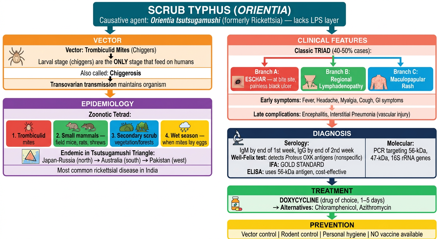
- Causative Agent - Orientia tsutsugamushi (lacks LPS layer, differs from Rickettsia)
- Vector - Trombiculid mites; only larval stage (chiggers) feed on humans → "Chiggerosis"; transovarian transmission
- Epidemiology - Zoonotic tetrad (mites + small mammals + scrub vegetation + wet season); Tsutsugamushi triangle; most common rickettsial disease in India
- Clinical Triad (40-50%) - Eschar + Regional lymphadenopathy + Maculopapular rash; early: fever/headache/myalgia; late: encephalitis/pneumonia
- Diagnosis - Serology (IgM wk 1, IgG wk 2), Weil-Felix (OXK), IFA (gold standard), ELISA (56-kDa), PCR
- Treatment - Doxycycline (DOC); alternatives: chloramphenicol, azithromycin
- Prevention - Vector/rodent control, hygiene; no vaccine available


Explain leptospirosis in form of a diagram in form of image from given data make it cplourful
A detailed, colorful medical flowchart/diagram explaining LEPTOSPIROSIS with all sections flowing top to bottom, clean white background, bold headers, connecting arrows between sections. SECTION 1 - TOP HEADER BOX (deep teal): "LEPTOSPIROSIS (Weil's Disease)" | Causative agent: Leptospira interrogans | 26 serogroups, 300+ serovars | L. biflexa = saprophyte (non-pathogenic) SECTION 2 - EPIDEMIOLOGY BOX (bright orange): Left side: "Mode of Transmission: ZOONOTIC (no human-to-human)" with bullet points: Indirect contact — water, moist soil, animal urine | Direct contact — urine/placenta of infected animals | Source: Rats, dogs, cattle, pigs Right side: "3 R's (Epidemiological Determinants):" with 3 colored bubbles: RODENTS (red) | RAINFALL (blue) | RICE FIELD (green) Bottom: "Seasonality: Rainy and post-monsoon | Risk groups: Agricultural workers, fishermen, sewer workers" India box (yellow): "India: Andaman hemorrhagic fever | Common serovars: L. Andamana, L. Pomona, L. Grippotyphosa, L. Hebdomadis, L. Javanica, L. Autumnalis, L. Canicola" SECTION 3 - PATHOGENESIS BOX (purple): Two phase arrows side by side: Phase 1 arrow (red): "SEPTICEMIC PHASE (Days 1-10)" → Entry via conjunctiva/oral mucosa/abraded skin → Bloodstream → Disseminates to brain, liver, lung, heart, kidney → Vascular damage to capillaries (hyaluronidase) Phase 2 arrow (blue): "IMMUNE PHASE (Days 10-30)" → Antibodies develop → Spirochetes disappear from blood → Antigen-antibody complexes in organs → Renal colonization: proximal tubular brush border → excreted in urine SECTION 4 - CLINICAL MANIFESTATIONS BOX (coral): Two columns with colored headers: Column A (green header): "MILD ANICTERIC FEBRILE ILLNESS (90%)" | Biphasic course | Stage 1 (Septicemic 3-10 days): Fever, Myalgia, Headache, Conjunctival suffusion, Abdominal pain, Pharyngeal erythema, Vomiting | Stage 2 (Immune 10-30 days): Meningitis, Uveitis, Optic neuritis, Chorioretinitis, Rash, Peripheral neuropathy | Isolation: Blood/CSF → then Urine | Susceptible to antibiotics Column B (red header): "WEIL'S DISEASE / Hepato-Renal-Hemorrhagic Syndrome (10%)" | Severe icteric illness | Stage 1: High-grade fever, Liver jaundice (5-10%), Hemorrhages (Pulmonary, Petechiae, GI, Conjunctival), Raised serum urea | Typical biphasic may be absent | Case fatality: 5-15% SECTION 5 - LABORATORY DIAGNOSIS BOX (dark navy blue): Three colored sub-boxes: Sub-box 1 (yellow): "MICROSCOPY" | Specimens: CSF, Blood (first 10 days), Urine (10-30 days) | Dark ground or phase contrast microscope | Silver impregnation staining | Spirally coiled bacilli with hooked ends (umbrella handle) Sub-box 2 (green): "CULTURE & ISOLATION" | 30°C for 4-6 weeks (slow growing) | Media: EMJH, Korthof's, Fletcher's | Animal inoculation: Hamsters and young guinea pigs Sub-box 3 (orange): "SEROLOGY" | Genus-specific: Macroscopic slide agglutination, Latex agglutination, ELISA, ICT | Serovar-specific: Microscopic Agglutination Test (MAT) | IgM appears early, peaks later | Molecular: PCR (16S/23S rRNA, IS1533 genes), PCR-RFLP, PFGE SECTION 6 - TREATMENT BOX (bright lime green): "TREATMENT" | Mild disease: Doxycycline / Ampicillin / Amoxicillin | Severe (Weil's): IV Penicillin G / Ceftriaxone | Prevention: Doxycycline prophylaxis, rodent control, protective gear, avoid contaminated water Use vibrant distinct colors for every section. Include small icons: a rat for epidemiology, a spiral bacterium for the causative agent, a kidney for Weil's disease, a microscope for lab diagnosis. All text clearly readable with large bold section headers.
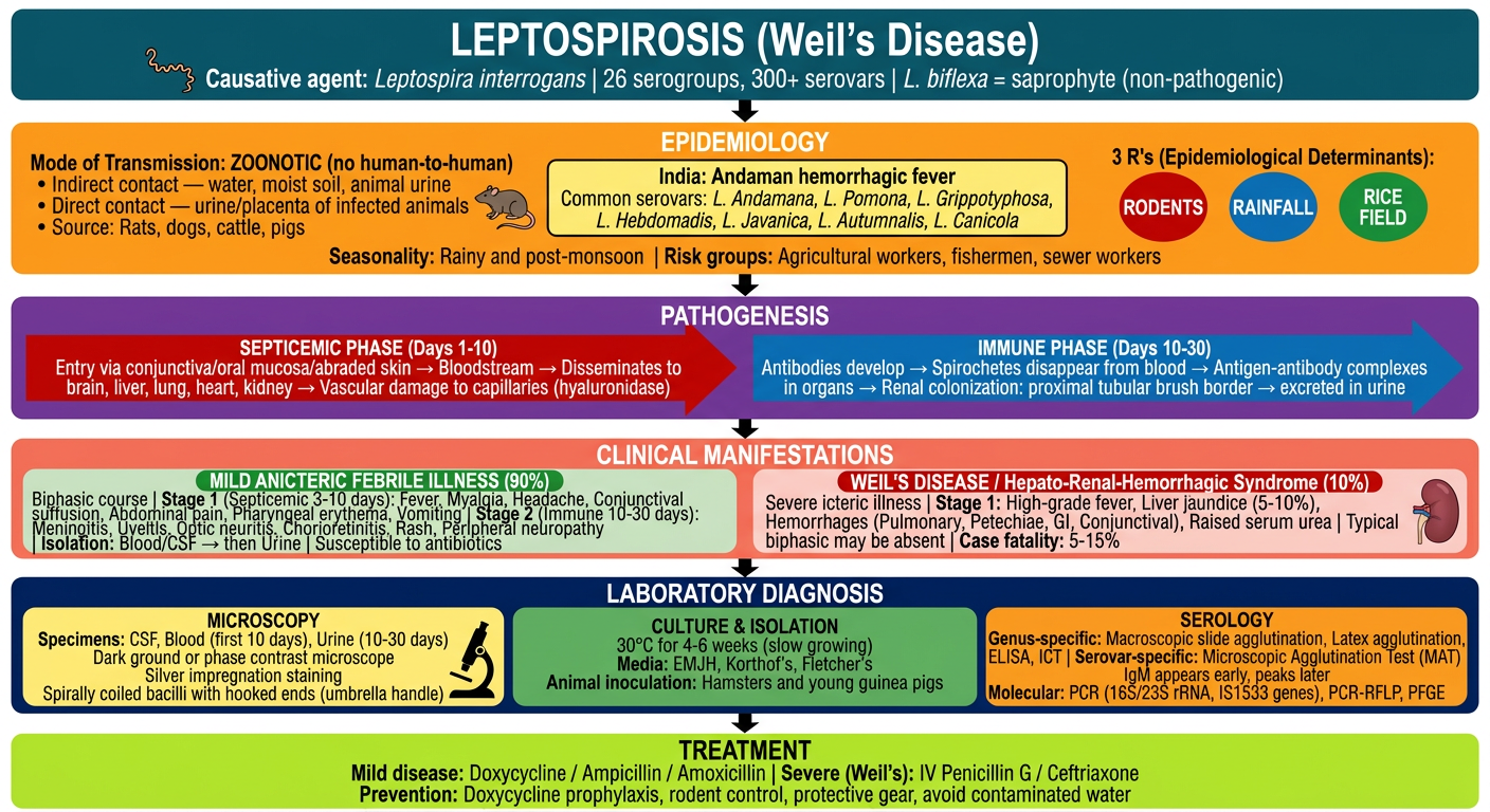
| Section | Key Points |
|---|---|
| Agent | L. interrogans (26 serogroups, 300+ serovars); L. biflexa = non-pathogenic |
| Epi (3 R's) | Rodents + Rainfall + Rice field; Zoonotic; no human-to-human spread |
| Pathogenesis | Phase 1 (Septicemic): entry via mucosa → bloodstream → organs |
| Clinical | Mild (90%): biphasic flu-like illness |
| Lab Dx | Microscopy (dark ground/silver stain) + Culture (EMJH/Korthof's, 30°C/4-6 wks) + MAT (gold standard serology) + PCR |
| Treatment | Mild: Doxycycline/Amoxicillin |
Make it less congested and remove inforamation about serogans
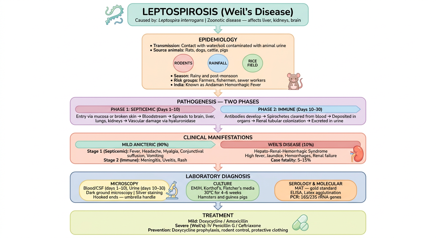
A clean, spacious, colorful medical flowchart for LEPTOSPIROSIS. White background. Large text. Plenty of white space between sections. Simple and easy to read. Flowing top to bottom with bold arrows connecting each section. BOX 1 - TOP (dark teal, wide): "LEPTOSPIROSIS" Caused by: Leptospira interrogans Zoonotic disease — affects liver, kidneys, and can cause death if untreated BOX 2 - (bright orange, wide): "EPIDEMIOLOGY" • Transmission: Contact with water/soil contaminated with animal urine • Source animals: Rats, dogs, cattle, pigs • 3 R's: Rodents | Rainfall | Rice field • Season: Rainy and post-monsoon • Risk groups: Farmers, fishermen, sewer workers • India: Known as Andaman Hemorrhagic Fever BOX 3 - (purple, wide, two side-by-side arrows inside): "PATHOGENESIS — TWO PHASES" Left arrow (red background): "PHASE 1: SEPTICEMIC (Days 1–10)" Entry via mucosa or broken skin → Bloodstream → Spreads to brain, liver, lungs, kidneys → Vascular damage via hyaluronidase Right arrow (blue background): "PHASE 2: IMMUNE (Days 10–30)" Antibodies develop → Spirochetes cleared from blood → Deposited in organs → Renal tubular colonization → Excreted in urine BOX 4 - (coral/salmon, wide, two columns inside): "CLINICAL MANIFESTATIONS" Left column (green header): "MILD ANICTERIC (90%)" Stage 1 (Septicemic): Fever, Headache, Myalgia, Conjunctival suffusion, Vomiting Stage 2 (Immune): Meningitis, Uveitis, Rash Right column (red header): "WEIL'S DISEASE (10%)" Hepato-Renal-Hemorrhagic Syndrome High fever, Jaundice, Hemorrhages (pulmonary, GI, conjunctival), Renal failure Case fatality: 5–15% BOX 5 - (navy blue, wide, three spaced sub-boxes): "LABORATORY DIAGNOSIS" Sub-box A (yellow): "MICROSCOPY" Specimens: Blood/CSF (days 1–10), Urine (days 10–30) Dark ground microscopy | Silver staining Hooked ends — umbrella handle appearance Sub-box B (green): "CULTURE" Media: EMJH, Korthof's, Fletcher's 30°C for 4–6 weeks Animal inoculation: Hamsters, guinea pigs Sub-box C (orange): "SEROLOGY & MOLECULAR" MAT — gold standard (serovar-specific) ELISA, Latex agglutination (genus-specific) PCR: 16S/23S rRNA genes BOX 6 - BOTTOM (bright lime green, wide): "TREATMENT" Mild: Doxycycline / Amoxicillin Severe (Weil's): IV Penicillin G / Ceftriaxone Prevention: Doxycycline prophylaxis, rodent control, protective clothing Style: Very spacious layout, large font, each box has plenty of padding, no text crowding, bold section titles, simple clean icons (rat icon near epidemiology, kidney icon near Weil's disease, microscope icon near lab diagnosis), vibrant distinct colors per section, wide boxes with breathing room.
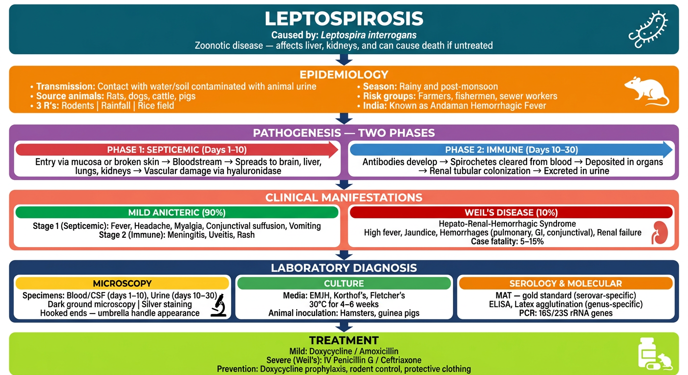
- No serogroup/serovar information - removed as requested
- More white space between each section so it's easier to read
- 6 clear sections flowing top to bottom: Agent → Epidemiology → Pathogenesis (2 phases) → Clinical Features → Lab Diagnosis → Treatment
- Distinct colors for each box so sections are easy to tell apart at a glance

Make it using pastel themed colours
A clean, spacious, PASTEL-themed medical flowchart for LEPTOSPIROSIS. White background. Soft pastel colors only throughout. Large readable text. Plenty of white space. Flowing top to bottom with soft arrows connecting each section. Same layout and content as described below but ALL colors must be soft pastels. BOX 1 - TOP (pastel teal/mint green header, dark text): "LEPTOSPIROSIS (Weil's Disease)" Caused by: Leptospira interrogans | Zoonotic disease — affects liver, kidneys, brain Small spiral bacteria icon on left BOX 2 - (pastel peach/light orange background, dark brown text): "EPIDEMIOLOGY" • Transmission: Contact with water/soil contaminated with animal urine • Source animals: Rats, dogs, cattle, pigs • 3 R's: three soft pastel bubbles — RODENTS (pastel red/pink) | RAINFALL (pastel blue) | RICE FIELD (pastel green) • Season: Rainy and post-monsoon • Risk groups: Farmers, fishermen, sewer workers • India: Known as Andaman Hemorrhagic Fever Small cute rat icon BOX 3 - (pastel lavender/soft purple background, two side-by-side arrow boxes inside): "PATHOGENESIS — TWO PHASES" Left arrow box (pastel rose/light pink): "PHASE 1: SEPTICEMIC (Days 1–10)" Entry via mucosa or broken skin → Bloodstream → Spreads to brain, liver, lungs, kidneys → Vascular damage via hyaluronidase Right arrow box (pastel sky blue): "PHASE 2: IMMUNE (Days 10–30)" Antibodies develop → Spirochetes cleared from blood → Deposited in organs → Renal tubular colonization → Excreted in urine BOX 4 - (pastel coral/light salmon background, two columns inside): "CLINICAL MANIFESTATIONS" Left column (pastel mint green header): "MILD ANICTERIC (90%)" Stage 1 (Septicemic): Fever, Headache, Myalgia, Conjunctival suffusion, Vomiting Stage 2 (Immune): Meningitis, Uveitis, Rash Right column (pastel red/rose header): "WEIL'S DISEASE (10%)" Hepato-Renal-Hemorrhagic Syndrome High fever, Jaundice, Hemorrhages, Renal failure Case fatality: 5–15% Small kidney icon BOX 5 - (pastel navy/soft steel blue background with light text, three spaced sub-boxes): "LABORATORY DIAGNOSIS" Sub-box A (pastel yellow): "MICROSCOPY" Blood/CSF (days 1–10), Urine (days 10–30) Dark ground microscopy | Silver staining Hooked ends — umbrella handle Microscope icon Sub-box B (pastel green): "CULTURE" EMJH, Korthof's, Fletcher's media 30°C for 4–6 weeks Hamsters and guinea pigs Sub-box C (pastel orange): "SEROLOGY & MOLECULAR" MAT — gold standard ELISA, Latex agglutination PCR: 16S/23S rRNA genes BOX 6 - BOTTOM (pastel lime/soft yellow-green background, dark text): "TREATMENT" Mild: Doxycycline / Amoxicillin Severe (Weil's): IV Penicillin G / Ceftriaxone Prevention: Doxycycline prophylaxis, rodent control, protective clothing Pill icon Overall style: ALL pastel palette — no bright or saturated colors anywhere. Soft muted tones. Generous padding and white space inside each box. Clean sans-serif font. Bold section titles in slightly darker shade of the pastel. Rounded corners on all boxes. Soft pastel arrows between sections.
- Pastel mint - title header
- Pastel peach - epidemiology
- Pastel lavender - pathogenesis (with soft pink & sky blue for the two phases)
- Pastel coral - clinical manifestations
- Soft steel blue - lab diagnosis (with pastel yellow, green & orange sub-boxes)
- Pastel lime - treatment


Explain pathogenesis of hiv form given data in form of flowchart and diagram in form of image use pastel themed colours
A clean, spacious, PASTEL-themed medical flowchart and diagram explaining the PATHOGENESIS OF HIV. White background. Soft pastel colors only. Large readable text. Generous white space between sections. Flowing top to bottom with soft rounded arrows. All boxes have rounded corners and gentle pastel fills. BOX 1 - TOP HEADER (pastel teal/mint, dark teal text): "PATHOGENESIS OF HIV" HIV enters via mucosa or broken skin → targets CD4+ T cells BOX 2 - (pastel peach background, dark text): "MODE OF TRANSMISSION" Three soft pastel sub-bubbles side by side: • SEXUAL (pastel pink) — 75% worldwide | Heterosexual commonest | Anal intercourse = higher risk | Risk: 0.1–1% per coitus • BLOOD TRANSFUSION (pastel red/rose) — 5% cases but HIGHEST risk (90–95%) • PERINATAL (pastel lavender) — Mother to fetus 20–40% | Maximum risk: recently infected mother or mother with AIDS | Breast milk also transmits • PERCUTANEOUS/MUCOSAL (pastel yellow) — Needle stick, drug abuse, tattooing BOX 3 - (pastel lavender/soft purple background): "RECEPTOR ATTACHMENT" Two columns inside: Left (pastel blue box): "MAIN RECEPTOR" gp120 binds CD4 receptor on helper T cells Also on: Monocytes, Macrophages, Langerhans cells, Astrocytes, Keratinocytes, Glial cells Right (pastel green box): "CO-RECEPTORS" CXCR4 — on T lymphocytes CCR5 — on macrophage lineage cells DC-SIGN — dendritic cell lectin receptor (facilitates transport to lymphoid organs) ⭐ CCR5 Delta 32 Mutation: Homozygous = completely resistant | Heterozygous = progression to AIDS delayed BOX 4 - (pastel sky blue background): "REPLICATION STEPS" — horizontal step-by-step flow with small soft arrows: Step 1 (pastel yellow pill): gp120 binds CD4 + co-receptor → Step 2 (pastel pink pill): gp41 mediates fusion with host cell membrane → Step 3 (pastel green pill): RNA genome enters cell | Reverse transcriptase makes DNA-RNA hybrid → Step 4 (pastel orange pill): ssDNA → dsDNA (RNase H degrades RNA) → Step 5 (pastel lavender pill): Pre-integration complex forms (linear dsDNA + gag matrix protein + integrase) — transported to nucleus → Step 6 (pastel blue pill): Viral integrase integrates dsDNA into host chromosome → called PROVIRUS → Step 7 (pastel rose pill): LATENCY established — HIV different from other latent viruses as it can replicate even in latent state and infect neighboring cells BOX 5 - (pastel coral/salmon background): "DISEASE PROGRESSION — NATURAL COURSE (5 stages)" Five sequential boxes connected by arrows: Stage 1 box (pastel red-pink): "ACUTE HIV / ACUTE RETROVIRAL SYNDROME" HIV carried to lymph nodes → multiplication in T cells → Primary viremia → Flu-like illness (50–75% patients, 3–6 weeks after infection) → Significant drop in CD4+ T cells Stage 2 box (pastel yellow): "ASYMPTOMATIC STAGE (CLINICAL LATENCY)" Adequate immune response within 1 month | CD8 T cells + neutralizing antibodies | Viremia drops, CD4 count normalizes | Clinical latency (NOT microbiological) | Virus persists in lymph nodes | Lasts months to 30 years | Once broken: rapid progression, death in 2 years if untreated Stage 3 box (pastel green): "PERSISTENT GENERALIZED LYMPHADENOPATHY (PGL)" 25–30% of infected asymptomatic people | Enlarged lymph nodes >1 cm in 2+ non-contiguous sites | Persists ≥3 months | Must distinguish from lymphoma Stage 4 box (pastel orange): "SYMPTOMATIC HIV (AIDS-RELATED COMPLEX)" CD4 falling | Constitutional symptoms: Unexplained diarrhea >1 month, weight loss >10%, fatigue, night sweats | Mild opportunistic infections: oral thrush Stage 5 box (pastel purple/mauve): "AIDS" CD4 <200 cells/μL | High viral load | Lymphoid tissue replaced by fibrous tissue | Opportunistic infections (PCP, Cryptococcus, CMV, MAC, HSV, lymphoma) | CNS lymphoma, HIV encephalopathy | Death if untreated BOX 6 - BOTTOM (pastel lime green, dark text): "TYPICAL PROGRESSORS (80–90%)" Median survival ~10 years without ART Rapid progressor: AIDS in 2–3 years (5–10%) Long-term non-progressor (LTNP): >10 years, CD4 >500, VL <5000 Elite controller: CD4 stable, VL <50 copies/mL Style: ALL soft pastel palette. Rounded boxes. Generous padding. Clean sans-serif font. Bold section headers in slightly darker shade of each pastel. Soft pastel arrows. Very readable and well-spaced. No crowding.
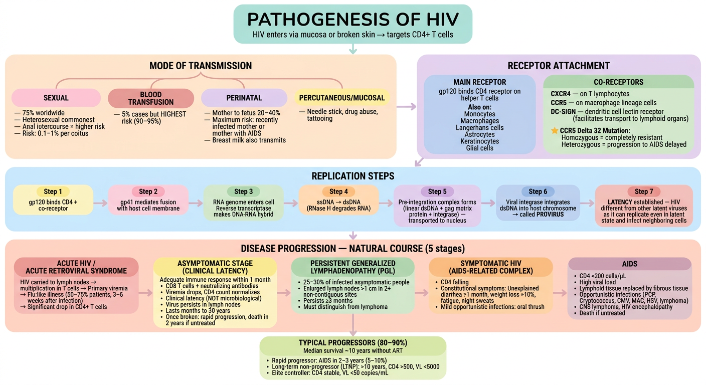
| Section | Key Content |
|---|---|
| Transmission | Sexual (75%) > Blood transfusion (5%, highest risk 90-95%) > Perinatal (20-40%) > Percutaneous |
| Receptor Attachment | gp120 → CD4 receptor + co-receptors (CXCR4 / CCR5) |
| Replication | gp120 binds → fusion → RNA enters → Reverse transcriptase → dsDNA → Pre-integration complex → PROVIRUS (integrase) → Latency |
| Stage 1 | Acute retroviral syndrome - flu-like illness, CD4 drops |
| Stage 2 | Asymptomatic/Clinical latency - lasts months to 30 years |
| Stage 3 | PGL - enlarged lymph nodes ≥3 months |
| Stage 4 | Symptomatic HIV (ARC) - diarrhea, weight loss, oral thrush |
| Stage 5 | AIDS - CD4 <200, opportunistic infections, death |


Make a flowchart about lab diagonsis of hiv in form of image in pastel themed colours
A clean, spacious, PASTEL-themed medical flowchart for LABORATORY DIAGNOSIS OF HIV. White background. Soft pastel colors only throughout. Large readable text. Generous white space. Rounded corners on all boxes. Flowing top to bottom with soft pastel arrows. BOX 1 - TOP HEADER (pastel teal/mint background, dark teal bold text): "LABORATORY DIAGNOSIS OF HIV" Subtitle: "Window Period: Antibodies appear 3–12 weeks after infection | IgG = consistent & long-lasting (detected by most assays)" Small virus icon on left SECTION A - (pastel peach/light orange background, dark text, bold header): "SPECIFIC TESTS FOR HIV" Three columns side by side connected below: Column 1 (pastel yellow rounded box): "1. SCREENING TESTS" (Antibody Detection) • ELISA — takes 2–3 hours - 3rd generation: detects HIV antibodies using recombinant/synthetic peptides - 4th generation: detects BOTH HIV antibodies + p24 antigen → reduces window period considerably - Types: Indirect ELISA, Competitive ELISA, Sandwich ELISA • Rapid/Simple Test — takes <30 minutes - Dot blot assay, Dipstick test - Immunochromatography (ICT / lateral flow) - Particle agglutination (latex, gelatin, RBCs) - Dipstick/Combtest ⭐ NACO recommends: Sensitivity ≥99.5%, Specificity ≥98% ⚠ Result of single screening test NEVER used as final interpretation — always confirm! Column 2 (pastel lavender/soft purple rounded box): "2. SUPPLEMENTAL TESTS" (Antibody Detection — Highly Specific) • Western Blot Assay (most commonly used, recommended by NACO) - Detects individual antibodies to antigenic fragments separately - Antibodies to: gag products (p55, p40, p24, p18), pol products (p65/66, p55/51, p31), env products (gp120, gp160, gp41) - Antigen-antibody complexes appear as DISTINCT BANDS on nitrocellulose strip - WHO criteria: ≥2 envelope bands (gp120, gp160 or gp41) ± gag/pol bands - CDC criteria: ≥2 bands from p24, gp120, gp160, gp41 • Line Immunoassay (LIA) Column 3 (pastel rose/light pink rounded box): "3. CONFIRMATORY TESTS" (Antigen/Nucleic Acid Detection) • p24 Antigen Detection - Detectable 12–26 days after infection, lasts 3–4 weeks - Elevated again in late/advanced AIDS - Less sensitive (antibody-antigen complex removes it from blood) - Uses: Diagnosis during window period, late AIDS/CNS disease, pediatric HIV, monitor progression, resolve equivocal Western blot - Antigen dissociation assay = better sensitivity • Viral RNA Detection — GOLD STANDARD ⭐ - RT-PCR (Reverse Transcriptase PCR) - Branched DNA assay - NASBA (Nucleic Acid Sequence-Based Amplification) - Real-time RT-PCR — for viral load estimation - Detects HIV 10–14 days after infection (earliest of all methods) - Most sensitive & specific | Best for window period | Viral load monitoring | Typing HIV-1 vs HIV-2 | Drug resistance detection • DNA PCR - Detects proviral DNA - Best for pediatric HIV diagnosis - Differentiates latent from active infection - Useful during window period • Viral Culture (Co-cultivation) - PBMCs co-cultured with donor PBMCs - Detected by viral RNA or antigen in culture - Time consuming (6+ weeks), expensive, research only SECTION B - (pastel sky blue background, dark text): "NON-SPECIFIC IMMUNOLOGICAL TESTS" Four soft pastel sub-bubbles in a row: • CD4+ T cell count (pastel green) — measured by flow cytometry | Useful for: assessing OI risk, starting ART, monitoring ART response • Hypergammaglobulinemia (pastel yellow) — Neopterin ↑ | β2-microglobulin ↑ | Soluble IL-2 receptor ↑ • Altered CD4:CD8 ratio (pastel orange) — CD4:CD8 ratio falls (normal >1, in AIDS <1) BOX BOTTOM - (pastel lime green, dark text): "IMPORTANT NOTES" • Consent + Confidentiality mandatory before testing (3Cs: Consent, Counseling, Confidentiality) • Positive result should NOT be written on report form — use "HIV positive" only in designated records • Counseling before and after test | Motivate to inform spouse/family Style: ALL soft pastel palette. Generous padding inside every box. Bold headers in slightly darker shade. Clean sans-serif font. Soft rounded arrows connecting all sections. No crowding. Very readable.
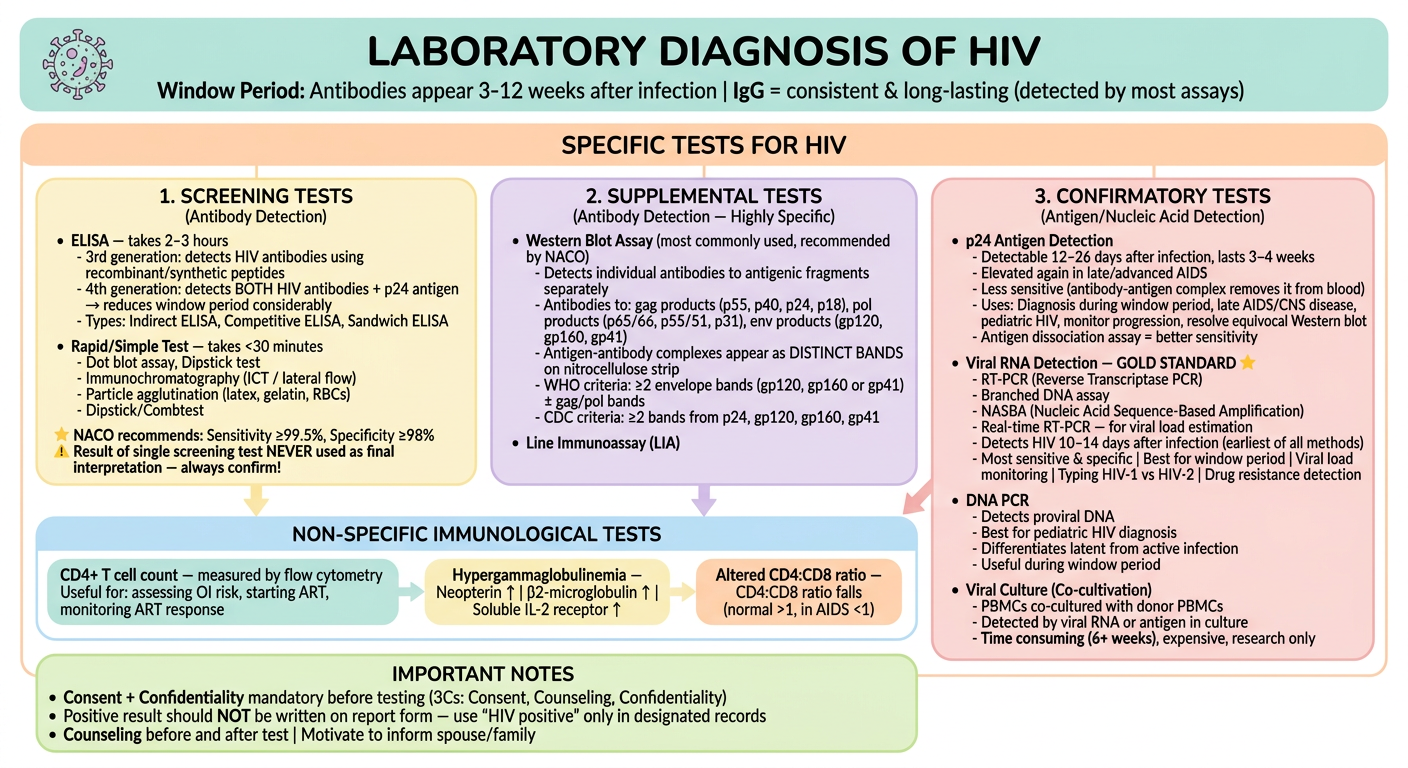
| Test Category | Key Points |
|---|---|
| Screening (Antibody) | ELISA (2–3 hrs) — 3rd gen (antibodies) / 4th gen (antibodies + p24 antigen, shorter window) |
| Supplemental (Antibody) | Western Blot — NACO recommended, detects bands against gag/pol/env proteins |
| Confirmatory (Antigen/NAT) | p24 Ag — detectable day 12–26 |
| Non-specific | CD4+ T cell count (flow cytometry) |
| Ethics | 3Cs — Consent, Confidentiality, Counseling mandatory before testing |


Explain naco stragegy in form of flowchart from given data in form of image make it colourful use pastel themed colurs
A clean, spacious, PASTEL-themed medical flowchart for "NACO STRATEGY FOR HIV DIAGNOSIS" (National AIDS Control Organization, India). White background. Soft pastel colors only. Large readable text. Rounded corners. Generous white space. Bold headers. TOP HEADER BOX (pastel teal/mint, bold dark teal text): "NACO STRATEGY FOR HIV DIAGNOSIS" Subtitle: "4 Strategic Algorithms | First test = HIGH SENSITIVITY | Second & Third tests = HIGH SPECIFICITY | Three tests use different principles/antigens | Same kit NOT repeated" Below header, four large side-by-side strategy boxes (each a different pastel color), then below each box show the decision flowchart for that strategy: ━━━━━━━━━━━━━━━━━━━━━━━━━━━━━━━━ STRATEGY I BOX (pastel yellow background, amber bold header): "STRATEGY I" Purpose: Transfusion & Transplantation Safety (Screening of blood donors in blood banks) Flowchart below: [A1 — Single Screening Test] ↓ ┌─────┴─────┐ REACTIVE NON-REACTIVE ↓ ↓ [Unit of [Report Blood is Negative — Destroyed] Blood Safe] ━━━━━━━━━━━━━━━━━━━━━━━━━━━━━━━━ STRATEGY IIA BOX (pastel blue background, blue bold header): "STRATEGY IIA" Purpose: Sentinel Surveillance (Estimate prevalence of HIV infection) Method: UAT — Unlinked Anonymous Testing (Blood specimens decoded of personal identifiers, NO informed consent) Flowchart below: [A1 — First Screening Test] ↓ ┌─────┴─────┐ A1 REACTIVE A1 NON-REACTIVE ↓ ↓ [A2 — Second [Report Screening Test] Negative] ↓ ┌──┴──┐ A2+ A2− ↓ ↓ Report Report Positive Negative ━━━━━━━━━━━━━━━━━━━━━━━━━━━━━━━━ STRATEGY IIB BOX (pastel lavender/soft purple background, purple bold header): "STRATEGY IIB" Purpose: Diagnosis of HIV/AIDS in SYMPTOMATIC patients Flowchart below: [A1 — First Screening Test] ↓ ┌─────┴─────┐ A1 REACTIVE A1 NON-REACTIVE ↓ ↓ [A2 — Second [Report Screening Test] Negative] ↓ ┌──────┴──────┐ A1+ A2+ A1+ A2− ↓ ↓ Report [A3 — Third Positive Screening Test] with post- ↓ test ┌────┴────┐ counseling A3+ A3− ↓ ↓ Report Indeterminate Positive (Repeat after 14–28 days / send for WB or RT-PCR) ━━━━━━━━━━━━━━━━━━━━━━━━━━━━━━━━ STRATEGY III BOX (pastel rose/light pink background, rose bold header): "STRATEGY III" Purpose: Diagnosis of ASYMPTOMATIC HIV patients, Antenatal screening, Pre-surgery screening Flowchart below: [A1 — First Screening Test] ↓ ┌─────┴─────┐ A1 REACTIVE A1 NON-REACTIVE ↓ ↓ [A2 — Second [Report Screening Test] Negative] ↓ A1+ A2+ ↓ [A3 — Third Screening Test] ↓ ┌──────┴──────┐ A1+A2+A3+ A1+A2+A3− or A1+A2−A3+ ↓ ↓ Report Indeterminate Positive Repeat after 14–28 days with post- Send to reference center test Western Blot or RT-PCR counseling ━━━━━━━━━━━━━━━━━━━━━━━━━━━━━━━━ BOTTOM NOTE BOX (pastel lime green, dark text): "IMPORTANT NOTES" • ⭐ CONSENT is mandatory before testing (EXCEPT Strategy IIA — UAT) • Supplemental/confirmatory tests ONLY when screening results are equivocal/indeterminate • Indeterminate (Strategy IIB & III): Repeat test after 14–28 days; send to reference center for Western Blot or RT-PCR confirmation Style: ALL soft pastel palette. Generous padding. Clean sans-serif font. Bold section headers. Soft rounded arrows in flowcharts. Green = positive/report negative path. Rose/red = reactive path. Very readable. No crowding. Well spaced layout.
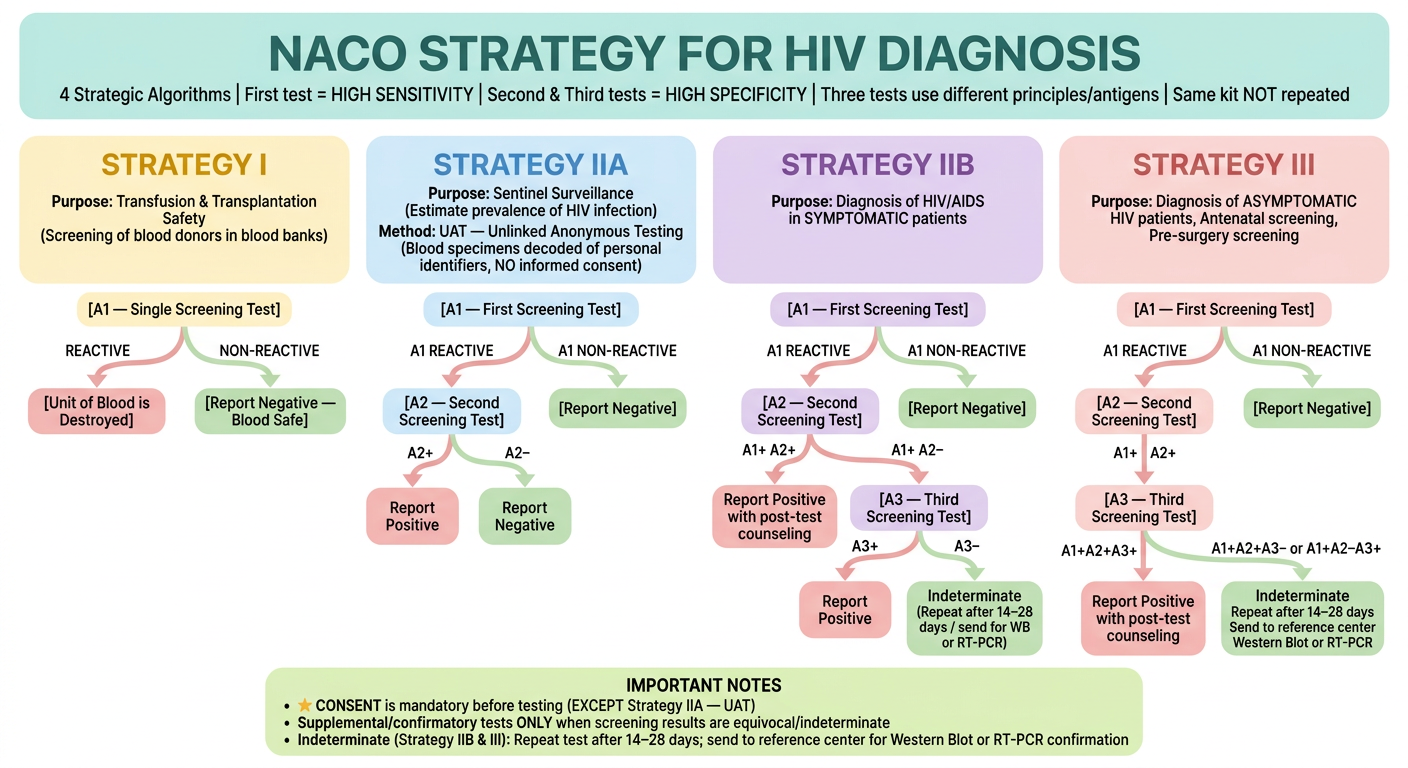
| Strategy | Purpose | Format | Key Point |
|---|---|---|---|
| Strategy I | Blood bank / transfusion safety | 1 test only | Reactive → destroy blood unit |
| Strategy IIA | Sentinel surveillance | 2 tests | UAT method — no consent needed, samples decoded |
| Strategy IIB | Symptomatic patients | 2–3 tests | A1+A2+ = report positive |
| Strategy III | Asymptomatic / antenatal / pre-surgery | 3 tests | ALL three must be reactive to report positive |
- 1st test = high sensitivity | 2nd & 3rd = high specificity
- Never repeat the same kit
- Supplemental/confirmatory tests only for equivocal results
- Indeterminate in IIB or III → repeat after 14–28 days or send for Western Blot / RT-PCR
- Consent mandatory for all except Strategy IIA (UAT)


Explain regarding denguw e with respect to given data in form of flowchart in form of image use pastel coloured theme
A clean, spacious, PASTEL-themed medical flowchart for DENGUE. White background. Soft pastel colors only. Large readable text. Rounded corners on all boxes. Generous white space. Bold headers. Flowing top to bottom with soft pastel arrows. BOX 1 - TOP HEADER (pastel teal/mint, bold dark teal text): "DENGUE FEVER" Vector: Aedes mosquito | Cause: Dengue virus (4 serotypes: DEN-1 to DEN-4) | Endemic: 100+ countries, 2.5 billion at risk | Tropical Southeast Asia & Western Pacific highest risk India: DEN-1 and DEN-2 widespread | Max cases: West Bengal, Tamil Nadu, Punjab, Kerala, Delhi BOX 2 - (pastel peach/light orange background): "PATHOGENESIS" Two columns inside: Left column (pastel yellow box): "PRIMARY INFECTION" First-time infection with any one serotype → Neutralizing antibodies produced (protective, lifelong against that serotype, short-term cross-protection against others) → Mild/self-limiting disease Right column (pastel rose box): "SECONDARY INFECTION (Different serotype)" NON-NEUTRALIZING antibodies (from 1st infection) are heterotypic → Instead of neutralizing the 2nd serotype, they ENHANCE entry into mononuclear cells → Release of cytokines → ANTIBODY DEPENDENT ENHANCEMENT (ADE) → More severe disease ⭐ ADE most severe: Serotype 1 followed by Serotype 2 ⭐ Serotype 2 = most dangerous overall BOX 3 - (pastel lavender background): "CLINICAL CLASSIFICATIONS" Two sub-sections side by side: Left (pastel blue box, bold header): "TRADITIONAL WHO 1997 CLASSIFICATION" Three downward-pointing boxes: Box A (pastel yellow): "1. DENGUE FEVER (DF)" Abrupt high fever (biphasic / break-bone / saddle-back fever) Severe frontal headache | Retro-orbital pain Maculopapular rash over chest and upper limbs Muscle and joint pain | Lymphadenopathy Loss of appetite, nausea, vomiting Box B (pastel orange): "2. DENGUE HEMORRHAGIC FEVER (DHF)" All DF features PLUS: High-grade continuous fever | Hepatomegaly Thrombocytopenia (platelet <1 lakh/mm³) Raised hematocrit (packed cell volume) by 20% Hemorrhages — Positive tourniquet test (>20 petechiae per sq inch in cubital fossa) | Spontaneous bleeding from skin, nose, mouth, gums Box C (pastel red/rose): "3. DENGUE SHOCK SYNDROME (DSS)" All DHF features PLUS shock: Rapid and weak pulse Narrow pulse pressure (<20 mmHg) or hypotension Cold and clammy skin | Restlessness Right (pastel green box, bold header): "WHO 2009 CLASSIFICATION" Two downward boxes: Box D (pastel mint): "DENGUE ± WARNING SIGNS" Probable Dengue: Live in/travel to endemic area + Fever + 2 of: Nausea/vomiting, Rash, Aches and pains, Tourniquet test positive, Leukopenia, Any warning sign Warning Signs (require strict observation): Abdominal pain or tenderness | Persistent vomiting | Clinical fluid accumulation | Mucosal bleed | Lethargy/restlessness | Liver enlargement >2 cm | Lab: Increase in HCT with rapid decrease in platelet count Box E (pastel coral): "SEVERE DENGUE" 1. Severe plasma leakage → Shock (DSS) | Fluid accumulation with respiratory distress 2. Severe bleeding (hemodynamic instability, may need transfusion) 3. Severe organ involvement: Liver AST/ALT ≥1000 IU/mL | CNS: Impaired consciousness | Heart and other organs BOX 4 - (pastel sky blue background): "FACTORS DETERMINING OUTCOME" Three pastel bubbles in a row: • INFECTING SEROTYPE (pastel pink) — Type 2 most dangerous • SEQUENCE OF INFECTION (pastel yellow) — Serotype 1 → Serotype 2 = most severe (DHF/DSS more common) • AGE (pastel green) — Children <12 years more prone to DHF and DSS BOX 5 - BOTTOM (pastel lime green, dark text): "DENGUE IN PREGNANCY" Perinatal transmission can occur | Peripartum infection → newborn: fever, thrombocytopenia, ascites, pleural effusions | Typically in first week of life Style: ALL soft pastel palette. Rounded boxes. Generous padding. Clean sans-serif font. Bold section headers in slightly darker shade. Soft arrows between sections. Very readable. Well spaced. No crowding.
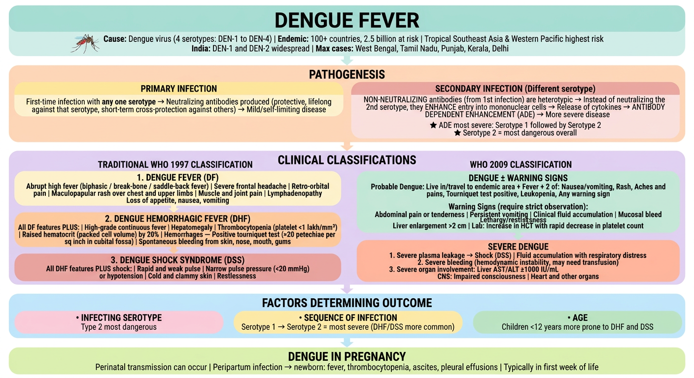
| Section | Key Points |
|---|---|
| Vector/Agent | Aedes mosquito |
| Pathogenesis | Primary = mild |
| DF (1997) | Biphasic/saddle-back fever, rash, retro-orbital pain, myalgia |
| DHF (1997) | DF + thrombocytopenia + raised hematocrit + hemorrhages (tourniquet test +ve) |
| DSS (1997) | DHF + shock (narrow pulse pressure, cold clammy skin) |
| WHO 2009 | Dengue ± warning signs → Severe dengue (plasma leakage / severe bleed / organ failure) |
| Outcome factors | Serotype 2 most dangerous |
| Pregnancy | Perinatal transmission possible — newborn: fever, thrombocytopenia, effusions |


Explain according to given data about dengue in form of flowchart in image form in shades of pink colour use images where possiblde
A clean, spacious, PINK-SHADED medical flowchart for LABORATORY DIAGNOSIS, TREATMENT & PREVENTION OF DENGUE. All shades of pink — from light blush to hot pink to rose to dusty pink to magenta. White background. Large readable text. Rounded corners. Generous white space. Bold headers. Decorative medical icons throughout. Flowing top to bottom. TOP HEADER BOX (deep rose/hot pink background, white bold text): "DENGUE — LAB DIAGNOSIS, TREATMENT & PREVENTION" Small illustrated Aedes mosquito icon on left. Small blood drop icon on right. ━━━━━━━━━━━━━━━ SECTION 1 — (light blush pink background, deep rose bold header): "🔬 LABORATORY DIAGNOSIS" Subtitle: "Outlined similar to other arboviruses" Three columns inside with pink-shade sub-boxes: COLUMN A (pale pink box with rose header): "NS1 ANTIGEN DETECTION" 🧪 Test formats: ELISA and ICT • Detectable from DAY 1 of fever • Remains positive up to 18 days • Highly specific — differentiates between flaviviruses • Can be specific to different dengue serotypes • Useful for EARLY detection of infection COLUMN B (medium pink box with magenta header): "ANTIBODY DETECTION" 🩸 Primary Infection: • Slow, low-titer response • IgM appears after 5 days of fever • IgM detectable within 90 days • IgG detectable at low titer by 14–21 days, then slowly rises 🩸 Secondary Infection: • IgG rises rapidly • IgG cross-reactive with many flaviviruses → false positives (yellow fever, JE) • IgM titer significantly low or undetectable 🩸 Past Infection: • Low IgG detectable for 60+ years even without symptoms ⭐ MAC-ELISA (IgM Antibody Capture ELISA): • RECOMMENDED serological test in India • Kits supplied by NIV, Pune • Principle: Double sandwich ELISA — captures human IgM on microtiter plate using anti-human IgM antibody → adds all 4 dengue serotype envelope protein antigens → avidin-biotin complex (ABC) enhances sensitivity • Limitation: Cross-reactivity with other flaviviruses 🔬 Neutralization Tests: • Plaque reduction test, microneutralization test • Most specific serologic tests but cumbersome COLUMN C (dusty rose box with deep pink header): "VIRAL DETECTION" 🧬 Virus Isolation: • Detectable in blood from day −1 to +5 days of symptoms • Inoculation into mosquito cell lines (C6/36, AP61) or mouse • Research purpose only, reference centers 🧬 Molecular Method (RT-PCR): • Detection of viral RNA (3'-UTR region) by Real-time RT-PCR • Most sensitive (80–90%) and specific (95%) • Detects serotypes + viral load quantification • Viral RNA detectable from day −1 to +5 days • Negative PCR after day 5 = "indeterminate" → needs serological confirmation after day 5 🧬 Genotype Detection: • Each serotype comprises several genotypes • Detected by molecular typing • Total 13 genotypes: DENV-1 (3), DENV-2 (2), DENV-3 (4), DENV-4 (4) 💉 Rapid Diagnostic Tests (RDT): • ICT for IgM antibodies or NS1 antigen • Poor sensitivity and specificity • Govt of India 2016: Positive RDT = "probable diagnosis" — MUST be confirmed by ELISA ━━━━━━━━━━━━━━━ SECTION 2 — (medium rose/salmon-pink background, dark rose bold header): "💊 TREATMENT" Small pill icon on left. • NO specific antiviral therapy available • Treatment is SYMPTOMATIC and SUPPORTIVE: ✅ Replacement of plasma losses ✅ Correction of electrolyte and metabolic disturbances ✅ Platelet transfusion if needed ━━━━━━━━━━━━━━━ SECTION 3 — (hot pink background, white bold header): "🛡️ PREVENTION" Two columns: Left column (light pink box): "DENGUE VACCINE — CYD-TDV (Dengvaxia)" 💉 Licensed for human use since 2015 | Developed by Sanofi Pasteur • Chimeric Yellow Fever-Dengue Live-Attenuated Tetravalent Dengue Vaccine (CYD-TDV) • Uses live attenuated Yellow Fever 17D virus as vector with all 4 dengue serotype target genes integrated by recombinant technique • Age: 9–45 years • Schedule: 3 injections of 0.5 mL subcutaneously at 6-month intervals • Efficacy: ~80% against hospitalized dengue illness • WHO recommends in high-burden countries (seroprevalence >70%) • ⚠️ WHO: Use ONLY in previously infected individuals — if given to dengue-naive person → HIGHER RISK of severe dengue on subsequent DENV infection • Contraindications: Allergic reactions, immunodeficient individuals (e.g. HIV), pregnant and breastfeeding women • Currently approved: Mexico, Philippines, Brazil, Indonesia, Thailand, Singapore • India: NOT yet available due to safety issues • Indian trials ongoing: TetraVax-DV (live-attenuated), DSV4 (tetravalent subunit vaccine) Right column (pale pink box): "OTHER PREVENTION" 🦟 Mosquito control measures (vector control) • Elimination of Aedes breeding sites • Insecticides, larvicides • Personal protection: nets, repellents, full-sleeve clothing ━━━━━━━━━━━━━━━ BOTTOM NOTE BOX (blush pink, dark rose text): "KEY TIMELINE SUMMARY" Day −1 to +5: Virus isolation / PCR positive | NS1 Ag: Day 1 to Day 18 | IgM: from Day 5 | IgG (primary): Day 14–21 | IgG (secondary): rises rapidly from day 1 | IgG (past): detectable 60+ years Style: ALL shades of pink from pale blush to hot pink to rose to magenta. Rounded boxes. Generous padding. Bold pink-shade headers. Soft pink arrows. Small icons: mosquito, blood drop, microscope, syringe, pill, DNA helix. Very readable. Well spaced. No crowding.
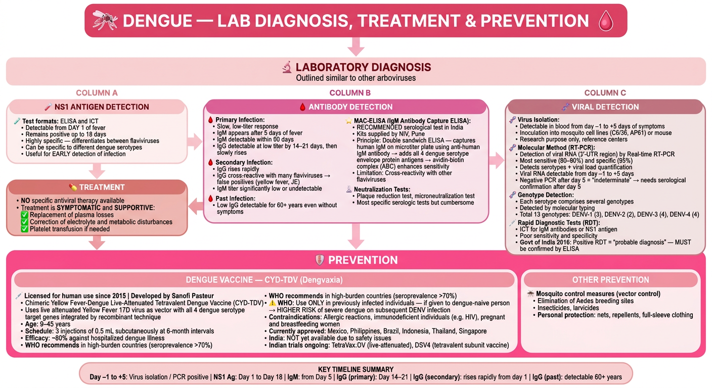
| Test | When Positive | Key Point |
|---|---|---|
| NS1 Antigen | Day 1 – Day 18 | Early detection, highly specific |
| Viral RNA (RT-PCR) | Day −1 to +5 | Most sensitive (80–90%), gold standard early |
| IgM (MAC-ELISA) | From Day 5 | Recommended in India (NIV Pune kits) |
| IgG | Primary: Day 14–21 / Secondary: rises rapidly | Past infection detectable 60+ years |
| RDT | Rapid but poor sensitivity | Positive RDT = probable only, confirm by ELISA |
- Age 9–45 yrs | 3 doses at 6-month intervals
- ⚠️ Only for previously infected individuals - naive persons at higher risk of severe dengue
- Not available in India yet due to safety concerns

Explain above data about malaria in form of flowchart in form of image use image where possible.use pastel shades of gren and yellow for colours
A clean, spacious medical flowchart for MALARIA — COMPLICATIONS, CHRONIC COMPLICATIONS & SPECIAL SITUATIONS. All colors in soft pastel shades of GREEN and YELLOW only — from pale mint to sage green to soft lime, and from cream yellow to butter yellow to golden yellow. White background. Large readable text. Rounded corners. Generous white space. Bold headers. Small medical icons where appropriate. TOP HEADER BOX (sage green background, dark forest green bold text): "MALARIA — COMPLICATIONS, CHRONIC FORMS & SPECIAL SITUATIONS" Small illustrated mosquito icon on left. Small red blood cell icon on right. Note: "P. falciparum = most acute & severe | More complications than other malaria" ━━━━━━━━━━━━━━━ SECTION 1 — (pale yellow background, olive green bold header): "⚡ ACUTE COMPLICATIONS OF P. FALCIPARUM MALARIA" Small brain icon on top right. Eight complication boxes arranged in a 2x4 grid, each in alternating pastel mint green and pastel yellow: Box 1 (mint green): "🧠 CEREBRAL MALARIA" Most dangerous complication of falciparum malaria Plugging of brain capillaries by sequestered parasitized RBCs → vascular occlusion → cerebral anoxia (Details in Chapter 75) Box 2 (pale yellow): "🌡️ PERNICIOUS MALARIA" Characterized by: Blackwater fever, algid malaria, septicemic malaria Box 3 (mint green): "🩸 BLACK WATER FEVER" Sudden intravascular hemolysis → hemoglobinuria → dark urine Occurs after quinine treatment in P. falciparum-infected patients Autoimmune mechanism: Antibodies vs parasitized & quininized RBCs → immunocomplex → complement-mediated destruction of both parasitized AND non-parasitized RBCs ⚠️ Triangle warning icon Box 4 (pale yellow): "❄️ ALGID MALARIA" Cold, clammy skin Hypotension Peripheral circulatory failure Profound shock → DIC Box 5 (mint green): "🦠 SEPTICEMIC MALARIA" High-grade fever Dissemination of parasite to various organs → Multi-organ failure Box 6 (pale yellow): "🫁 PULMONARY EDEMA / ARDS" Severe falciparum in adults Non-cardiogenic pulmonary edema Often aggravated by over-hydration Does NOT respond to antimalarials Mortality >80% Box 7 (mint green): "📉 HYPOGLYCEMIA" Poor prognosis marker Problematic in children and pregnant women Following quinine therapy Due to: Erythrocyte sequestration in renal microvasculature Box 8 (pale yellow): "🫘 RENAL FAILURE" Acute tubular necrosis More common in adults than children Due to erythrocyte sequestration in renal microvasculature Three more boxes below: Box 9 (sage green): "💉 BLEEDING / DIC" Significant bleeding — gums, nose, intestine With or without evidence of disseminated intravascular coagulation Immune complex involvement Box 10 (butter yellow): "🟡 SEVERE JAUNDICE" More common in adults than children Hemolysis + hepatocyte injury + cholestasis Box 11 (mint green): "🔴 SEVERE NORMOCHROMIC NORMOCYTIC ANEMIA" Hematocrit <15% or hemoglobin <5 g/dL With parasitemia >10⁵/μL (>2%) Box 12 (pale yellow): "⚗️ ACIDOSIS" Accumulation of organic acids like lactic acid ━━━━━━━━━━━━━━━ SECTION 2 — (soft lime green background, dark green bold header): "🔄 CHRONIC COMPLICATIONS" Two side-by-side boxes: Left box (pastel yellow): "1. TROPICAL SPLENOMEGALY SYNDROME (Hyperreactive Malarial Syndrome)" Also called: Hyperreactive malarial splenomegaly Endemic in Africa & Asia (including India) Immune response to repeated malaria → abnormal immunologic response → excessive polyclonal B-cell activation → splenomegaly Responds well to antimalarial chemoprophylaxis Right box (pastel mint): "2. QUARTAN MALARIAL NEPHROPATHY" Due to chronic P. malariae infection (rarely P. knowlesi) Immune complex deposition → injury to renal glomeruli → nephrotic syndrome Immune complex deposition on glomerular basement membrane Box below center (sage green): "3. BURKITT'S LYMPHOMA (Association)" Malaria-induced severe immune suppression in African children Provokes Epstein-Barr virus (EBV) infection → develops Burkitt's lymphoma ━━━━━━━━━━━━━━━ SECTION 3 — (pastel golden yellow background, olive bold header): "🌿 MALARIA IN SPECIAL SITUATIONS" Three columns: Column A (mint green box): "💉 TRANSFUSION MALARIA" Transmitted by: Blood transfusion, needle stick injury, organ transplantation Clinical features and management similar to naturally acquired infection (mosquito-borne) but differs: • Infective form = intraerythrocytic forms (merozoites, trophozoites, schizonts) — NOT gametocytes • NO pre-erythrocytic stage → NO relapse • Incubation period often short • Radical chemotherapy with primaquine UNNECESSARY (no relapse) Column B (pale yellow box): "🤰 MALARIA IN PREGNANCY" Increases risk of fetal distress Premature labor Low birth weight Stillbirth Pregnant women particularly vulnerable to: • Severe anemia • Hypoglycemia • Acute pulmonary edema Column C (mint green box): "👶 MALARIA IN CHILDREN" Nearly 1 million children die of falciparum malaria/year in endemic countries Common complications in children: • Convulsions • Coma • Hypoglycemia • Metabolic acidosis • Severe anemia Unusual in children (compared to adults): • Jaundice • Acute renal failure • Acute pulmonary edema ━━━━━━━━━━━━━━━ BOTTOM BOX (soft sage green, dark green text): "🛡️ IMMUNITY AGAINST MALARIA" Both innate and acquired immunity contribute to resistance against malaria PRIMP (primigravida): Undergoes frequent antigenic variation — this helps parasite in evading host immune response Style: ONLY pastel greens and yellows. Mint green, sage green, pale yellow, butter yellow, lime green, cream. Rounded boxes with generous padding. Clean sans-serif font. Small relevant icons: mosquito, brain, blood drop, syringe, baby, pregnant woman silhouette. Soft arrows connecting sections. Very readable. No crowding.
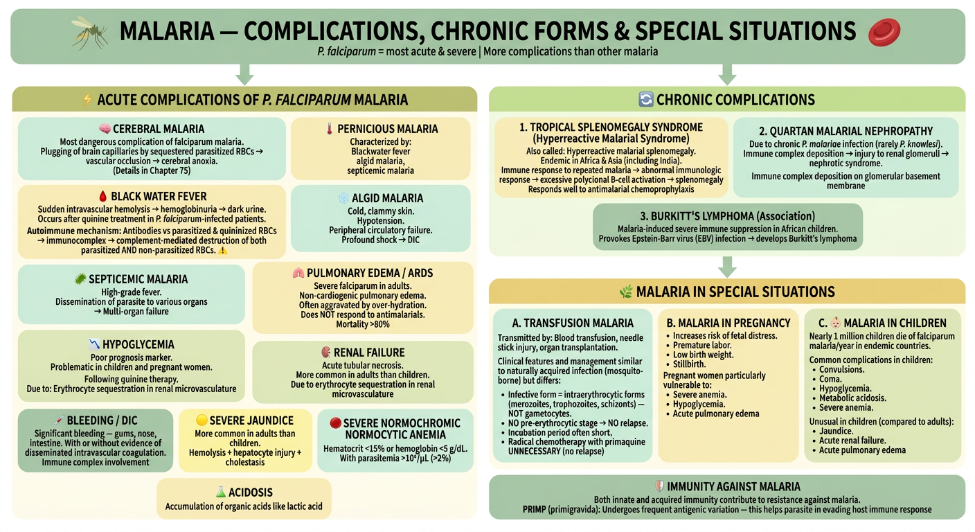
| Complication | Key Feature |
|---|---|
| Cerebral malaria | Brain capillary plugging → cerebral anoxia |
| Black water fever | Dark urine, autoimmune hemolysis (quinine + P. falciparum) → destroys both parasitized & non-parasitized RBCs |
| Algid malaria | Cold, clammy, shock → DIC |
| Septicemic malaria | Multi-organ dissemination |
| Pulmonary edema/ARDS | Non-cardiogenic, mortality >80%, don't over-hydrate |
| Hypoglycemia | Children & pregnant women, post-quinine |
| Renal failure | Adults > children, tubular necrosis |
| Bleeding/DIC, Jaundice, Anemia, Acidosis | Severe falciparum markers |


Explain above data about malaria in form of flowchart in form of image use image where possible.use pastel shades of gren and yellow for colours
A clean, spacious medical flowchart for MALARIA — PATHOGENESIS & CLINICAL FEATURES. All colors exclusively in soft pastel shades of GREEN and YELLOW — pale mint, sage green, soft lime, cream yellow, butter yellow, golden yellow. White background. Large readable text. Rounded corners. Generous white space. Bold headers. Small cute medical icons throughout. TOP HEADER BOX (sage green background, dark forest green bold text, wide): "MALARIA — PATHOGENESIS & CLINICAL FEATURES" Small mosquito icon 🦟 left | Small red blood cell icon right Two species: Benign malaria (P. vivax, P. ovale, P. malariae, P. knowlesi) | Malignant = P. falciparum ━━━━━━━━━━━━━ SECTION 1 (pale lemon yellow background, olive green bold header): "🔄 FEBRILE PAROXYSM" Fever comes intermittently depending on species: Two yellow sub-bubbles side by side: Left bubble (butter yellow): "72-hour cycle" → P. malariae (Quartan fever) Right bubble (mint green): "48-hour cycle" → P. vivax, P. ovale, P. falciparum (Tertian fever) Three-stage horizontal flow with arrows between (each stage a rounded box): Stage 1 (pale mint): "❄️ COLD STAGE" — Patient feels very cold, shivering Stage 2 (pastel yellow): "🔥 HOT STAGE" — High-grade fever, flushed face, skin hot and dry Stage 3 (sage green): "💧 SWEATING STAGE" — Fever comes down with profuse sweating, patient feels better Note below: "Classic triad may not always be present (maturation of populations of parasites at different times) | In P. falciparum: fever more irregular or continuous with marked prostration, headache, nausea" ━━━━━━━━━━━━━ SECTION 2 (soft lime green background, dark green bold header): "🩸 ANEMIA" Patient develops normocytic normochromic anemia Three cause boxes in a row: Box A (pale yellow): "Parasite-induced RBC destruction" — Lysis of RBC due to release of merozoites Box B (mint green): "Splenic removal" — Removal of both infected RBC and uninfected RBC coated with immune complexes Box C (butter yellow): "Bone marrow suppression" — Leading to decreased RBC production ━━━━━━━━━━━━━ SECTION 3 (pastel yellow background, olive bold header): "🫁 SPLENOMEGALY" After few weeks of febrile paroxysms → spleen gets enlarged and becomes palpable Due to: Massive proliferation of macrophages that engulf parasitized and non-parasitized coated RBCs Note: Significant feature in all malaria species ━━━━━━━━━━━━━ SECTION 4 (sage green background, dark forest green bold header, IMPORTANT section): "⚠️ P. FALCIPARUM — MALIGNANT TERTIAN MALARIA" Subtitle: "Pathogenesis is DIFFERENT from other species" Two columns: Left column (pale mint box): "SEQUESTRATION OF PARASITES" Important feature of P. falciparum pathogenesis Ability to SEQUESTER (hold back) parasites in blood vessels of deep visceral organs — brain, kidney etc. → Blockage of vessels → congestion and hypoxia of internal organs Sequestration mediated by: 1. CYTOADHERENCE (pastel yellow inner box): Binding of infected erythrocytes to endothelial cells Mediated by PfEMP-1 (P. falciparum erythrocyte membrane protein-1) PfEMP-1 binds to specific receptors on vascular endothelium of deep organs 2. ROSETTING (mint inner box): PfEMP-1 also helps bind infected RBCs to uninfected RBCs → process called ROSETTING Result (golden yellow box): Since parasites are sequestered in deep vessels → they AVOID frequent spleen passages → ESCAPE splenic clearance → survive and multiply Right column (butter yellow box): "WHY P. FALCIPARUM IS MORE DANGEROUS" • Only species that causes severe/cerebral malaria • Sequestration causes hypoxia in vital organs • PfEMP-1 undergoes frequent antigenic variation → helps parasite evade host immune response (PFEMP-1 = rosetting) • Causes ALL complications: Cerebral malaria, Black water fever, Algid malaria, Septicemic malaria, ARDS, Hypoglycemia, Renal failure, DIC, Severe anemia, Acidosis • Mnemonic on chart: C-Pneu-BAR: Cerebral, Pernicious, neurology, ARDS, Blackwater, Algid, Renal ━━━━━━━━━━━━━ SECTION 5 (pale yellow background, sage green bold header): "🔬 BENIGN MALARIA — KEY FEATURES" Mild in nature | Caused by all 4 species | Triad: Febrile paroxysm + Anemia + Splenomegaly P. knowlesi: sometimes shows dot ring forms and gametocytes (ROTs) detected using molecular methods ━━━━━━━━━━━━━ BOTTOM BOX (soft lime green, dark green text): "🛡️ IMMUNITY AGAINST MALARIA" Both innate AND acquired immunity contribute to resistance against malaria PRIMP (primigravida) undergoes frequent antigenic variation — helps parasite evade host immune response Style: ONLY pastel greens and yellows. Mint, sage, lime, pale yellow, butter yellow, golden yellow. Rounded boxes with generous padding. Clean sans-serif font. Small icons: mosquito, red blood cells, brain, spleen, thermometer, blood drop. Soft green/yellow arrows. Very readable. No crowding. Well spaced.
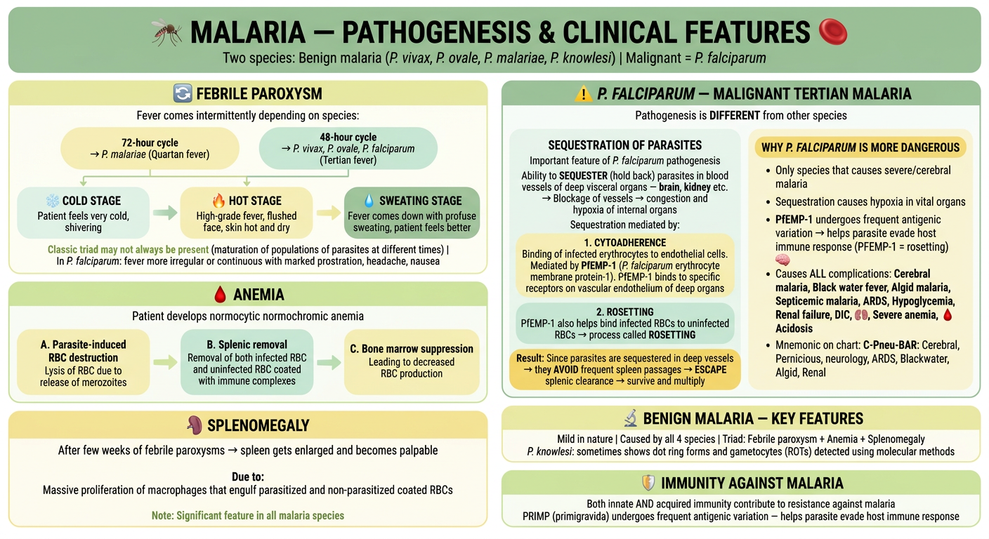
| Section | Key Points |
|---|---|
| Febrile Paroxysm | 72-hr cycle = P. malariae |
| Anemia | Parasite destroys RBCs + Spleen removes immune-coated RBCs + Bone marrow suppression |
| Splenomegaly | Macrophage proliferation engulfing parasitized & coated RBCs |
| P. falciparum (Malignant) | Sequestration via PfEMP-1 → Cytoadherence to endothelium + Rosetting → blocks vessels → hypoxia → multi-organ failure |
| Why most dangerous | PfEMP-1 antigenic variation → evades immunity |
| Benign malaria triad | Febrile paroxysm + Anemia + Splenomegaly |
- Mediates cytoadherence to endothelial cells
- Mediates rosetting (infected RBCs bind uninfected RBCs)
- Undergoes antigenic variation → immune evasion
Reaarange beningn malaria key features before explaining about febrile parooxym making bening malaria as main heading and febrile paroxym anemia and spleenomegaly as sub headings under it aur add note about chronic complication of malaria from given data
A clean, spacious medical flowchart for MALARIA — PATHOGENESIS & CLINICAL FEATURES. All colors exclusively in soft pastel shades of GREEN and YELLOW — pale mint, sage green, soft lime, cream yellow, butter yellow, golden yellow. White background. Large readable text. Rounded corners. Generous white space. Bold headers. Small cute medical icons throughout. TOP HEADER BOX (sage green background, dark forest green bold text, wide): "MALARIA — PATHOGENESIS & CLINICAL FEATURES" Small mosquito icon 🦟 left | Small red blood cell icon right Two species: Benign malaria (P. vivax, P. ovale, P. malariae, P. knowlesi) | Malignant = P. falciparum ━━━━━━━━━━━━━ LARGE SECTION A (soft lime green background, dark green bold header — MAIN SECTION): "🟢 BENIGN MALARIA" Subtitle: "Mild in nature | Caused by all 4 species | Classic Triad: Febrile Paroxysm + Anemia + Splenomegaly" P. knowlesi: dot ring forms and gametocytes detected by molecular methods (ROTs) Three sub-sections inside BENIGN MALARIA, each clearly labeled as a sub-heading: SUB-HEADING 1 (pale lemon yellow rounded box inside, olive green sub-header): "↳ 🌡️ FEBRILE PAROXYSM" Fever comes intermittently depending on species: Two yellow mini-bubbles side by side: Left (butter yellow): "72-hour cycle" → P. malariae (Quartan fever) Right (mint green): "48-hour cycle" → P. vivax, P. ovale, P. falciparum (Tertian fever) Three-stage horizontal flow with arrows: [❄️ COLD STAGE] → [🔥 HOT STAGE] → [💧 SWEATING STAGE] Cold: Patient feels very cold, shivering Hot: High-grade fever, flushed face, skin hot and dry Sweating: Fever comes down, profuse sweating, patient feels better Note: "Classic triad may not always be present | In P. falciparum: fever more irregular or continuous with marked prostration, headache, nausea" SUB-HEADING 2 (pale mint rounded box inside, sage sub-header): "↳ 🩸 ANEMIA" Normocytic normochromic anemia Three cause boxes in a horizontal row: • Parasite-induced RBC destruction — lysis due to merozoite release • Splenic removal — infected + uninfected immune-coated RBCs removed • Bone marrow suppression — decreased RBC production SUB-HEADING 3 (butter yellow rounded box inside, olive sub-header): "↳ 🫁 SPLENOMEGALY" After few weeks of febrile paroxysms → spleen enlarges and becomes palpable Due to: Massive macrophage proliferation engulfing parasitized and non-parasitized coated RBCs Significant feature in all malaria species ━━━━━━━━━━━━━ LARGE SECTION B (sage green background, dark forest green bold header — IMPORTANT): "⚠️ P. FALCIPARUM — MALIGNANT TERTIAN MALARIA" Subtitle: "Pathogenesis DIFFERENT from other species — Most severe form" Two columns inside: Left column (pale mint box): "SEQUESTRATION OF PARASITES" P. falciparum ability to SEQUESTER parasites in deep visceral organ blood vessels (brain, kidney) → Blockage → congestion and hypoxia Mediated by two mechanisms: 1. CYTOADHERENCE (yellow inner box): PfEMP-1 binds infected RBCs to vascular endothelium of deep organs 2. ROSETTING (mint inner box): PfEMP-1 binds infected RBCs to uninfected RBCs Result (golden yellow box): Parasites avoid spleen → escape splenic clearance → survive and multiply Right column (butter yellow box): "WHY MORE DANGEROUS" • PfEMP-1 undergoes frequent antigenic variation → evades host immunity • Causes ALL serious complications: Mnemonic C-Pneu-BAR: C — Cerebral malaria P — Pernicious malaria (Blackwater fever) neu — Neurological (ARDS/Pulmonary edema) B — Blackwater fever / Bleeding DIC A — Algid malaria (shock) R — Renal failure + Hypoglycemia, Severe anemia, Acidosis, Jaundice ━━━━━━━━━━━━━ SECTION C (pastel golden yellow background, olive bold header): "📋 CHRONIC COMPLICATIONS OF MALARIA" Three boxes in a row: Box 1 (mint green rounded): "1. 🌿 TROPICAL SPLENOMEGALY SYNDROME" Also called: Hyperreactive Malarial Splenomegaly (HMS) • Endemic in tropical Africa & Asia (including India) • Due to abnormal immunologic response to repeated malaria infections • Elevated IgM → polyclonal B-cell activation → massive splenomegaly • Responds well to antimalarial chemoprophylaxis (proguanil) Box 2 (pale yellow rounded): "2. 🫘 QUARTAN MALARIAL NEPHROPATHY" Chronic complication of P. malariae (rarely P. knowlesi) • Immune complex injury to renal glomeruli • Results in: Nephrotic syndrome • Immune complexes deposited on glomerular basement membrane Box 3 (sage green rounded): "3. 🔬 BURKITT'S LYMPHOMA (Association)" Malaria-induced severe immunosuppression in African children • Provokes Epstein-Barr virus (EBV) reactivation • → Develops Burkitt's lymphoma ━━━━━━━━━━━━━ BOTTOM BOX (soft lime green, dark green bold text): "🛡️ IMMUNITY AGAINST MALARIA" Both innate AND acquired immunity contribute to resistance PRIMP (primigravida): undergoes frequent antigenic variation → parasite evades host immune response Style: ONLY pastel greens and yellows. Mint, sage, lime, pale yellow, butter yellow, golden yellow. Rounded boxes with generous padding. Clean sans-serif font. Small icons: mosquito, RBC, brain, spleen, thermometer, kidney. Soft arrows. Very readable. Well spaced. No crowding. Benign Malaria section is clearly the first major section after the header.
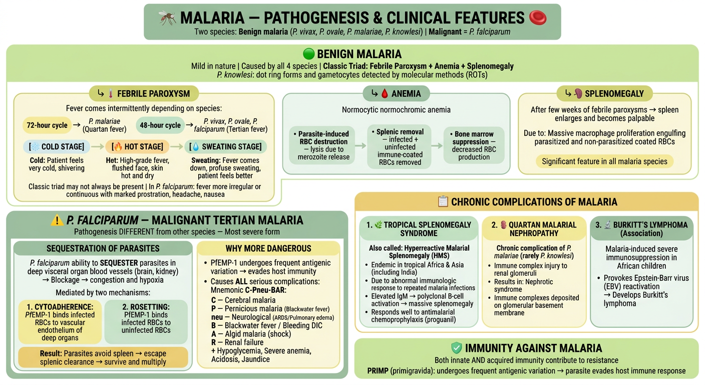
- ↳ Febrile Paroxysm (Cold → Hot → Sweating stages | 48hr vs 72hr cycles)
- ↳ Anemia (3 causes: parasite destruction + splenic removal + bone marrow suppression)
- ↳ Splenomegaly (macrophage proliferation)
- Sequestration via PfEMP-1 (Cytoadherence + Rosetting)
- C-Pneu-BAR complications mnemonic
- Tropical Splenomegaly Syndrome (HMS) — polyclonal B-cell activation, responds to proguanil
- Quartan Malarial Nephropathy — P. malariae → immune complex → nephrotic syndrome
- Burkitt's Lymphoma association — immunosuppression → EBV reactivation

Explain immunity against malaria in form of flowchart from given data in form of image make it colourful use pastel shades of indigo and orange for colours
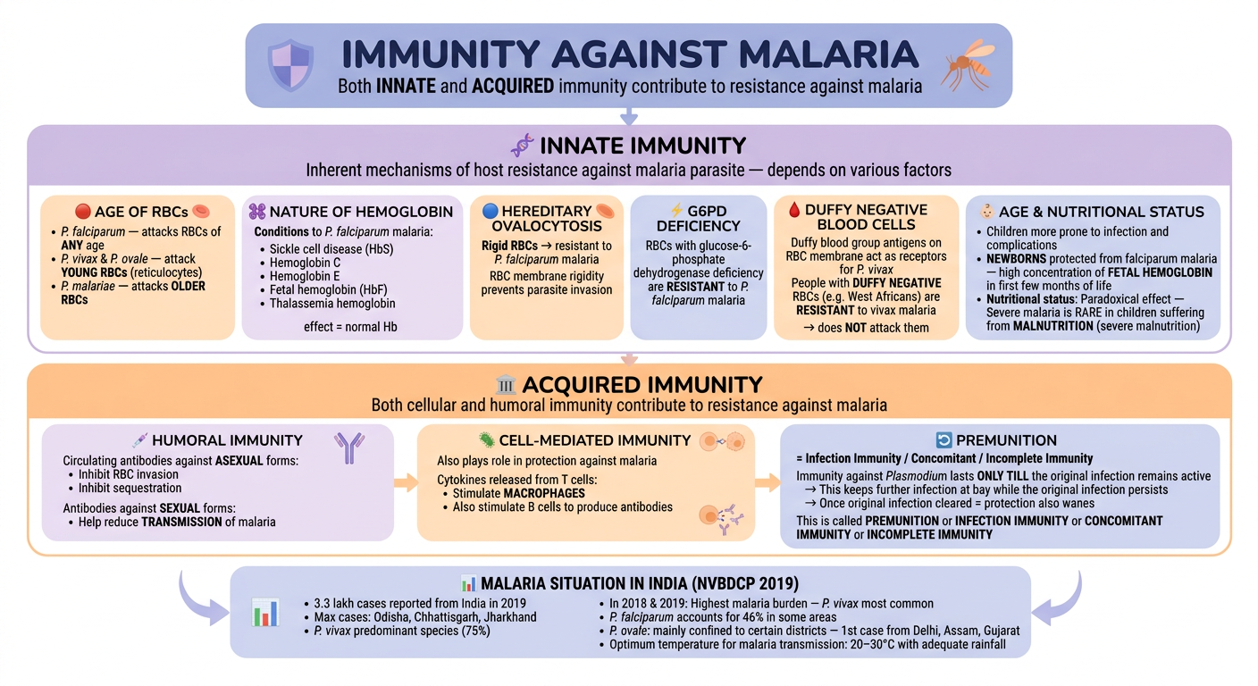
A clean, spacious medical flowchart for IMMUNITY AGAINST MALARIA. All colors exclusively in soft pastel shades of INDIGO/PURPLE and ORANGE — pale lavender, soft periwinkle, dusty indigo, light purple, and pastel peach, soft orange, warm apricot, light tangerine. White background. Large readable text. Rounded corners. Generous white space. Bold headers. Small medical icons throughout. TOP HEADER BOX (soft indigo/periwinkle background, dark indigo bold text, wide): "IMMUNITY AGAINST MALARIA" Subtitle: "Both INNATE and ACQUIRED immunity contribute to resistance against malaria" Small shield icon on left 🛡️ | Small mosquito icon on right 🦟 ━━━━━━━━━━━━━ LARGE SECTION A (pale lavender/soft indigo background, dark purple bold header): "🧬 INNATE IMMUNITY" Subtitle: "Inherent mechanisms of host resistance against malaria parasite — depends on various factors" Six sub-boxes arranged in 2 rows of 3, alternating pastel lavender and pastel peach/orange: Sub-box 1 (pale peach/apricot): "🔴 AGE OF RBCs" • P. falciparum — attacks RBCs of ANY age • P. vivax & P. ovale — attack YOUNG RBCs (reticulocytes) • P. malariae — attacks OLDER RBCs Small RBC icon Sub-box 2 (soft lavender): "🧫 NATURE OF HEMOGLOBIN" Resistant to P. falciparum malaria: • Sickle cell disease (HbS) • Hemoglobin C • Hemoglobin E • Fetal hemoglobin (HbF) • Thalassemia hemoglobin Note: "effect = normal Hb" Sub-box 3 (pastel orange): "🔵 HEREDITARY OVALOCYTOSIS" Rigid RBCs → resistant to P. falciparum malaria RBC membrane rigidity prevents parasite invasion Sub-box 4 (pale periwinkle): "⚡ G6PD DEFICIENCY" RBCs with glucose-6-phosphate dehydrogenase deficiency are RESISTANT to P. falciparum malaria Sub-box 5 (pastel peach): "🩸 DUFFY NEGATIVE BLOOD CELLS" Duffy blood group antigens on RBC membrane act as receptors for P. vivax People with DUFFY NEGATIVE RBCs (e.g. West Africans) are RESISTANT to vivax malaria Note: "does NOT attack them" Sub-box 6 (soft indigo): "👶 AGE & NUTRITIONAL STATUS" • Children more prone to infection and complications • NEWBORNS protected from falciparum malaria — high concentration of FETAL HEMOGLOBIN in first few months of life • Nutritional status: Paradoxical effect — Severe malaria is RARE in children suffering from MALNUTRITION (severe malnutrition) ━━━━━━━━━━━━━ LARGE SECTION B (soft peach/warm orange background, dark orange bold header): "🏛️ ACQUIRED IMMUNITY" Subtitle: "Both cellular and humoral immunity contribute to resistance against malaria" Three boxes in a row: Box 1 (pale lavender rounded): "💉 HUMORAL IMMUNITY" Circulating antibodies against ASEXUAL forms: • Inhibit RBC invasion • Inhibit sequestration Antibodies against SEXUAL forms: • Help reduce TRANSMISSION of malaria Small antibody/Y-shape icon Box 2 (pastel orange rounded): "🦠 CELL-MEDIATED IMMUNITY" Also plays role in protection against malaria Cytokines released from T cells: • Stimulate MACROPHAGES • Also stimulate B cells to produce antibodies T cell and macrophage interaction icon Box 3 (soft indigo rounded): "🔄 PREMUNITION" = Infection Immunity / Concomitant / Incomplete Immunity Immunity against Plasmodium lasts ONLY TILL the original infection remains active → This keeps further infection at bay while the original infection persists → Once original infection cleared = protection also wanes This is called PREMUNITION or INFECTION IMMUNITY or CONCOMITANT IMMUNITY or INCOMPLETE IMMUNITY ━━━━━━━━━━━━━ BOTTOM NOTE BOX (soft lavender/periwinkle, dark indigo text): "📊 MALARIA SITUATION IN INDIA (NVBDCP 2019)" • 3.3 lakh cases reported from India in 2019 • Max cases: Odisha, Chhattisgarh, Jharkhand • P. vivax predominant species (75%) • In 2018 & 2019: Highest malaria burden — P. vivax most common • P. falciparum accounts for 46% in some areas • P. ovale: mainly confined to certain districts — 1st case from Delhi, Assam, Gujarat • Optimum temperature for malaria transmission: 20–30°C with adequate rainfall Style: ONLY pastel indigo/lavender/purple AND pastel orange/peach/apricot. No other colors. Rounded boxes with generous padding. Clean sans-serif font. Bold section headers in darker shade. Soft arrows. Small icons: shield, mosquito, RBC, antibody Y shape, T cell, brain. Very readable. Well spaced. No crowding.
| Factor | Protection Against |
|---|---|
| Age of RBCs | P. vivax/ovale → reticulocytes only |
| Hemoglobin type | HbS (sickle cell), HbC, HbE, HbF, Thalassemia → resistant to P. falciparum |
| Hereditary ovalocytosis | Rigid RBCs → P. falciparum cannot invade |
| G6PD deficiency | Resistant to P. falciparum |
| Duffy negative | West Africans → resistant to P. vivax (no receptor for parasite) |
| Fetal Hb + malnutrition | Newborns protected |
- Humoral — antibodies vs asexual forms (block invasion + sequestration) | vs sexual forms (reduce transmission)
- Cell-mediated — T cells → cytokines → activate macrophages + B cells
- Premunition (Infection Immunity) — immunity lasts ONLY while original infection is active; wanes once cleared


Explain given data above malaria in form of flowchadrt in form of image use image where possible use pastel shades of yellow and red for colours
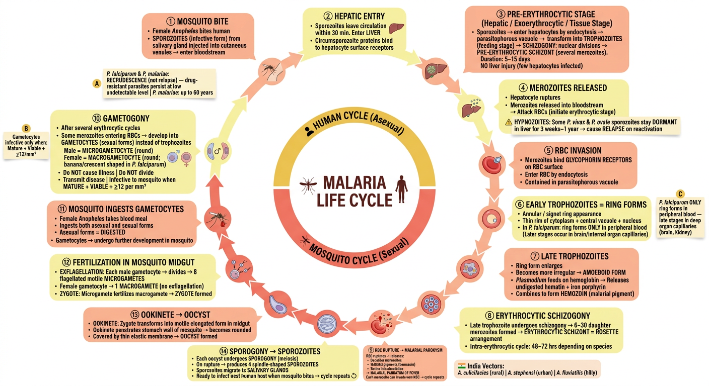
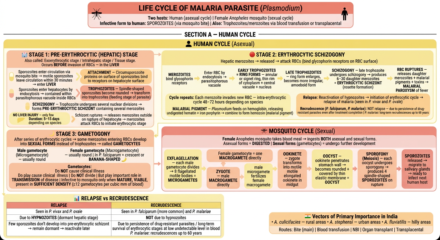
A clean, spacious medical flowchart for LIFE CYCLE OF MALARIA PARASITE. All colors exclusively in soft pastel shades of YELLOW and RED — pale lemon yellow, butter yellow, golden yellow, soft coral, pastel rose, light salmon, warm apricot, dusty red. White background. Large readable text. Rounded corners. Generous white space. Bold headers. Small medical icons throughout. Circular life cycle diagram style with linear explanations below. TOP HEADER BOX (soft coral/warm red background, dark red bold text): "LIFE CYCLE OF MALARIA PARASITE (Plasmodium)" Two hosts: Human (asexual cycle) | Female Anopheles mosquito (sexual cycle) Infective form to human: SPOROZOITES (via mosquito bite) | Also: Trophozoites/merozoites via blood transfusion or transplacental Small mosquito icon 🦟 on left | Small human silhouette icon on right ━━━━━━━━━━━━━ SECTION A — HUMAN CYCLE (pale lemon yellow background, golden yellow bold header): "👤 HUMAN CYCLE (Asexual)" Three stages labeled clearly with arrows between them: STAGE 1 BOX (pastel coral/salmon, bold red header): "🏥 STAGE 1: PRE-ERYTHROCYTIC (HEPATIC) STAGE" Also called: Exoerythrocytic stage / Intrahepatic stage / Tissue stage Occurs BEFORE invasion of RBCs — in the LIVER Step-by-step flow (soft yellow arrow boxes): Step 1 (pale yellow pill): "Sporozoites enter circulation via mosquito bite → motile sporozoites leave circulation within 30 minutes → enter LIVER" Step 2 (apricot pill): "ATTACHMENT — Circumsporozoite proteins on surface of sporozoites bind to receptors on hepatocyte surface" Step 3 (pale yellow pill): "Sporozoites enter hepatocytes by endocytosis → contained within parasitophorous vacuole inside RBCs" Step 4 (coral pill): "TROPHOZOITES — Spindle-shaped sporozoites become rounded → transform into trophozoites (feeding stage of parasite)" Step 5 (pale yellow pill): "SCHIZOGONY — Trophozoite undergoes several nuclear divisions → forms PRE-ERYTHROCYTIC SCHIZONT containing several merozoites" Step 6 (apricot pill): "Schizont ruptures → releases merozoites outside on rupture of hepatocyte → merozoites attack RBCs to initiate erythrocytic stage" Key notes in yellow bubbles: • "NO LIVER INJURY — only few hepatocytes infected; as only few hepatocytes are infected by Plasmodium, hepatic damage does not occur" • "Duration: 5–15 days depending on species" • "HYPNOZOITES: Some sporozoites of P. vivax and P. ovale do NOT develop further — remain dormant as HYPNOZOITES for 3 weeks to 1 year → cause RELAPSE" • "Relapse: Reactivation of hypnozoites → initiation of erythrocytic cycle → relapse of malaria (seen in P. vivax and P. ovale)" • "Recrudescence (P. falciparum, P. malariae): NOT relapse — due to persistence of drug-resistant parasites even after treatment completion | P. malariae: long-term recrudescences up to 60 years" STAGE 2 BOX (butter yellow background, warm red bold header): "🔴 STAGE 2: ERYTHROCYTIC SCHIZOGONY" Hepatic merozoites → released → attack RBCs (bind glycophorin receptors on RBC surface) Step-by-step horizontal flow: [MEROZOITES bind glycophorin receptors] → [Enter RBC by endocytosis → parasitophorous vacuole] → [EARLY TROPHOZOITES = RING FORMS — annular or signet ring, thin rim of cytoplasm + central vacuole + nucleus] → [LATE TROPHOZOITES — ring form enlarges, becomes more irregular, amoeboid form] → [SCHIZOGONY — late trophozoite undergoes schizogony → produces 6–30 daughter merozoites = ERYTHROCYTIC SCHIZONT (rosette formation)] → [RBC RUPTURES — releases daughter merozoites + malarial pigments + toxins → MALARIAL PAROXYSM of fever] Key notes: • "Cycle repeats: Each merozoite invades new RBC — intra-erythrocytic cycle 48–72 hours depending on species" • "MALARIAL PIGMENT — Plasmodium feeds on hemoglobin, releasing undigested hematin + iron porphyrin → combine to form hemozoin (malarial pigment)" • "P. falciparum SPECIAL: Later stages of erythrocytic cycle occur in capillaries of brain and internal organs → ONLY RING FORMS found in peripheral blood (NOT late trophozoites and schizonts)" STAGE 3 BOX (pale coral/salmon background, dark red bold header): "🌙 STAGE 3: GAMETOGONY" After series of erythrocytic cycles → some merozoites entering RBCs develop into SEXUAL FORMS instead of trophozoites → called GAMETOCYTES Two mini-boxes side by side: Left (pale yellow): "Male gametocyte (Microgametocyte)" — usually round Right (apricot): "Female gametocyte (Macrogametocyte)" — usually round | In P. falciparum = crescent or BANANA-SHAPED Note (coral box): "Gametocytes: Do NOT cause clinical illness | Do NOT divide | But play important role in TRANSMISSION of disease | Infective to mosquito only when MATURE, VIABLE, present in SUFFICIENT DENSITY (≥12 gametocytes per cubic mm of blood)" ━━━━━━━━━━━━━ SECTION B — MOSQUITO CYCLE (pastel rose/soft red background, dark red bold header): "🦟 MOSQUITO CYCLE (Sexual)" Female Anopheles mosquito takes blood meal → ingests BOTH asexual and sexual forms Asexual forms = DIGESTED | Sexual forms (gametocytes) = undergo further development Horizontal step flow (alternating yellow and coral mini-boxes): [EXFLAGELLATION — each male gametocyte divides → 8 flagellated motile bodies = MICROGAMETES] → [Female gametocyte = one MACROGAMETE directly] → [ZYGOTE — male microgamete fertilizes female macrogamete] → [OOKINETE — zygote transforms into motile elongated ookinete in midgut] → [OOCYST — ookinete penetrates stomach wall → becomes rounded + covered by thin elastic membrane = OOCYST] → [SPORОГONY (Meiosis) — each oocyst undergoes sporogony → produces 4 spindle-shaped SPOROZOITES on rupture] → [SPOROZOITES released → migrate to salivary glands → ready to infect next human host] ━━━━━━━━━━━━━ BOTTOM TABLE BOX (pale lemon yellow background, dark red text, two columns): "📊 RELAPSE vs RECRUDESCENCE" Left column (coral header): RELAPSE | Seen in P. vivax and P. ovale | Due to HYPNOZOITES (dormant hepatic stage) | Few sporozoites don't develop into pre-erythrocytic schizont → remain dormant → reactivate later Right column (yellow header): RECRUDESCENCE | Seen in P. falciparum (more common) and P. malariae | NOT due to hypnozoites | Due to persistence of drug-resistant parasites / long-term survival of erythrocytic stages at low undetectable level in blood | P. malariae: recrudescences up to 60 years INDIA VECTORS NOTE (soft apricot box): "🇮🇳 Vectors of Primary Importance in India" • A. culicifacies — rural areas • A. stephensi — urban areas • A. fluviatilis — hilly areas Routes: Bite (main) | Blood transfusion | NBI | Organ transplant | Transplacental Style: ONLY pastel yellows and reds/corals. Pale lemon, butter yellow, golden yellow, soft coral, pastel rose, warm apricot, light salmon. Rounded boxes. Generous padding. Clean bold font. Small icons: mosquito, liver, RBC, banana shape for P. falciparum gametocyte, human silhouette. Soft arrows. Very readable. Well spaced. No crowding.
| Stage | Location | Key Events |
|---|---|---|
| Pre-erythrocytic (Hepatic) | Liver | Sporozoites → Trophozoites → Pre-erythrocytic schizont → Merozoites |
| Erythrocytic Schizogony | RBCs | Merozoites bind glycophorin → Ring forms → Late trophozoites → Schizont (rosette, 6–30 merozoites) → RBC rupture → fever paroxysm |
| Gametogony | RBCs | Some merozoites → gametocytes |
- Relapse (P. vivax/ovale) = hypnozoites reactivate
- Recrudescence (P. falciparum/malariae) = drug-resistant parasites persist; P. malariae can recrudesce up to 60 years
- India vectors: A. culicifacies (rural) | A. stephensi (urban) | A. fluviatilis (hilly)
Try to put all above data other than difference between relapse and recurdescence in form of a circular cycle with each steps occurring in correct order do not omit any information from the flowchart
A detailed, circular life cycle diagram of Malaria parasite in pastel yellow and red/coral color theme. White background. The cycle flows CLOCKWISE in a large circle with TWO ARCS — the TOP HALF of the circle represents the HUMAN CYCLE and the BOTTOM HALF represents the MOSQUITO CYCLE. Each step is a rounded box connected by curved arrows following the circle. In the CENTER of the circle, write "MALARIA LIFE CYCLE" with a small mosquito icon and human silhouette icon. The circle has ~14 numbered steps arranged clockwise as rounded boxes connected by curved arrows. Each step alternates between pale lemon yellow and soft coral/apricot background. All text is dark, bold, readable. Steps are well-spaced with enough room for text inside each box. LABEL THE TOP ARC (above center): "👤 HUMAN CYCLE (Asexual)" in golden yellow banner LABEL THE BOTTOM ARC (below center): "🦟 MOSQUITO CYCLE (Sexual)" in coral/red banner CLOCKWISE STEPS starting from TOP-LEFT going RIGHT: STEP 1 (coral box, top-left of circle): "① MOSQUITO BITE" Female Anopheles bites human SPOROZOITES (infective form) from salivary gland injected into cutaneous venules → enter bloodstream STEP 2 (yellow box, top of circle): "② HEPATIC ENTRY" Sporozoites leave circulation within 30 min Enter LIVER Circumsporozoite proteins bind to hepatocyte surface receptors STEP 3 (coral box, top-right): "③ PRE-ERYTHROCYTIC STAGE" (Hepatic / Exoerythrocytic / Tissue Stage) Sporozoites → enter hepatocytes by endocytosis → parasitophorous vacuole → transform into TROPHOZOITES (feeding stage) → SCHIZOGONY: nuclear divisions → PRE-ERYTHROCYTIC SCHIZONT (several merozoites) Duration: 5–15 days NO liver injury (few hepatocytes infected) STEP 4 (yellow box, right of circle — branching note): "④ MEROZOITES RELEASED" Hepatocyte ruptures Merozoites released into bloodstream → Attack RBCs (initiate erythrocytic stage) ⚠️ HYPNOZOITES: Some P. vivax & P. ovale sporozoites stay DORMANT in liver for 3 weeks–1 year → cause RELAPSE on reactivation STEP 5 (coral box, right side): "⑤ RBC INVASION" Merozoites bind GLYCOPHORIN RECEPTORS on RBC surface Enter RBC by endocytosis Contained in parasitophorous vacuole STEP 6 (yellow box, lower-right): "⑥ EARLY TROPHOZOITES = RING FORMS" Annular / signet ring appearance Thin rim of cytoplasm + central vacuole + nucleus In P. falciparum: ring forms ONLY in peripheral blood (Later stages occur in brain/internal organ capillaries) STEP 7 (coral box, bottom-right): "⑦ LATE TROPHOZOITES" Ring form enlarges Becomes more irregular → AMOEBOID FORM Plasmodium feeds on hemoglobin → Releases undigested hematin + iron porphyrin → Combines to form HEMOZOIN (malarial pigment) STEP 8 (yellow box, bottom): "⑧ ERYTHROCYTIC SCHIZOGONY" Late trophozoite undergoes schizogony → 6–30 daughter merozoites formed → ERYTHROCYTIC SCHIZONT = ROSETTE arrangement Intra-erythrocytic cycle: 48–72 hrs depending on species STEP 9 (coral box, bottom-left): "⑨ RBC RUPTURE → MALARIAL PAROXYSM" RBC ruptures → releases: • Daughter merozoites • Malarial pigments (hemozoin) • Toxins into circulation → MALARIAL PAROXYSM OF FEVER Each merozoite can invade new RBC → cycle repeats STEP 10 (yellow box, left side — branch): "⑩ GAMETOGONY" After several erythrocytic cycles Some merozoites entering RBCs → develop into GAMETOCYTES (sexual forms) instead of trophozoites Male = MICROGAMETOCYTE (round) Female = MACROGAMETOCYTE (round; banana/crescent shaped in P. falciparum) Do NOT cause illness | Do NOT divide Transmit disease | Infective to mosquito when MATURE + VIABLE + ≥12 per mm³ STEP 11 (coral box, lower-left — now in MOSQUITO ARC): "⑪ MOSQUITO INGESTS GAMETOCYTES" Female Anopheles takes blood meal Ingests both asexual and sexual forms Asexual forms = DIGESTED Gametocytes → undergo further development in mosquito STEP 12 (yellow box, bottom-left mosquito arc): "⑫ FERTILIZATION IN MOSQUITO MIDGUT" EXFLAGELLATION: Each male gametocyte → divides → 8 flagellated motile MICROGAMETES Female gametocyte → 1 MACROGAMETE (no exflagellation) ZYGOTE: Microgamete fertilizes macrogamete → ZYGOTE formed STEP 13 (coral box, bottom mosquito arc): "⑬ OOKINETE → OOCYST" OOKINETE: Zygote transforms into motile elongated form in midgut Ookinete penetrates stomach wall of mosquito → becomes rounded Covered by thin elastic membrane → OOCYST formed STEP 14 (yellow box, bottom-right mosquito arc — connects back to Step 1): "⑭ SPOROGONY → SPOROZOITES" Each oocyst undergoes SPOROGONY (meiosis) On rupture → produces 4 spindle-shaped SPOROZOITES Sporozoites migrate to SALIVARY GLANDS Ready to infect next human host when mosquito bites → cycle repeats ↺ ADD SMALL SIDE NOTE BOXES floating near the circle (in pale yellow): Note A (near Step 3): "P. falciparum & P. malariae: RECRUDESCENCE (not relapse) — drug-resistant parasites persist at low undetectable level | P. malariae: up to 60 years" Note B (near Step 10): "Gametocytes infective only when: Mature + Viable + ≥12/mm³" Note C (near Step 6): "P. falciparum ONLY ring forms in peripheral blood — late stages in deep organ capillaries (brain, kidney)" INDIA VECTORS small box (pale apricot, outside circle bottom): "🇮🇳 India Vectors: A. culicifacies (rural) | A. stephensi (urban) | A. fluviatilis (hilly)" Style: Pastel yellow and coral/red only. Circular layout. Clockwise arrows. Bold text in each step box. Clear section banners for Human vs Mosquito cycle. Small relevant icons: liver icon near step 3, RBC near step 5, ring form icon near step 6, mosquito near step 11, fertilization icon near step 12. Generous spacing. Very readable. Professional medical diagram style.