
Discuss the role of calcium ion as a second messenger
Reading File
Searching sources...
Finding Sources
Finding Sources
Good results. Now let me read the most relevant sections in parallel.
Reading File
Finding Sources
Reading File
Reading File
Reading File
Reading File
Now let me also fetch a couple of relevant diagrams and look for calcium's role in muscle contraction and immune signaling.
Finding Sources
Finding Sources
Loading Image
I now have comprehensive material from authoritative textbooks. Let me compile the full answer.
Calcium Ion (Ca²⁺) as a Second Messenger
Introduction
Second messengers are intracellular signaling molecules that relay signals received at the cell surface to effector proteins inside the cell. Ca²⁺ is one of the most versatile and ubiquitous second messengers in biology, operating alongside cAMP and the products of phospholipid breakdown. A single hormone may activate more than one second messenger system simultaneously. - Guyton and Hall Textbook of Medical Physiology, p. 912
1. The Concept of Second Messengers
When a peptide hormone or neurotransmitter binds its receptor on the cell surface, it cannot cross the plasma membrane. Instead, it causes the formation or release of an intracellular signal - the second messenger - that then triggers the cell's biochemical response. Ca²⁺, cAMP, and diacylglycerol (DAG) are the three principal second messengers. - Guyton and Hall, p. 912
2. Sources of Intracellular Calcium
Ca²⁺ enters the cytoplasm from two major sources:
- Extracellular influx - through voltage-gated Ca²⁺ channels (opened by changes in membrane potential) or ligand-gated Ca²⁺ channels (opened by hormone-receptor interaction).
- Intracellular stores - primarily the endoplasmic reticulum (ER) and, to a lesser extent, mitochondria. These stores are mobilized by the second messenger inositol trisphosphate (IP3). - Guyton and Hall, p. 613–619
The resting cytoplasmic Ca²⁺ concentration is very low: 10⁻⁸ to 10⁻⁷ mol/L. Upon stimulation it rises to 10⁻⁶ to 10⁻⁵ mol/L, which is the threshold for activating calmodulin and other calcium-binding proteins. - Guyton and Hall, p. 619
3. The Phospholipase C - IP3 - Ca²⁺ Cascade
This is the main pathway by which many hormones and neurotransmitters mobilize Ca²⁺ as a second messenger.
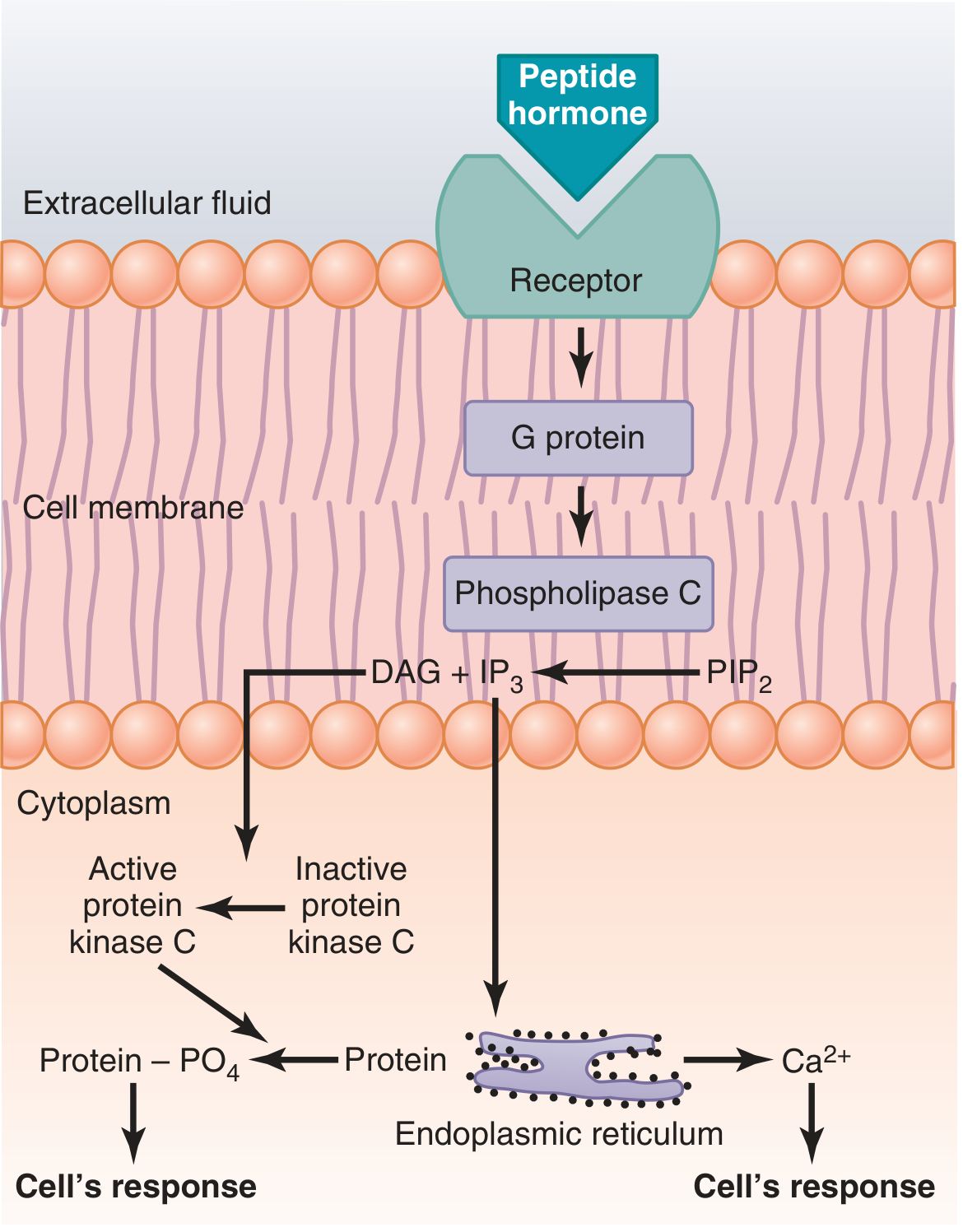
Steps:
- A hormone (e.g., epinephrine at α-receptors, angiotensin II, oxytocin) binds its G protein-coupled receptor (GPCR).
- The Gq protein activates phospholipase C (PLC), which cleaves membrane phosphatidylinositol 4,5-bisphosphate (PIP2) into two second messengers: IP3 and DAG.
- IP3 (a soluble molecule) diffuses to the ER membrane, binds the IP3 receptor (a Ca²⁺ channel on the ER surface), and opens it - releasing Ca²⁺ from the ER lumen into the cytoplasm.
- The released Ca²⁺ then acts as a second messenger in its own right, activating calmodulin and other calcium-binding proteins.
- DAG remains in the membrane and activates protein kinase C (PKC), which phosphorylates many target proteins. - Guyton and Hall, p. 608–610; Basic Medical Biochemistry, p. 948
4. Calcium-Induced Calcium Release (CICR)
Ca²⁺ can amplify its own release in a positive-feedback manner. Once cytoplasmic Ca²⁺ rises, it binds the ryanodine receptor - another integral protein in the ER/sarcoplasmic reticulum (SR) membrane. This receptor-channel opens in response to cytoplasmic Ca²⁺ (not IP3), releasing further stored Ca²⁺. This mechanism, called CICR, is especially important in cardiac and skeletal muscle for excitation-contraction coupling. - Eric Kandel, Principles of Neural Science, 6th ed., p. 355
5. Calmodulin - The Principal Calcium Sensor
The primary intracellular effector of Ca²⁺ is calmodulin, a small (17 kDa) cytoplasmic protein with four Ca²⁺-binding sites. When three or four sites are occupied, calmodulin undergoes a conformational change that allows it to bind and regulate dozens of target enzymes. - Guyton and Hall, p. 617
Key Calmodulin-Dependent Effects
| Effector Activated | Physiological Outcome |
|---|---|
| Myosin light chain kinase (MLCK) | Smooth muscle contraction |
| Phosphorylase kinase | Glycogen breakdown (glycogenolysis) |
| CaM kinase (CaMK I, II, IV) | Phosphorylation of diverse substrates; synaptic plasticity; memory |
| Calcineurin (a phosphatase) | Dephosphorylation of NFAT; nuclear translocation; immune gene transcription |
| Adenylyl cyclase (certain isoforms) | Cross-talk with cAMP pathway |
- Guyton and Hall, p. 617; Firestein & Kelley's Textbook of Rheumatology; Kandel, Principles of Neural Science
6. Calcium/Calmodulin-Dependent Protein Kinase (CaMK)
CaM kinase is a multimeric enzyme in which each subunit contains both a regulatory and a catalytic domain. In the absence of Ca²⁺/calmodulin, the regulatory domain autoinhibits the catalytic domain. When the Ca²⁺/calmodulin complex binds, the catalytic domain is unmasked and the kinase phosphorylates serine/threonine residues on target proteins.
A particularly important feature is autophosphorylation: once activated, CaMK phosphorylates itself, converting it into a constitutively active form that remains active even after Ca²⁺ levels fall back to basal. This mechanism underlies long-term changes in synaptic strength and memory consolidation. - Kandel, Principles of Neural Science, p. 354–355
7. Role of Ca²⁺ in Muscle Contraction
- Skeletal muscle: Ca²⁺ released from the SR upon action potential binds troponin C (structurally and functionally similar to calmodulin), causing the troponin-tropomyosin complex to shift and expose actin binding sites for myosin.
- Smooth muscle: Ca²⁺/calmodulin activates MLCK, which phosphorylates myosin light chains, enabling cross-bridge cycling and contraction.
- Cardiac muscle: Ca²⁺ entry through L-type channels triggers CICR via ryanodine receptors (RyR2) in the SR, generating the large Ca²⁺ transient needed for systolic contraction. - Basic Medical Biochemistry, p. 949; Barash, Clinical Anesthesia
8. Role of Ca²⁺ in Glycogen Metabolism
Epinephrine acting on hepatic α1-receptors triggers the PIP2 pathway, raising cytoplasmic Ca²⁺. Ca²⁺:
- Activates phosphorylase kinase (via calmodulin subunit) → activates glycogen phosphorylase → glycogenolysis ↑
- Activates CaM synthase kinase → phosphorylates and inhibits glycogen synthase → glycogen synthesis ↓
- Together with DAG activates protein kinase C → further phosphorylation of glycogen synthase at different serine residues
This makes Ca²⁺ synergistic with cAMP/glucagon in promoting hepatic glucose mobilization during stress. - Basic Medical Biochemistry, p. 948–949
9. Role of Ca²⁺ in Immune Cell Activation (T cells, Mast Cells)
In T-cell activation, TCR signaling activates PLCγ, which generates IP3 and DAG from PIP2. IP3 releases ER Ca²⁺ into the cytoplasm. This Ca²⁺:
- Binds calmodulin → activates calcineurin (a serine/threonine phosphatase)
- Calcineurin dephosphorylates NFAT (nuclear factor of activated T cells), exposing its nuclear localization signal
- NFAT translocates to the nucleus and drives transcription of IL-2 and other cytokine genes
This is the pathway blocked by the immunosuppressants cyclosporine and tacrolimus (FK506), which inhibit calcineurin. - Firestein & Kelley's Textbook of Rheumatology; Sabiston Textbook of Surgery
10. PKC - The Other Branch of the PLC Pathway
While Ca²⁺/calmodulin handles one arm of the PLC cascade, DAG activates protein kinase C (PKC). PKC itself requires Ca²⁺ for full activation (along with DAG). Once active, PKC phosphorylates numerous proteins, including:
- IκB kinase (IKK) → activates NF-κB → pro-inflammatory and survival gene transcription
- AP-1 transcription factors
- Various growth factor-related targets
This illustrates a convergence between the Ca²⁺ and DAG signals. - Firestein & Kelley's Rheumatology, p. 9290
11. Role in Secretion and Neurotransmitter Release
Transient rises in intracellular Ca²⁺ are key second-messenger signals for secretory responses:
- In neurons: Ca²⁺ influx through presynaptic voltage-gated Ca²⁺ channels triggers vesicle fusion (exocytosis) and neurotransmitter release.
- In enterocytes: Transient increases in intracellular Ca²⁺ act as key second-messenger signals for secretory responses. - Sleisenger and Fordtran's Gastrointestinal and Liver Disease
- In pancreatic acinar cells: Ca²⁺ regulates amylase secretion.
- In mast cells: Ca²⁺ and DAG activate PKC to drive degranulation and histamine release. - Cellular and Molecular Immunology
12. Signal Termination
To prevent runaway activation, cytoplasmic Ca²⁺ is rapidly cleared by:
- SERCA pumps (sarco/endoplasmic reticulum Ca²⁺-ATPase) - pump Ca²⁺ back into the ER
- Plasma membrane Ca²⁺-ATPase (PMCA) - extrude Ca²⁺ out of the cell
- Na⁺/Ca²⁺ exchanger (NCX) - particularly important in cardiac muscle
- Mitochondrial uptake - buffers large Ca²⁺ loads
Calmodulin also releases Ca²⁺ as concentrations fall, rapidly reversing enzyme activation. Because Ca²⁺ is sequestered so quickly by calcium-binding proteins and pumped out of the cytoplasm, it can act highly locally as a spatially-confined messenger. - Katzung's Basic and Clinical Pharmacology, 16th ed.; Kandel, p. 355
Summary
| Feature | Detail |
|---|---|
| Resting [Ca²⁺]i | 10⁻⁸ - 10⁻⁷ mol/L |
| Activating [Ca²⁺]i | 10⁻⁶ - 10⁻⁵ mol/L |
| Main sources | ER (via IP3R or RyR), extracellular (via voltage/ligand-gated channels) |
| Primary sensor | Calmodulin (4 binding sites) |
| Key effectors | CaM kinase, calcineurin, MLCK, phosphorylase kinase, PKC |
| Functions | Muscle contraction, secretion, glycogenolysis, gene transcription, neurotransmission, immune activation, memory |
| Termination | SERCA, PMCA, NCX, mitochondrial uptake |
Ca²⁺ is unique among second messengers in its spatial versatility - it can act as a highly localized microdomainal signal (e.g., at the presynaptic terminal) or as a cell-wide wave (as in the fertilization response), depending on the cell type and stimulus intensity.
This is a shared conversation. Sign in to Orris to start your own chat.