
Is tinia capitis happens due to tinia cruris by same same hand hand used for the scratch the groin to scratch the scalp and what is the treatment for tinia capitis
tinea capitis treatment griseofulvin 2025 guidelines
tinea capitis scalp ringworm fungal infection
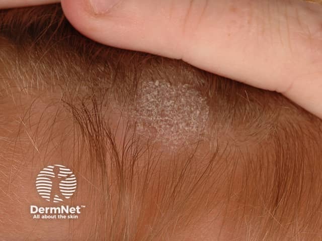
This is a high-resolution clinical photograph of a localized scalp lesion suspicious for tinea capitis (scalp ringworm). Modality: Clinical photography; technique: macro close-up digital image with diffuse, realistic lighting; no dermatoscopic pattern or fluorescence. Anatomical location: Scalp, especially near the temporal hairline; integumentary involvement. The lesion appears as an annular plaque with a grey to pale scale-coated surface and a distinct active border of peripheral scaling. Central clearing is evident, with hair thinning and mild perifollicular scaling. Surrounding skin shows normal pigmentation and mild dryness. Pattern is localized/unilateral. Differential diagnoses include dermatophyte infection (tinea capitis), seborrheic dermatitis, discoid lupus erythematosus, psoriasis capitis, and eczema. Clinically significant for fungal infection; warrants fungal testing (KOH microscopy, culture) and antifungal therapy. Management may involve systemic antifungals (e.g., griseofulvin, terbinafine in children) or topical agents for limited involvement; assess for hair shaft breakage and monitor resolution. This image is valuable for education on ringworm morphology, differential diagnosis, and clinical decision-making in pediatric dermatology. Clinical follow-up and dermoscopy may further delineate perifollicular inflammation and guide antifungal choice.
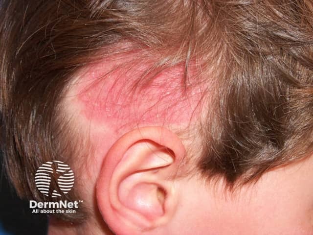
Imaging modality: Clinical photography of the scalp using standard external photography. The image depicts the left temporal/parietal scalp in a pediatric patient, showing a circular to oval erythematous patch with fine scaling and hair shedding compatible with tinea capitis (scalp ringworm). Visual features include a well-demarcated plaque, superficial crusting at the margins, and accentuated perifollicular erythema around affected follicles. Hair within the lesion appears sparse or broken, and surrounding skin is normal or mildly irritated, with no deep dermal involvement evident. The underlying pathophysiology is dermatophyte invasion of keratinized structures, most commonly Microsporum or Trichophyton species, leading to inflammatory alopecia and patchy hair loss. Clinically this appearance supports a differential diagnosis that includes tinea capitis, seborrheic dermatitis, and inflammatory scalp conditions; however, the combination of erythema, scaling, and patchy alopecia favors a fungal etiology. Diagnostic significance lies in recognizing a contagious dermatophyte infection requiring mycologic confirmation (KOH preparation, fungal culture, or Wood lamp examination when indicated). Potential clinical uses include guiding early antifungal therapy, infection control counseling, and educational demonstration of scalp dermatophytosis for medical training. This image serves as a case-based reference for dermatology, pediatrics, infectious disease education, and clinical dermatopathology.
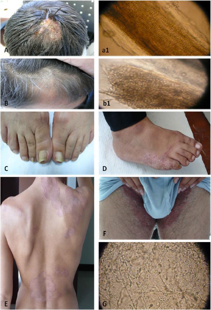
This composite educational image illustrates various clinical and microscopic manifestations of dermatophytosis. Panels A and B display Tinea Capitis: (A) demonstrates Endothrix infection with a visible patch of broken hair shafts and scalp inflammation, while (B) shows Ectothrix infection characterized by circular alopecia with fine gray scaling. Panels a1 and b1 provide corresponding microscopic views of hair shafts, illustrating fungal spores inside and outside the shaft, respectively. Panel C shows Tinea Unguium (onychomycosis) with yellowing and thickening of the toenails. Panel D illustrates Tinea Pedis (athlete's foot) presenting as erythematous scaling on the lateral foot and interdigital spaces. Panel E depicts Tinea Corporis (ringworm) on the back, showing classic annular, erythematous plaques with raised, scaly borders and central clearing. Panel F demonstrates Tinea Cruris (jock itch) with a confluent erythematous rash in the bilateral groin area. Finally, Panel G shows a high-power microscopic view of fungal hyphae and spores. This collection serves as a diagnostic reference for dermatological fungal infections across different anatomical regions.
Tinea Capitis and Its Relation to Tinea Cruris - Can One Cause the Other?
Your Question: Is Tinea Capitis Caused by Scratching the Groin (Tinea Cruris) and Then the Scalp?
- Tinea cruris is most commonly caused by Trichophyton rubrum, T. interdigitale, or Epidermophyton floccosum - and predominantly affects adults.
- Tinea capitis predominantly affects children and is most commonly caused by T. tonsurans (in the US/UK) or Microsporum canis (from pets) - and these organisms are different fungi from those causing tinea cruris.
- The groin-to-scalp autoinoculation route is therefore possible but not the common mode of acquiring tinea capitis. More typical transmission occurs via direct contact with infected children, sharing combs/hats/pillowcases, or from infected animals (especially cats).
Treatment of Tinea Capitis
First-Line Systemic Agents
| Drug | Dose | Duration | Best For |
|---|---|---|---|
| Griseofulvin (microsize) | 20-25 mg/kg/day with fatty food | 6-8 weeks | Microsporum spp. (first choice) |
| Griseofulvin (ultramicrosize) | 10-15 mg/kg/day | 6-8 weeks | Microsporum spp. |
| Terbinafine | 3-6 mg/kg/day (weight-based: 250 mg if >40 kg; 125 mg if 20-40 kg; 62.5 mg if <20 kg) | 3-6 weeks | Trichophyton spp. (especially T. tonsurans) |
- Griseofulvin is preferred for Microsporum infections (efficacy ~88% for Microsporum).
- Terbinafine is preferred for Trichophyton tonsurans infections and has a shorter course.
- A 2026 review (Tyson et al.) confirmed griseofulvin is effective against both genera, while terbinafine is reliably effective against Trichophyton but less so against Microsporum.
Second-Line Agents
- Fluconazole: 3-6 mg/kg/day for 3-6 weeks, OR 6 mg/kg/week for 8-12 weeks (useful as liquid formulation)
- Itraconazole: 5 mg/kg/day for 2-3 weeks (use with caution - reports of cardiac effects)
- Infants <2 years: Fluconazole preferred by many practitioners
Adjunctive (Topical) Therapy
- Selenium sulfide 2.5% shampoo or ketoconazole 2% shampoo - leave on scalp for 5 minutes, 2-4x weekly
- All household members should also use antifungal shampoo to cut transmission
Special Situations
- Prompt oral antifungals are most important.
- Short-course oral corticosteroids (prednisone 1-2 mg/kg/day for ~1 week) may reduce the inflammatory response and scarring risk.
- Culture of kerion may be false negative - treat empirically.
- Widespread vesicular or papulosquamous eruption on trunk/limbs can occur during treatment - this is an immune reaction, not a drug reaction.
- Continue antifungal treatment; it resolves as the infection clears.
Hygiene and Prevention
- Combs, brushes, and hats should be thoroughly cleaned; natural bristle brushes should be discarded.
- Avoid sharing headgear, towels, or pillowcases.
- Treat infected household contacts and pets (especially cats carrying M. canis).
- Hand hygiene is important - to directly answer your question, washing hands thoroughly after touching any infected area before touching the scalp will reduce autoinoculation risk.