

IMG-20260712-WA0043.jpg
Answer all the questions
I'll analyze the image first, then read the medical library skill and answer all 13 questions comprehensively.
analyse_media
Reading File
Now I'll search the medical library for key topics across all 13 questions in parallel:
Finding Sources
Finding Sources
Finding Sources
Finding Sources
Finding Sources
Finding Sources
Finding Sources
Finding Sources
Reading File
Finding Sources
Reading File
Reading File
I have enough material from the textbooks and my knowledge base. Let me now fetch the key limbic system image and compile comprehensive answers for all 13 questions.
Loading Image
Loading Image
Here are comprehensive answers to all 13 questions from your Central Nervous System - Group A paper:
CENTRAL NERVOUS SYSTEM - GROUP A: Complete Answers
Q1. Components of Limbic System | Vegetative Functions of Hypothalamus | Reward and Punishment [3+5+4]
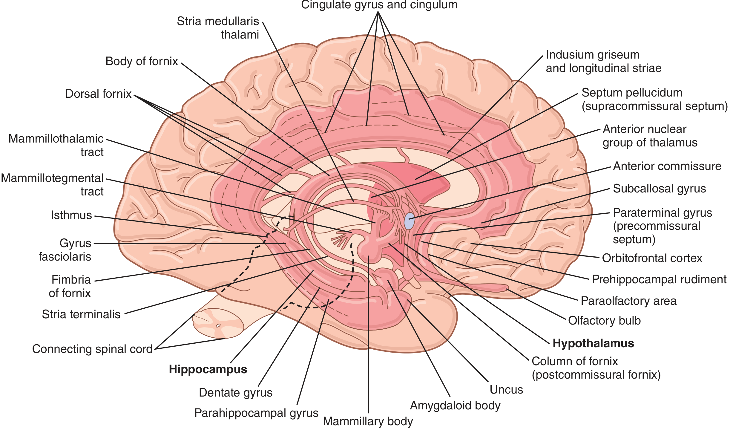
Components of the Limbic System (Diagram + Text)
The limbic system is the "border system" of the brain, forming a ring of paleocortex and deep nuclei around the diencephalon.
Diagram of the Limbic System:
Key position of Hypothalamus within the Limbic System:
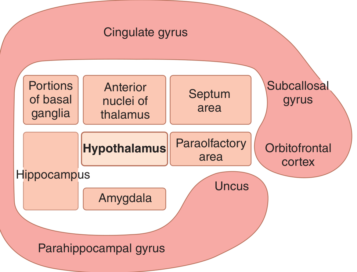
Components include:
Cortical components (Limbic cortex - ring on medial/ventral hemisphere):
- Cingulate gyrus (above corpus callosum)
- Parahippocampal gyrus
- Subcallosal/subcingulate gyrus
- Orbitofrontal cortex
- Paraolfactory area (uncus)
Subcortical components:
- Hypothalamus (central key structure)
- Hippocampus and dentate gyrus
- Amygdala (amygdaloid body)
- Anterior nuclei of thalamus
- Portions of basal ganglia (septum area)
- Mammillary bodies
- Fornix (connecting tract: fimbria, body, columns)
- Mammillothalamic tract
- Stria terminalis
(Guyton and Hall, Medical Physiology)
Vegetative Functions of the Hypothalamus (5 marks)
The hypothalamus controls the body's internal milieu - these are its vegetative (autonomic + endocrine) functions:
| Function | Mechanism |
|---|---|
| Temperature regulation | Anterior hypothalamus: cooling (sweating, vasodilation); Posterior: heating (shivering, vasoconstriction) |
| Thirst and water balance | Osmoreceptors detect plasma osmolarity; stimulate thirst and ADH (vasopressin) release from posterior pituitary |
| Hunger and feeding | Lateral hypothalamus = "hunger center"; Ventromedial nucleus = "satiety center" |
| Cardiovascular control | Posterior/lateral areas increase BP and HR; anterior area decreases them |
| Gastrointestinal regulation | Controls GI motility, gastric secretion (via parasympathetic) |
| Sleep-wake cycle | Suprachiasmatic nucleus governs circadian rhythm |
| Endocrine control | Releases releasing hormones (TRH, CRH, GnRH, GHRH, somatostatin) into hypophyseal portal blood controlling anterior pituitary |
| Posterior pituitary | Cell bodies of neurons that secrete ADH (antidiuretic hormone) and Oxytocin reside in supraoptic and paraventricular nuclei respectively |
| Sexual behavior | Preoptic area controls sexual drive |
| Emotional expression | Together with amygdala, mediates rage, fear, and pleasure responses |
Role of Hypothalamus in Reward and Punishment (4 marks)
-
When certain areas of the hypothalamus and limbic system are stimulated by implanted electrodes, animals self-stimulate repeatedly (thousands of times per hour) - these are reward/pleasure areas. The medial forebrain bundle (MFB) and lateral hypothalamus are the primary reward areas. Dopaminergic pathways (mesolimbic/mesocortical) are the key neurotransmitter system.
-
Reward areas: Lateral hypothalamus, medial forebrain bundle, septal nuclei, ventral tegmental area (VTA), nucleus accumbens. Stimulation produces feelings of euphoria, satisfaction, and motivation for repetitive behavior.
-
Punishment areas: Periventricular zone of hypothalamus, posterior hypothalamus, central gray of mesencephalon. Stimulation produces fear, escape behavior, aversion.
-
Physiological role: The reward-punishment system reinforces survival behaviors (eating, drinking, reproduction). The dopamine mesolimbic pathway (VTA → nucleus accumbens) is the neurobiological basis of reward. Drugs of abuse (cocaine, opioids) hijack this system.
-
Interactions: The amygdala feeds information about emotional significance of stimuli; the prefrontal cortex modulates reward decisions. Punishment stimulation suppresses eating, drinking, and reproductive behaviors; reward stimulation amplifies them.
Q2. Functional Divisions of Cerebellum | Connections | Functions | Clinical Manifestations [2+3+3+4]
Functional Divisions (2 marks)
The cerebellum has 3 functional (longitudinal) divisions:
| Division | Anatomical Equivalent | Main Input | Function |
|---|---|---|---|
| Vestibulocerebellum (Archicerebellum) | Flocculonodular lobe | Vestibular apparatus | Balance, eye movements, posture |
| Spinocerebellum (Paleocerebellum) | Vermis + intermediate zone | Spinal cord (proprioception) | Regulation of ongoing movement, muscle tone |
| Cerebrocerebellum (Neocerebellum) | Lateral hemispheres | Cerebral cortex (via pons) | Planning, initiation, timing of skilled voluntary movements |
To-and-Fro Connections (Diagram) (3 marks)
Afferent (Input to Cerebellum):
- Vestibulocerebellar tract (from vestibular nuclei/apparatus → flocculonodular lobe)
- Spinocerebellar tracts: Dorsal (Clarke's column, T1-L2) and Ventral (Gowers' tract) - ipsilateral and bilateral respectively
- Corticopontocerebellar pathway: Cerebral cortex → ipsilateral pontine nuclei → crosses → contralateral cerebellar hemisphere (main input to neocerebellum)
- Olivocerebellar tract: Inferior olivary nucleus → all parts of cerebellum (via climbing fibers - carry "error signals")
- Reticulocerebellar, cuneocerebellar tracts
Efferent (Output from Cerebellum) - all via deep cerebellar nuclei:
Cerebellar Cortex
       ↓
Deep Cerebellar Nuclei (Dentate, Emboliform, Globose, Fastigial)
       ↓              ↓
Dentate nucleus    Fastigial nucleus
       ↓              ↓
Thalamus (VL)    Vestibular nuclei
       ↓              ↓
Motor cortex     Spinal cord (via VST/reticulospinal)
(voluntary movement)     (posture/balance)
- Dentate → (via Superior cerebellar peduncle, crosses) → VL thalamus → Motor cortex
- Fastigial → (via inferior peduncle) → Vestibular nuclei, reticular formation
Functions of the Cerebellum (3 marks)
- Coordination of voluntary movement (smooth, accurate)
- Maintenance of posture and equilibrium
- Regulation of muscle tone
- Planning and timing of movements
- Coordination of eye movements (vestibuloocular reflex)
- Motor learning (e.g., learning to ride a bicycle)
- Correction of movement errors (compares intended vs. actual movement)
Clinical Manifestations of Cerebellar Lesion (4 marks)
Mnemonic: DANISH
| Sign | Description |
|---|---|
| Dysdiadochokinesia | Inability to perform rapid alternating movements |
| Ataxia (gait) | Wide-based, staggering, "drunken" gait; falls toward side of lesion |
| Nystagmus | Horizontal nystagmus, with fast phase toward lesion side |
| Intention tremor | Tremor that worsens as limb approaches target (worse at end of movement) |
| Scanning dysarthria | Slow, slurred, monotonous speech ("staccato speech") |
| Hypotonia | Decreased muscle tone on side of lesion |
Additional signs: Past-pointing (dysmetria), Rebound phenomenon (Holmes' rebound), truncal ataxia, decomposition of movement, cerebellar ataxia.
- Neocerebellum lesion: Limb ataxia, intention tremor, dysmetria, dysarthria
- Vestibulocerebellum lesion: Truncal ataxia, gait ataxia, nystagmus, equilibrium disturbances
- Spinocerebellum lesion: Hypotonia, gait ataxia
Q3 & Q4. Basal Ganglia - Nuclei, Connections, Functions | Parkinsonism [7+5 / 2+3+3+4]
Components/Nuclei of Basal Ganglia (2 marks)
The basal ganglia are a group of subcortical nuclei:
Striatum:
- Caudate nucleus
- Putamen
- (Together = neostriatum, the main input nucleus)
Pallidum (Globus Pallidus):
- Globus Pallidus externa (GPe)
- Globus Pallidus interna (GPi) - main output nucleus
Other components:
- Subthalamic nucleus (STN)
- Substantia nigra pars compacta (SNc) - dopaminergic input
- Substantia nigra pars reticulata (SNpr) - output
Connections/Pathways (3 marks)
Direct Pathway (excitatory net effect on movement):
Cortex → Striatum (putamen) --[inhibit]-→ GPi/SNpr
                                              ↓ [less inhibition]
                                          Thalamus (VL/VA) → Cortex
                                          (MORE movement facilitated)
Neurotransmitters: Striatum uses GABA; D1 receptors in striatum facilitate direct pathway.
Indirect Pathway (inhibitory net effect):
Cortex → Striatum --[inhibit]-→ GPe --[less inhibit]-→ STN
                                                         ↓ [excites]
                                                        GPi → [inhibits] Thalamus
                                                        (LESS movement)
D2 receptors in striatum inhibit the indirect pathway (normally facilitates movement when dopamine is present).
Dopaminergic Input (Nigrostriatal pathway):
- SNc → Striatum via nigrostriatal tract
- Dopamine activates D1 (direct pathway, excitatory) and inhibits D2 (indirect pathway, facilitatory)
- Net result: dopamine facilitates movement
Functions of Basal Ganglia (3 marks)
- Control of voluntary movements - inhibition of unwanted movements, scaling of movement amplitude
- Regulation of muscle tone (inhibit excess tone)
- Procedural learning and habit formation
- Executive functions (working memory, attention, planning)
- Emotional processing (limbic loop)
- Ocular motor control (saccadic eye movements)
Features of Parkinsonism (3 marks)
Parkinson's disease results from degeneration of dopaminergic neurons in the substantia nigra pars compacta, causing loss of dopamine input to striatum. This leads to overactivation of the indirect pathway → excessive inhibition of thalamus → reduced cortical activation → bradykinesia.
Cardinal Features (TRAP):
- Tremor - resting "pill-rolling" tremor (4-6 Hz), reduced with voluntary movement
- Rigidity - "lead-pipe" or "cogwheel" rigidity (uniform resistance throughout range)
- Akinesia/Bradykinesia - slowness/poverty of movement; reduced arm swing, mask-like face (hypomimia), micrographia
- Postural instability - loss of righting reflexes, falls, stooped posture (flexed neck, trunk, knees)
Other features: Shuffling gait with festination, freezing episodes, seborrhea, drooling, dysarthria/hypophonia, dementia (later), autonomic dysfunction, depression.
Treatment/Remedy for Parkinsonism (4-5 marks)
Pharmacological:
| Drug | Mechanism |
|---|---|
| Levodopa + Carbidopa | L-DOPA is precursor; carbidopa blocks peripheral decarboxylation, more L-DOPA reaches brain; converted to dopamine - gold standard |
| Dopamine agonists (Pramipexole, Ropinirole, Bromocriptine) | Directly stimulate D2 receptors |
| MAO-B inhibitors (Selegiline, Rasagiline) | Block dopamine breakdown |
| COMT inhibitors (Entacapone) | Reduce peripheral L-DOPA degradation |
| Amantadine | Dopamine release enhancer, NMDA antagonist (also reduces dyskinesias) |
| Anticholinergics (Benztropine, Trihexyphenidyl) | Restore dopamine-ACh balance in striatum; mainly for tremor |
Surgical:
- Deep Brain Stimulation (DBS) of subthalamic nucleus or GPi - most effective surgical option
- Thalamotomy (for tremor)
- Pallidotomy (for rigidity/dyskinesia)
Rehabilitation: Physiotherapy, speech therapy, occupational therapy
Q5. Synapse | Synaptic Potential | Ionic Basis | Properties [1+5+6]
Definition of Synapse (1 mark)
A synapse is the functional junction between two neurons (or between a neuron and an effector cell) where a signal is transmitted from one cell (presynaptic) to the next (postsynaptic). Most synapses in the CNS are chemical synapses.
Synaptic Potential (5 marks)
Synaptic potentials are graded potentials generated in the postsynaptic membrane in response to neurotransmitter binding.
Types:
-
EPSP (Excitatory Postsynaptic Potential)
- Produced by excitatory neurotransmitters (glutamate, acetylcholine)
- Ligand-gated channels open → Na⁺ influx (+K⁺ efflux) → membrane depolarization
- Local, graded depolarization (makes membrane more positive, toward -55 mV threshold)
- Summation (spatial + temporal) → action potential if threshold reached
-
IPSP (Inhibitory Postsynaptic Potential)
- Produced by inhibitory neurotransmitters (GABA, glycine)
- Cl⁻ channels open (Cl⁻ influx) or K⁺ channels open (K⁺ efflux) → hyperpolarization
- Makes membrane more negative (e.g., -75 mV) → harder to fire
Ionic Basis of EPSP:
- Neurotransmitter binds ionotropic receptor (e.g., AMPA receptor for glutamate)
- Combined Na⁺/K⁺ channel opens; Na⁺ equilibrium potential (+60 mV) draws Na⁺ in
- Reversal potential ~0 mV
- Net result: depolarization from resting -70 mV toward threshold -55 mV
Ionic Basis of IPSP:
- GABA-A receptor: Cl⁻ channel opens; Cl⁻ equilibrium potential (-65 mV) causes Cl⁻ influx → hyperpolarization
- GABA-B receptor (metabotropic): K⁺ channels open → hyperpolarization
Properties of Synapse (6 marks)
| Property | Explanation |
|---|---|
| One-way conduction | Neurotransmitter released only from presynaptic terminal; receptors only on postsynaptic membrane |
| Synaptic delay | 0.3-0.5 ms (time for vesicle fusion, diffusion, receptor binding); this is the basis of reaction time |
| Summation | Spatial: simultaneous input from multiple presynaptic neurons. Temporal: rapid repeated firing from one neuron |
| Facilitation | Repeated stimulation increases amplitude of postsynaptic response (due to Ca²⁺ accumulation) |
| Fatigue | With repeated stimulation, synaptic transmission decreases due to depletion of neurotransmitter vesicles |
| Post-tetanic potentiation | Increased response after a period of intense stimulation; due to residual Ca²⁺ in presynaptic terminal |
| Convergence | Many presynaptic neurons synapsing on one postsynaptic neuron (summation of inputs) |
| Divergence | One presynaptic neuron synapsing on many postsynaptic neurons (amplification) |
| Excitability changes | Anoxia, drugs, disease alter synaptic transmission |
| Susceptibility to hypoxia | Synapses fail before nerve fibers during oxygen deprivation |
| Pharmacological sensitivity | Drugs can facilitate (strychnine blocks glycine receptors) or inhibit (benzodiazepines enhance GABA) |
Q6. Pyramidal Tract - Origin, Course, Termination | Babinski Sign [10+2]
Origin
The pyramidal (corticospinal) tract originates from:
- Primary motor cortex (precentral gyrus, Area 4) - ~30%
- Premotor cortex (Area 6) - ~30%
- Somatosensory cortex (Areas 1, 2, 3, 5, 7) - ~40%
- Total: ~1 million fibers per tract; ~3% are large (Betz cells from Area 4)
Course
Motor Cortex (Areas 4, 6)
    ↓ (Internal capsule - posterior limb,
       genu for corticobulbar fibers)
    ↓
Cerebral peduncle (middle 3/5 = basis pedunculi)
    ↓
Pons (fibers dispersed by pontine nuclei)
    ↓
Medullary pyramid (fibers re-converge)
    ↓
Pyramidal decussation (at junction of medulla and spinal cord)
85-90% cross → Lateral corticospinal tract (crossed)
10-15% stay → Anterior corticospinal tract (uncrossed, cross later at each spinal level)
    ↓
Spinal cord - descends in lateral and anterior funiculi
Corticobulbar fibers (for cranial nerve motor nuclei) leave at each level of the brain stem. Most are bilateral EXCEPT lower facial nucleus (contralateral only) and hypoglossal (predominantly contralateral).
Termination
- Lateral CST: Terminates in dorsal horn (laminae IV-VI) and ventral horn (lamina IX) - synapse on alpha motor neurons (direct) and interneurons (indirect)
- Anterior CST: Crosses in anterior white commissure at respective segmental level → ventral horn motor neurons
- Controls voluntary, skilled, fine movements especially of distal extremities
Babinski Sign (2 marks)
- Normal response: Plantar flexion of all toes when lateral sole is stroked (L5-S1 reflex)
- Babinski sign (positive/abnormal): Extension (dorsiflexion) of big toe + fanning (abduction) of other toes
- Significance: Indicates damage to the corticospinal (pyramidal) tract at any level above S1
- In infants under ~18 months: physiologically present (incomplete myelination of CST)
- Mechanism: Release of primitive spinal reflex from higher cortical control due to UMN lesion
- Other names: Extensor plantar response; Chaddock, Oppenheim, Gordon signs are equivalent tests
Q7. Muscle Tone | Types of Hypertonia | Differences [2+5+2+3]
Definition of Muscle Tone (2 marks)
Muscle tone (tonus) is the continuous partial contraction of muscle that is maintained even at rest. It is the resistance to passive stretch. Normal tone provides:
- Posture maintenance
- Readiness for movement
- Protection of joints
It is maintained by the stretch reflex arc (Ia afferent → spinal cord → alpha motor neuron) under constant supraspinal modulation.
How Muscle Tone is Maintained (5 marks)
Peripheral mechanism:
- Muscle spindles (intrafusal fibers) constantly monitor muscle length
- Ia afferents from nuclear bag/chain fibers → spinal cord → monosynaptic excitation of alpha motor neurons → maintain basal contraction
- Gamma motor neurons keep intrafusal fibers taut, maintaining spindle sensitivity
Supraspinal control:
- Facilitatory inputs (increase tone): Lateral vestibulospinal tract (LVST), reticulospinal (pontine/medial), corticospinal
- Inhibitory inputs (decrease tone): Cortical (basal ganglia, cerebellum via thalamus), dorsal reticulospinal (medullary)
- Balance of these determines resting tone
Decerebrate rigidity (section at midbrain level) → loss of inhibitory cortical/cerebellar influence → LVST + pontine reticulospinal overactive → marked extensor (antigravity) hypertonia - proves facilitatory > inhibitory at this level
Types of Hypertonia (2 marks)
- Spasticity (UMN/pyramidal lesion)
- Rigidity (extrapyramidal/basal ganglia lesion)
- Gegenhalten (Paratonia) - involuntary resistance that varies with force of examiner (frontal lobe lesion)
Differences Between Spasticity and Rigidity (3 marks)
| Feature | Spasticity | Rigidity |
|---|---|---|
| Lesion | Pyramidal/corticospinal tract (UMN) | Extrapyramidal (basal ganglia, e.g., Parkinson's) |
| Mechanism | Loss of descending inhibition → increased stretch reflex excitability | Loss of dopamine → imbalance of direct/indirect pathways → increased alpha motor neuron activity |
| Distribution | Flexors in arm, extensors in leg (antigravity muscles) | Both flexors and extensors equally (generalized) |
| Type of resistance | Velocity-dependent (increases with speed of stretch); "clasp-knife" quality (gives way suddenly) | Velocity-independent; uniform throughout range ("lead-pipe") or intermittent ("cogwheel" - tremor superimposed) |
| Clasp-knife | Present (Golgi tendon organ activation) | Absent |
| Reflexes | Hyperreflexia + Babinski sign | Normal or slightly increased reflexes |
| Clonus | May be present | Absent |
| Examples | Stroke (hemiplegia), spinal cord injury, MS | Parkinson's disease |
Q8. Pathway of Pain Sensation | Stress Analgesia [2+5+2+3]
What is Pain?
Pain is an unpleasant sensory and emotional experience associated with actual or potential tissue damage (IASP definition). Types: Acute (nociceptive), chronic, neuropathic.
Neural Pathway for Pain Sensation (with Diagram) (5 marks)
Receptors: Free nerve endings (nociceptors) in skin, viscera, muscles - respond to mechanical, thermal, chemical stimuli.
First-order neurons (Peripheral):
- Fast pain (sharp, localized): A-delta fibers (myelinated, 6-30 m/s) → detect sharp, pricking pain
- Slow pain (burning, aching): C fibers (unmyelinated, 0.5-2 m/s) → detect burning, aching, chronic pain
At Spinal Cord:
- Enter via dorsal root → Lissauer's tract (posterolateral funiculus) → Dorsal Horn (Substantia Gelatinosa, Rexed laminae I, II, V)
- Synapse on second-order neurons → Gate Control here (modulation by large fiber input)
Second-order neurons:
- Cross the midline in anterior white commissure (within 1-2 spinal segments)
- Ascend in Anterolateral (spinothalamic) tract in the contralateral lateral funiculus
Third-order neurons (Thalamus → Cortex):
Spinal cord (laminae I, II, V)
    ↓ (crosses midline)
Anterolateral/Spinothalamic tract
    ↓
Thalamus:
  - VPL (Ventral Posterolateral): fast/discriminative pain
  - Intralaminar nuclei: slow/diffuse pain, arousal
    ↓
Primary Somatosensory Cortex (SI, SII): localization, intensity
Anterior Cingulate Cortex: emotional/affective component
Insular Cortex: autonomic response
Visceral pain: Travels via sympathetic afferents (C fibers) → enters cord at multiple levels → referred pain (e.g., cardiac pain referred to left arm via T1-T4 segments - Hilton's law/dermatome overlap)
Stress Analgesia and Physiological Basis (2+3 marks)
Stress Analgesia: The reduction or absence of pain perception during extreme stress, fear, or injury (e.g., soldiers not feeling wounds during battle, athletes continuing despite injury).
Physiological Basis - Descending Pain Modulation System:
Endogenous opioid system:
- Stress activates hypothalamus → releases beta-endorphin, enkephalins, dynorphins
- These act on mu, delta, kappa opioid receptors in the dorsal horn, PAG, and raphe nuclei
The Analgesic (Descending Inhibitory) Pathway:
Stress/Emotion/Cortex
    ↓
Periaqueductal Gray (PAG) - midbrain
    ↓ (endorphins, enkephalins)
Nucleus Raphe Magnus (raphe nuclei) - medulla
    ↓ (Serotonin - 5-HT)
Dorsal horn (laminae I, II)
    ↓ also Locus coeruleus (Norepinephrine)
Gate control: Presynaptic inhibition of C fiber terminals
             + Inhibition of second-order neurons
Result: Reduced pain transmission
Gate Control Theory (Melzack & Wall, 1965):
- Large-diameter fibers (A-beta, touch/pressure) activate inhibitory interneurons (substantia gelatinosa) → "close the gate" for C fiber (pain) transmission
- Small C fibers "open the gate"
- Descending control can close the gate
Neurotransmitters involved: Enkephalin, Serotonin, Norepinephrine, Beta-endorphin, GABA
Q9. Cerebellum - Neural Connections | Functional Divisions | Smooth Movement | Abnormalities [3+3+3+3]
(Covered extensively in Q2 above; additional detail below)
Neural Connections in Cerebellum (3 marks)
Internal Circuit:
- Mossy fibers (from spinal cord, pons) → Granule cells → Parallel fibers → Purkinje cell dendrites
- Climbing fibers (from inferior olivary nucleus) → directly synapse on Purkinje cells (1:1 relationship) - carry "error signals"
- Purkinje cells (main output of cortex) → inhibitory (GABA) → Deep cerebellar nuclei
- Basket and Stellate cells (inhibitory interneurons) → inhibit Purkinje cells (lateral inhibition)
- Golgi cells (inhibitory) → feedback inhibition of granule cells
Deep Cerebellar Nuclei (output):
- Dentate (largest) - neocerebellum → to VL thalamus
- Emboliform + Globose (= Interpositus) - spinocerebellum → to red nucleus (rubrospinal)
- Fastigial - vestibulocerebellum → to vestibular nuclei
How Cerebellum Helps in Smooth and Coordinated Movement (3 marks)
-
Comparator function: Cerebellum receives an efference copy of the motor command (from cortex via pons) AND actual movement feedback (from proprioceptors via spinocerebellar tracts). It compares intended vs. actual movement and sends corrective signals.
-
Timing and coordination: Coordinates the timing and sequencing of muscle contractions across multiple joints; ensures agonist-antagonist pairs work smoothly.
-
Predictive control: Neocerebellum uses internal models to predict movement outcome and pre-correct, reducing oscillation.
-
Regulation of tone: Spinocerebellum adjusts muscle tone via vestibulospinal/reticulospinal tracts.
-
Motor learning: Long-term depression (LTD) of parallel fiber-Purkinje cell synapses (when activated together with climbing fiber error signals) is the cellular basis of cerebellar motor learning.
Abnormalities with Cerebellar Damage (3 marks)
(See Q2 clinical manifestations - DANISH)
Additional points:
- Lesion rule: Cerebellar hemisphere lesion produces ipsilateral signs (because cerebellum already receives from the opposite cortex via crossed cortico-ponto-cerebellar pathway)
- Cerebellar ataxia vs. sensory ataxia: Cerebellar ataxia - Romberg negative (doesn't worsen with eyes closed); sensory ataxia - Romberg positive
- Titubation: Rhythmic oscillatory tremor of head and trunk (midline/anterior lobe lesion)
Q10. Basal Ganglia - Components, Connections, Functions, Parkinsonism Treatment [2+3+3+3+5+2]
(Comprehensively covered in Q3/Q4 above)
Summary for quick reference:
- Components: Caudate, Putamen, GPe, GPi, STN, SNc, SNpr
- Pathways: Direct (facilitatory) and Indirect (inhibitory) - both modulated by dopamine from SNc
- Functions: Motor control, procedural learning, executive function, emotional processing
- Parkinsonism features: TRAP (Tremor-Rigidity-Akinesia-Postural instability)
- Treatment: Levodopa + Carbidopa (gold standard), dopamine agonists, MAO-B inhibitors, DBS surgery
Q11. Pain - Definition | Pathway | Inhibition of Transmission and Perception [2+5+5]
(Covered in Q8 above)
Additional detail on Inhibition of Transmission and Perception (5 marks):
Presynaptic inhibition at dorsal horn:
- Opioid receptors (mu) on presynaptic C fiber terminals → reduce Ca²⁺ influx → reduce substance P and glutamate release
- Enkephalinergic interneurons in lamina II activated by descending serotonergic/noradrenergic fibers
Postsynaptic inhibition:
- Glycine and GABA-mediated hyperpolarization of second-order neurons in dorsal horn
- Opioids increase K⁺ conductance → hyperpolarize postsynaptic neurons
Supraspinal modulation:
- Thalamic gate: Thalamic reticular neurons can filter pain signals before cortical perception
- Anterior cingulate cortex and prefrontal cortex provide cognitive/attentional modulation (distraction reduces pain perception)
- Placebo effect via endogenous opioid release
Pharmacological inhibition:
- NSAIDs: Inhibit cyclooxygenase → reduce prostaglandin (PGE2) synthesis → reduce sensitization of nociceptors (peripheral mechanism)
- Opioids: Activate PAG, rostral ventromedial medulla, and dorsal horn opioid receptors
- Local anesthetics: Block Na⁺ channels → prevent generation/propagation of action potentials in nociceptive fibers
- Tricyclic antidepressants/SNRIs: Enhance descending noradrenergic inhibition
Q12. Functional Divisions of Cerebellum | Principal Functions | Internal Circuit | Cerebellar Ataxia
(Functional divisions and internal circuit covered in Q2 and Q9)
Cerebellar Ataxia (important exam question)
Cerebellar ataxia is incoordination of voluntary movement due to cerebellar dysfunction.
Features:
- Gait: Wide-based, staggering, lurching gait; cannot walk tandem (heel-to-toe)
- Limb: Intention tremor (worsens near target), dysmetria (past-pointing), dysdiadochokinesia
- Speech: Scanning/staccato dysarthria
- Eye: Nystagmus (horizontal, most common; fast phase ipsilateral to lesion in hemisphere lesion)
- Tone: Hypotonia
- Romberg: Negative (balance worse with eyes open too - distinguishes from sensory ataxia)
- Truncal ataxia: Unable to sit/stand steadily (anterior lobe/midline lesion)
Causes: Stroke (PICA territory = lateral medullary or cerebellar), multiple sclerosis, alcohol (anterior vermis), hereditary (spinocerebellar ataxias, Friedreich's ataxia), tumors, paraneoplastic.
Q13. Stretch Reflex | Receptor | Reciprocal Innervation | Renshaw Cell Inhibition [2+6+2+2]
Stretch Reflex (2 marks)
The stretch reflex (myotatic reflex) is a monosynaptic spinal reflex in which stretch of a muscle leads to its own contraction.
Arc:
- Stimulus: Muscle stretch (e.g., tap on tendon)
- Receptor: Muscle spindle (nuclear bag and nuclear chain intrafusal fibers)
- Afferent: Ia (primary) afferents (fastest conducting, from both bag and chain fibers)
- Center: Spinal cord (ventral horn - monosynaptic)
- Efferent: Alpha motor neuron
- Effector: Same (stretched) muscle contracts
Example: Knee jerk (patellar reflex - L3,L4), ankle jerk (S1, S2), biceps jerk (C5,C6).
Muscle Spindle Receptor (6 marks)
The muscle spindle is the receptor for the stretch reflex.
Structure:
- Fusiform capsule embedded parallel to extrafusal (main) muscle fibers
- Contains intrafusal fibers (2 types):
- Nuclear bag fibers (large, nuclei clustered in equatorial bag; 2 types: dynamic bag1, static bag2) - respond to rate of change of stretch (dynamic sensitivity)
- Nuclear chain fibers (smaller, nuclei in chain; 3-5 per spindle) - respond to static length (static sensitivity)
Innervation:
- Afferent:
- Ia (primary) afferents: Annulospiral endings on all intrafusal fibers; respond to both dynamic and static stretch (most sensitive)
- II (secondary) afferents: Flower-spray endings mainly on chain fibers; respond to static stretch (length only)
- Efferent (Fusimotor):
- Dynamic gamma motor neurons: Innervate nuclear bag1 fibers → increase dynamic sensitivity
- Static gamma motor neurons: Innervate bag2 and chain fibers → maintain tonic sensitivity
Diagram:
       Alpha MN  ←────────────────────────────────
       (extrafusal)                                 |
                                                    |
Extrafusal fiber ====[SPINDLE]====                  |
                       ↕                           |
               Ia afferent ────────────────────────→ Spinal cord
               II afferent                          (Vc horn)
                       ↕
               Gamma MN (intrafusal tone)
Gamma-loop (fusimotor system):
- Supraspinal centers (cortex, cerebellum, reticular formation) continuously activate gamma neurons
- This keeps intrafusal fibers taut during alpha motor neuron-driven contraction
- Prevents spindle "unloading" so sensitivity is maintained throughout movement
- Alpha-gamma co-activation ensures continuous spindle monitoring during voluntary contraction
Reciprocal Innervation (2 marks)
When the stretch reflex activates an agonist muscle, the antagonist muscle is simultaneously inhibited - this is reciprocal innervation (Sherrington's law of reciprocal innervation).
Mechanism:
- Ia afferent from stretched muscle → spinal cord
- Excites alpha motor neurons of agonist (direct monosynaptic)
- Also activates Ia inhibitory interneurons → which synapse on and inhibit alpha motor neurons of antagonist
- Result: Agonist contracts, antagonist relaxes → smooth coordinated movement
Example: When quadriceps (agonist) contracts during knee jerk, hamstrings (antagonist) are simultaneously inhibited.
Renshaw Cell Inhibition (2 marks)
Renshaw cells are inhibitory interneurons in the ventral horn of the spinal cord that provide recurrent collateral inhibition of alpha motor neurons.
Circuit:
Alpha Motor Neuron
    ↓ (axon collateral - releases ACh)
Renshaw Cell (in lamina VII)
    ↓ (releases Glycine)
Back onto same Alpha Motor Neuron (inhibits it)
    + Nearby Alpha Motor Neurons (lateral inhibition)
    + Ia Inhibitory interneurons (disinhibits antagonist)
Significance:
- Negative feedback: Limits excessive motor neuron firing (prevents tetanic muscle contraction from runaway positive feedback)
- Lateral inhibition: Sharpens motor output by focusing activity to specific motor neuron pools (enhances contrast)
- Rhythmic patterns: Contributes to rhythmic locomotor patterns
- Pharmacology: Strychnine blocks glycine receptors → removes Renshaw inhibition → tetanic convulsions; Tetanus toxin blocks glycine release → same effect
---All 13 questions have been comprehensively answered. Here is a quick summary of what was covered:
Quick Reference Summary - All 13 Questions Answered:
| Q# | Topic | Key Points |
|---|---|---|
| 1 | Limbic system + Hypothalamus | Components (hippocampus, amygdala, cingulate gyrus, fornix, hypothalamus); Vegetative functions (temp, thirst, hunger, endocrine); Reward (MFB, dopamine) & Punishment (periventricular zone) |
| 2 | Cerebellum divisions, connections, functions, lesion | Vestibulocerebellum, Spinocerebellum, Cerebrocerebellum; DANISH signs of lesion |
| 3 | Basal ganglia nuclei + Parkinsonism features + treatment | Striatum, GP, STN, SN; Direct/indirect pathways; TRAP; L-DOPA, DBS |
| 4 | Basal ganglia components + Parkinson's disease | Same as Q3 with pathway diagram |
| 5 | Synapse, synaptic potential, ionic basis, properties | EPSP (Na⁺ influx), IPSP (Cl⁻ influx); 10 key properties |
| 6 | Pyramidal tract + Babinski sign | Origin (areas 4,6), internal capsule, pyramidal decussation, CST; Babinski = UMN lesion sign |
| 7 | Muscle tone + hypertonia types + differences | Spasticity (pyramidal, clasp-knife, velocity-dependent) vs Rigidity (extrapyramidal, lead-pipe, cogwheel) |
| 8 | Pain pathway + stress analgesia | A-delta/C fibers → spinothalamic tract → thalamus → cortex; PAG → Raphe → Enkephalin system |
| 9 | Cerebellar neural connections, coordinated movement, abnormalities | Mossy/climbing fibers, Purkinje cells, deep nuclei; comparator function; ipsilateral signs |
| 10 | BG components + Parkinsonism features + treatment | Full summary covering Q3/4 |
| 11 | Pain definition + pathway + inhibition | Gate control theory; endogenous opioids; NSAIDs, opioids, tricyclics |
| 12 | Cerebellar divisions + internal circuit + ataxia | Cerebellar ataxia: wide-based gait, intention tremor, Romberg negative |
| 13 | Stretch reflex + muscle spindle + reciprocal innervation + Renshaw | Ia afferent → monosynaptic reflex; Renshaw = recurrent collateral inhibition via glycine |
Sources: Guyton and Hall Medical Physiology (14th ed.), Costanzo Physiology (7th ed.) - Guyton and Hall, pp. 733, 550, 575-577; Costanzo, pp. 77-78
This is a shared conversation. Sign in to Orris to start your own chat.