
Perforator marking in brachial plexus injury
Reading File
Reading File
Finding Sources
Finding Sources
Searching PubMed
"brachial plexus injuries" AND perforator flap
Searching the Web
brachial plexus injury reconstruction perforator marking flap perforator
Finding Sources
Finding Sources
Searching the Web
free functional gracilis transfer brachial plexus injury perforator Doppler marking technique
If you mean free functional gracilis muscle transfer (FFGT) for late or complete brachial plexus injury:
- Perforator marking is not routinely needed when harvesting gracilis as a muscle-only functional transfer.
- The key marking is the dominant gracilis vascular pedicle, usually entering the deep surface of the muscle about 8-12 cm below the pubic tubercle, in the proximal third of the thigh.
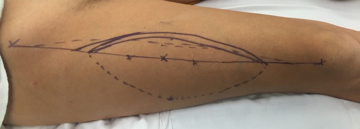
- Mark the gracilis line from the pubic tubercle to the medial tibial condyle. The skin incision is centered over its proximal half, with Doppler used to help localize the vascular pedicle if anatomy is uncertain.
- If taking a skin paddle for flap monitoring or soft-tissue cover, use handheld Doppler to map usable musculocutaneous perforators around the proximal to middle third, then design the paddle around a confirmed perforator.
- For transfer/inset, place 5-cm interval marks on the gracilis while in situ, then reproduce them after transfer. This helps maintain appropriate resting length and tension, rather than serving as perforator markings. One reported technique sets elbow flexion transfer tension with the elbow at approximately 45 degrees of flexion and forearm supinated.
For brachial plexus reconstruction, gracilis free-muscle transfer is principally used to restore elbow flexion, and may be innervated by spinal accessory or intercostal nerve. Rockwood and Green's Fractures in Adults, 10th ed, lines 2576-2580. A 2024 meta-analysis found that free functional gracilis transfer commonly restored at least antigravity elbow flexion, but results depend on patient factors, construct, and donor nerve choice, as summarized in this systematic review.
If by “perforator marking” you mean a different flap, such as ALT, TDAP, or a specific recipient-site marking, tell me which flap and I will give the exact surface landmarks.
Show the surface landmarks for ALT perforator marking
Reading File
Finding Sources
Finding Sources
Searching Images
anterolateral thigh flap perforator surface markings line anterior superior iliac spine patella diagram
Searching PubMed
"anterolateral thigh flap" AND perforator
Searching the Web
anterolateral thigh flap surface marking ASIS lateral patella perforator landmark Doppler
ALT perforator surface marking
-
Position the patient supine, with the hip neutral and knee extended.
-
Identify and mark:
- ASIS: anterior superior iliac spine
- Superolateral border of patella
-
Draw the ALT axis line from the ASIS to the superolateral patellar border. This approximates the intermuscular plane between rectus femoris medially and vastus lateralis laterally and overlies the descending branch territory of the lateral circumflex femoral system.
-
Mark the standard ABC perforator points:
| Point | Surface position |
|---|---|
| B | Midpoint of the ASIS-patella line, then 1.5 cm lateral to it |
| A | 5 cm proximal to B |
| C | 5 cm distal to B |
- Use a handheld Doppler to confirm the perforator(s), particularly around B and within about a 6-cm circle centered on the line midpoint. Center the skin paddle on the strongest reliable signal, not merely on a theoretical point.
       ASIS
        ●
         \
          \       A  ●  (5 cm proximal to B)
           \
            \      B  ●  midpoint + 1.5 cm lateral
             \
              \     C  ●  (5 cm distal to B)
               \
                ●  Superolateral patellar border
Practical points
- The midpoint is a useful starting landmark, but perforator location and course vary.
- Doppler identifies a likely cutaneous exit point but cannot reliably tell whether the vessel is musculocutaneous or septocutaneous. In larger, obese, scarred, or previously operated thighs, duplex ultrasonography or CTA mapping can be useful.
- Begin flap design only after confirming a usable perforator intraoperatively. The ALT flap is commonly based on perforators from the descending branch of the lateral circumflex femoral artery. Sabiston Textbook of Surgery, lines 1045-1050.
The ASIS-to-superolateral-patella line and the ABC system are described in this ALT flap technical guide and the surface-marking study.
This is a shared conversation. Sign in to Orris to start your own chat.