Now I have all the information needed. Let me compile the comprehensive response.
Low Back Pain — Complete Clinical Overview
1. DEFINITION & EPIDEMIOLOGY
Low back pain (LBP) is pain localized between the inferior costal margin and the inferior gluteal folds, with or without referred leg pain (sciatica). It is one of the most prevalent and disabling conditions globally.
- >70% of people experience LBP significant enough to limit daily activities at some point in their life
- Peak prevalence in the 45–64 year age group with no significant gender difference
- Accounts for the greatest number of work-related disability days worldwide
- ~25% of acute LBP cases develop into chronic LBP (symptoms >3 months)
- Recurrence rate: ~50% within 1 year even after resolution
2. ANATOMY & BIOMECHANICS
Lumbar Spine Anatomy
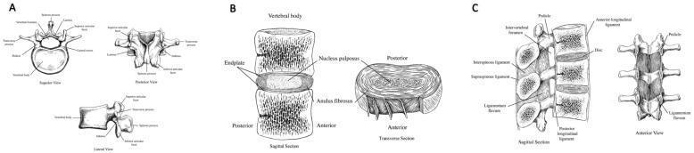
The lumbar spine (L1–L5) consists of vertebral bodies, intervertebral discs (IVDs), facet joints, ligaments, and supporting musculature. Each IVD has:
- Nucleus pulposus — gelatinous central core, absorbs compressive loads
- Annulus fibrosus — concentric fibrocartilaginous rings, resists shear and torsion
- Cartilaginous endplates — interface between disc and vertebral body
Key pain-sensitive structures in the lumbar spine:
- Outer annulus fibrosus (sinuvertebral nerve)
- Posterior longitudinal ligament
- Facet joint capsules (medial branch of dorsal ramus)
- Vertebral periosteum, epidural veins, dura mater
- Paraspinal muscles and thoracolumbar fascia
Biomechanical Concepts
| Mechanism | Effect |
|---|
| Axial compression (heavy lifting, prolonged sitting) | Increased intradiscal pressure; nucleus pulposus forced posteriorly |
| Flexion loading | Posterior annular stress; facet joint distraction |
| Extension loading | Posterior element (facet joint, pars) compression |
| Repetitive twisting | Annular fiber fatigue, circumferential tears |
| Sustained postures | Creep deformation of disc and ligaments |
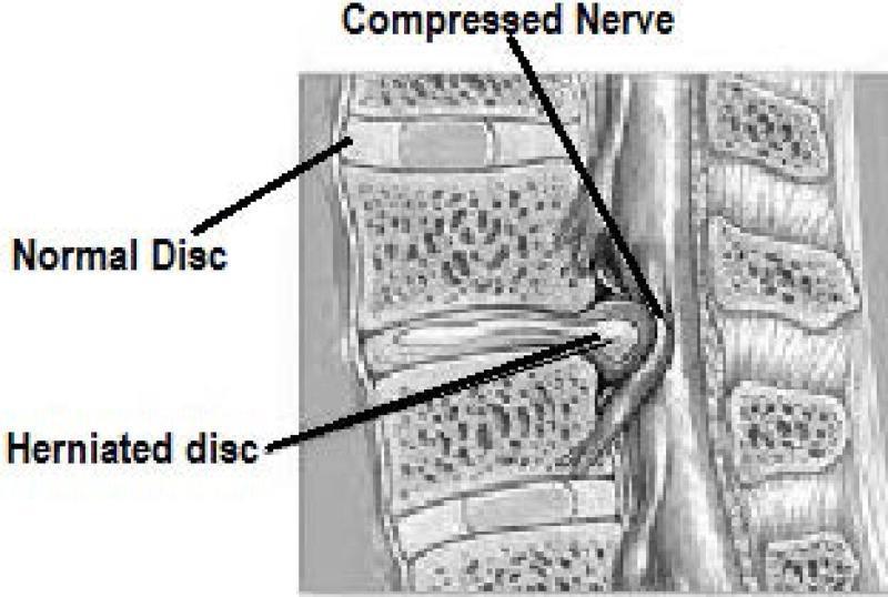
Disc herniation biomechanics: When intradiscal pressure exceeds annular tensile strength (especially in combined flexion + axial load), nucleus pulposus material protrudes or extrudes through the annular fibers, compressing adjacent nerve roots.
3. ETIOLOGY & CLASSIFICATION
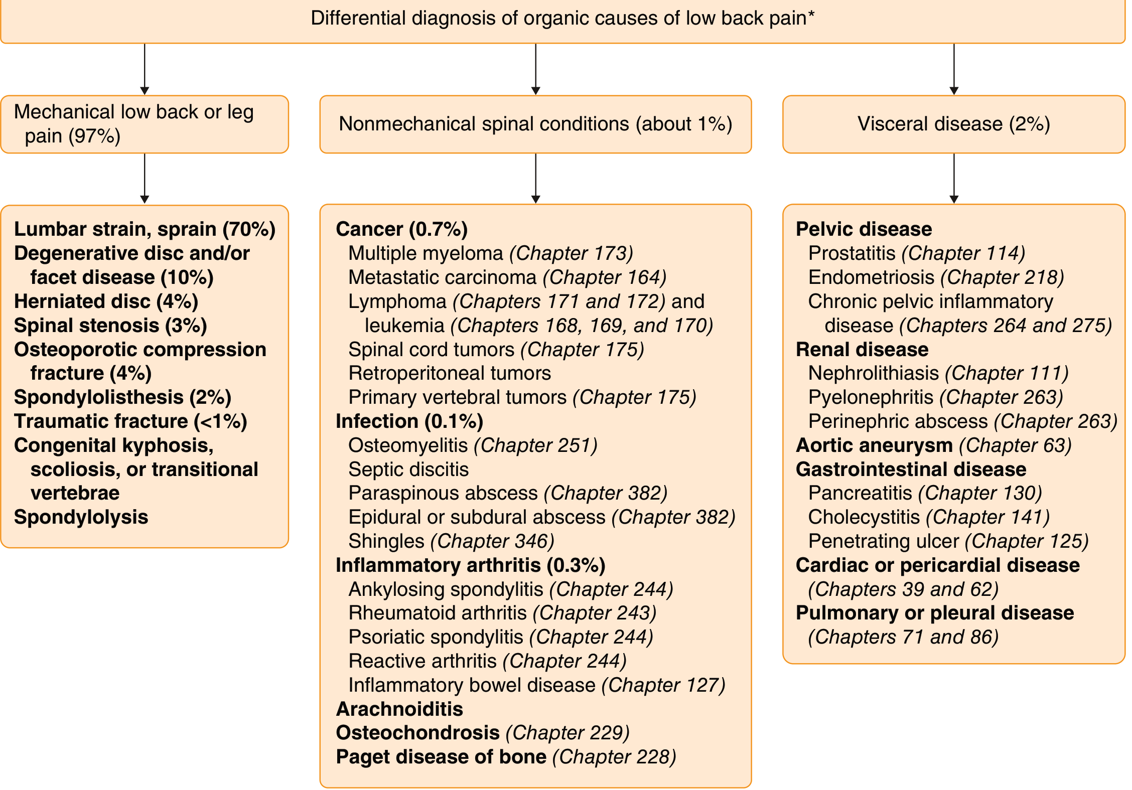
A. Mechanical (≈97% of cases)
| Category | Specific Conditions |
|---|
| Muscle/Ligament | Acute strain, chronic myofascial pain, piriformis syndrome |
| Disc pathology | Disc herniation (HNP), discogenic pain, disc degeneration (DDD) |
| Facet joint | Facet arthropathy (zygapophyseal joint pain) — 10–15% of axial LBP |
| Spondylosis | Degenerative changes: osteophytes, ligament hypertrophy, disc desiccation |
| Spinal stenosis | Central canal or foraminal narrowing (most common at L4–L5) |
| Spondylolysis / Spondylolisthesis | Pars interarticularis fracture; anterior vertebral slip (grades I–IV) |
| Sacroiliac joint | SI joint dysfunction — 20–35% of suspected lower axial LBP |
| Postural/Structural | Scoliosis, hyperlordosis, leg-length discrepancy |
B. Non-Mechanical / Serious (Red Flag) Causes
| System | Examples |
|---|
| Neoplastic | Primary bone tumour, spinal metastasis (breast, lung, prostate, thyroid, renal) |
| Infectious | Vertebral osteomyelitis, discitis, epidural abscess, TB of spine (Pott's disease) |
| Inflammatory | Ankylosing spondylitis, psoriatic arthropathy, reactive arthritis |
| Fracture | Osteoporotic compression fracture, traumatic fracture |
| Cauda equina syndrome | Large central disc herniation or tumour compressing cauda equina |
| Visceral referred pain | Aortic aneurysm, pancreatitis, renal calculi, pelvic pathology, endometriosis |
| Metabolic | Osteoporosis (Paget's disease), hyperparathyroidism |
Differential Diagnosis — Organic Causes of LBP
4. PATHOBIOLOGY OF DISC DEGENERATION & SPONDYLOSIS
Spondylosis is the universal degenerative cascade of the spine:
- Disc desiccation — loss of water content and proteoglycans from nucleus pulposus
- Loss of disc height — annular bulging, loss of elasticity of annulus fibrosus
- Osteophyte formation — marginal new bone growth at vertebral endplates
- Facet joint arthropathy — secondary degenerative change at zygapophyseal joints
- Ligamentum flavum hypertrophy — contributes to canal stenosis
- Spondylolisthesis — vertebral slip due to combined instability
Genetic predisposition: IVD degeneration has heritability estimates of 35–75%. Congenital or acquired spondylolysis (pars interarticularis fracture bilaterally) promotes spondylolisthesis development.
5. SIGNS & SYMPTOMS
Acute LBP (< 6 weeks)
- Sudden-onset pain following lifting, twisting, or minor trauma
- Restricted range of motion (especially flexion)
- Paraspinal muscle spasm and guarding
- Pain relieved by rest; worsened by movement and prolonged postures
Chronic LBP (> 3 months)
- Persistent, often bilateral aching pain
- Associated with psychosocial factors (depression, anxiety, catastrophizing)
- Poor sleep, reduced activity tolerance, functional disability
Radiculopathy (Sciatica)
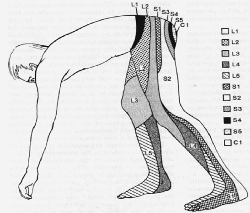
- L4 root: Pain radiating to medial leg; weakness of knee extension (quadriceps); reduced patellar reflex
- L5 root: Pain to dorsal foot and big toe; weakness of ankle/great toe dorsiflexion (foot drop); no reflex change typically
- S1 root: Pain to lateral foot and heel; weakness of plantarflexion; reduced/absent Achilles reflex
Lumbar Dermatomes
Neurological Signs by Root Level
| Level | Pain | Sensory Loss | Motor Weakness | Reflex Lost |
|---|
| L1–L2 | Groin, anterior thigh | Anterior thigh | Hip flexion | — |
| L3 | Anterior thigh to knee | Medial thigh | Knee extension (quad) | — |
| L4 | Medial leg to ankle | Medial leg | Ankle dorsiflexion | Patellar (↓) |
| L5 | Lateral leg, dorsal foot | Dorsal foot/big toe | EHL, foot dorsiflexion | — |
| S1 | Posterior thigh, heel, lateral foot | Lateral foot/heel | Plantarflexion | Achilles (absent) |
| S2–S4 | Perianal, saddle | Saddle region | Sphincter weakness | Anal wink (absent) |
Neurogenic Claudication (Spinal Stenosis)
- Bilateral buttock/leg pain, heaviness, paresthesias — worsened by walking/standing, relieved by sitting or forward flexion (the "shopping cart sign")
- Must be distinguished from vascular claudication (improved by standing still; peripheral pulses absent)
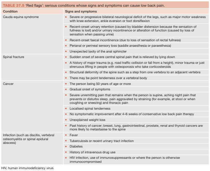
6. RED FLAGS — Warning Signs of Serious Disease
Key red flags from Goldman-Cecil Medicine:
| Red Flag | Possible Cause |
|---|
| History of cancer | Spinal metastasis |
| Unexplained weight loss, fever | Malignancy / infection |
| Age < 20 or > 50 at first episode | Atypical aetiology |
| Trauma history | Fracture |
| IV drug use / immunosuppression | Spinal epidural abscess |
| Bladder/bowel dysfunction + saddle anaesthesia | Cauda equina syndrome (surgical emergency) |
| Progressive bilateral neurological deficit | Cauda equina / cord compression |
| Pain unrelieved by rest; worse at night | Malignancy |
| Osteoporosis + corticosteroid use | Compression fracture |
Cauda equina syndrome is a neurosurgical emergency requiring MRI and urgent decompression within hours to prevent permanent incontinence and paralysis.
7. CLINICAL EXAMINATION
Observation & Inspection
- Spinal alignment: scoliosis, loss of lumbar lordosis, lateral shift
- Posture: antalgic lean, flexed posture
- Muscle wasting, atrophy of glutei/calf
- Skin: rashes (herpes zoster, psoriasis), scars, café-au-lait spots
Palpation
- Spinous process tenderness: fracture, infection, tumour (percussion test)
- Paraspinal tenderness: muscle spasm; trigger points
- SI joint tenderness: sacroiliitis
- Sciatic notch tenderness: piriformis syndrome
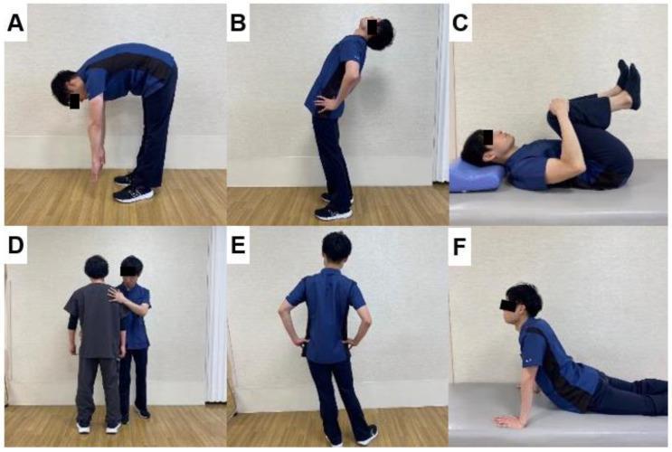
Range of Motion
| Movement | Normal | LBP Limitation |
|---|
| Lumbar flexion | Fingertips reach floor | Restricted, pain on flexion |
| Lumbar extension | 25–30° | Restricted in stenosis |
| Lateral flexion | 25° each side | Asymmetric restriction |
| Rotation | 30° each side | Pain with torsion |
Neurological Examination
- Motor: Hip flexors, knee extensors, ankle dorsiflexors, toe extensors, plantarflexors — graded 0–5/5 (MRC scale)
- Sensory: Light touch and pinprick across L1–S4 dermatomes
- Reflexes: Patellar (L4), Achilles (S1), plantar response
- Perineal sensation and anal tone: Mandatory if cauda equina suspected
Special / Provocative Tests
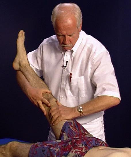
1. Straight Leg Raise (SLR / Lasègue's Sign)
Technique: Supine patient — passive hip flexion with knee extended. Positive if radicular pain reproduces at 30–70°.
- Sensitivity: 85–91% | Specificity: 26–52% for disc herniation
- Bowstring sign: Adding ankle dorsiflexion to positive SLR increases specificity
- Crossed SLR: Raising the unaffected leg provokes pain in the affected leg — Sensitivity 23–34%, Specificity 86–90% (highly specific for disc extrusion)
2. Femoral Nerve Stretch (Reverse SLR)
- Patient prone; examiner maximally flexes knee → reproduces anterior thigh pain
- Positive = L2, L3, or L4 root involvement
3. FABER Test (Patrick's Test)
- Flexion-Abduction-External Rotation of hip
- Positive = SI joint or hip pathology
4. FADIR Test
- Flexion-Adduction-Internal Rotation — tests for hip pathology / piriformis syndrome
5. Slump Test
- Seated: slumping forward + neck flexion + knee extension
- More sensitive than SLR for dural/neural tension
6. Waddell's Signs (Psychosocial / Non-organic LBP)
- Superficial/non-anatomical tenderness
- Axial loading / rotation causes pain
- Distracted SLR differs from supine SLR
- Widespread sensory/motor changes
- Overreaction during examination
(≥3 positive = significant psychosocial component)
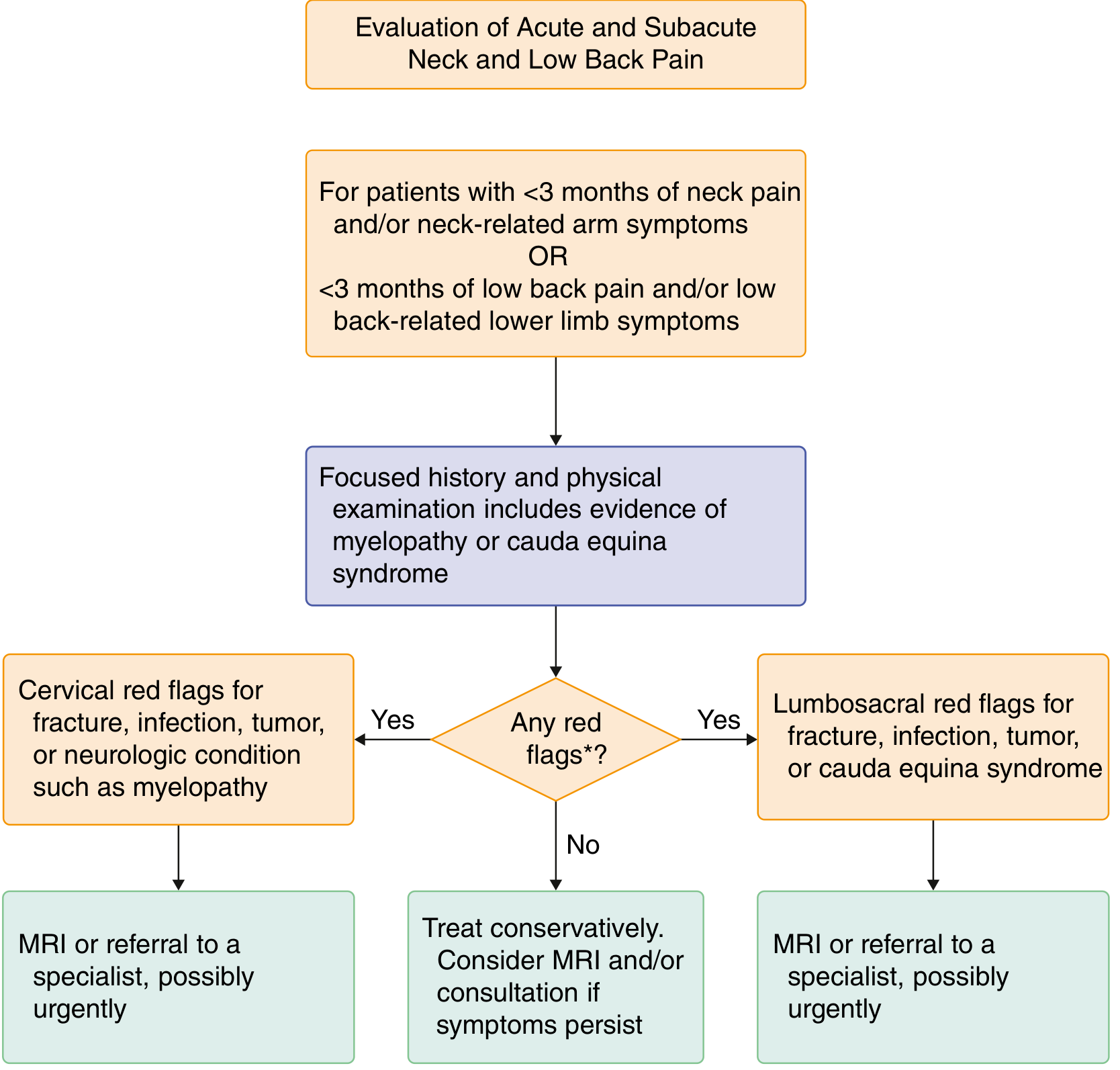
Clinical Examination Algorithm
8. INVESTIGATIONS
When to Investigate
- Without red flags: No immediate imaging needed. Wait 4–6 weeks.
- With red flags / persistent symptoms / progressive neurology: Investigate promptly.
Imaging
| Modality | Indication | Key Findings |
|---|
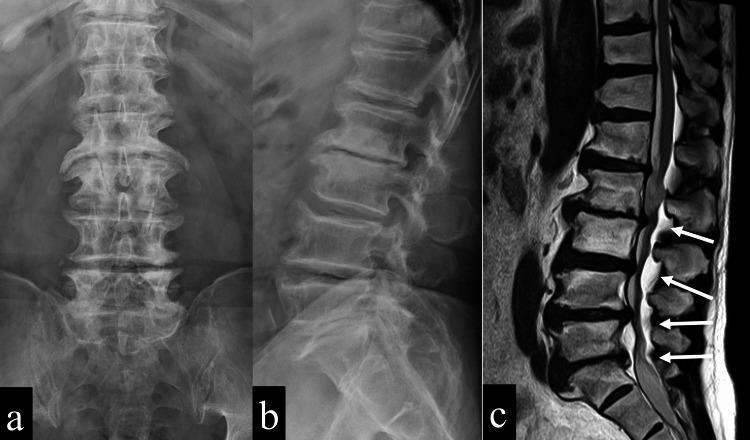
| Plain X-ray | Initial screening for fracture, spondylolisthesis, scoliosis, alignment | Disc space narrowing, osteophytes, vertebral slippage |
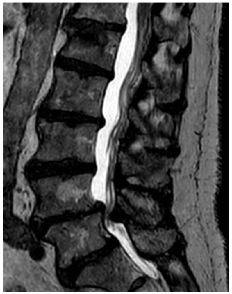
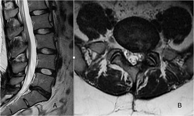
| MRI | Test of choice for most LBP with neurological features | Disc herniation, cord/nerve compression, infection, tumour, stenosis |
| CT scan | When MRI contraindicated; bone detail (pars defects, stenosis, post-surgery) | Bony anatomy superior |
| CT myelography | When both CT and MRI needed; post-surgical changes | Nerve root compression |
| Bone scan / SPECT | Stress fracture (spondylolysis), occult metastasis | Uptake at pars / vertebra |
| DEXA | Suspected osteoporosis | Bone mineral density |
Caution: MRI abnormalities are common in asymptomatic individuals — disc protrusion, disc desiccation, facet arthropathy, and foraminal stenosis are frequently incidental. Clinical correlation is mandatory.
Laboratory Tests
- ESR, CRP: Infection, inflammatory spondyloarthropathy, malignancy
- CBC: Infection, anaemia from malignancy
- HLA-B27: Ankylosing spondylitis
- Serum calcium, ALP: Metastatic disease, Paget's disease
- PSA: Prostatic metastasis
- Blood cultures: When spinal infection suspected
Electrodiagnostic Studies
- EMG/NCS: Useful to confirm and localise radiculopathy; helps exclude peripheral nerve disorders (plexopathy, CTS); becomes abnormal only 4 weeks after nerve injury onset
- Not indicated for pure axial LBP without neurological symptoms
9. TREATMENT
Principles (Biopsychosocial Model)
Contemporary management integrates biological, psychological, and social factors. Most acute LBP resolves within 6 weeks with conservative measures. The therapeutic hierarchy: nonpharmacologic first → pharmacologic → interventional → surgical.
A. ACUTE LBP Management
Nonpharmacologic (First Line)
- Activity modification: Continue routine activities as tolerated — bed rest is not recommended
- Heat application: Topical heat packs reduce acute paraspinal muscle spasm
- Education and reassurance: Positive prognosis; self-management strategies
- Manual therapy: Short-term benefit for acute pain; spinal manipulation (chiropractic/osteopathic)
Pharmacologic
| Drug Class | Example | Notes |
|---|
| NSAIDs | Ibuprofen 600 mg TDS × 2–3 weeks | First-line; anti-inflammatory + analgesic |
| Skeletal muscle relaxants | Cyclobenzaprine 5–10 mg TDS; Tizanidine 2–6 mg TDS; Carisoprodol 250–350 mg TDS × few weeks | Short-term benefit for spasm; sedating |
| Paracetamol | 1 g QDS | Not recommended for LBP — no significant analgesia demonstrated |
| Systemic corticosteroids | — | Not effective for LBP |
| Opioids | Use only for severe acute pain, short duration, case-by-case | No long-term benefit; risks addiction, respiratory depression |
B. CHRONIC LBP Management
Nonpharmacologic (Central Role)
- Physiotherapy/exercise — Core-strengthening, aerobic exercise, stretching (see Section 10)
- Cognitive-Behavioural Therapy (CBT): Targets fear avoidance, catastrophizing, pain coping
- Mindfulness-Based Stress Reduction (MBSR)
- Interdisciplinary pain rehabilitation programs: Best outcomes for chronic refractory LBP
Pharmacologic
| Drug | Notes |
|---|
| NSAIDs | Ongoing use with gastroprotection (PPI) |
| Duloxetine (SNRI) 60 mg OD | Shown effective for chronic LBP |
| Tricyclic antidepressants | Low-dose amitriptyline — sleep + pain |
| Gabapentinoids | Insufficient evidence for axial or radicular LBP |
| Opioids | Last resort; no demonstrated long-term benefit; significant harm risk |
Interventional Procedures
| Procedure | Indication |
|---|
| Epidural corticosteroid injection (transforaminal or interlaminar) | Lumbar radiculopathy, disc herniation |
| Medial branch nerve blocks | Diagnostic for facet joint pain |
| Radiofrequency ablation (RFA) | Confirmed facetogenic pain (after positive medial branch blocks) — months of relief |
| SI joint injection | Confirmed sacroiliac joint pain |
| Intradiscal procedures | Discogenic pain (limited evidence) |
| Spinal cord stimulation | Refractory back and leg pain |
Surgery
Indicated for:
- Disc herniation with radiculopathy not responding to 6+ weeks conservative management
- Cauda equina syndrome (surgical emergency)
- Lumbar spinal stenosis with severe claudication + neurological deficit
- Spinal instability: fracture, tumour, infection
- Spondylolisthesis with progressive neurological deficit
Lumbar fusion for chronic non-radicular LBP: only modestly better than no treatment in RCTs; NOT superior to an active physical therapy program (Goldman-Cecil Medicine).
10. PHYSIOTHERAPY & REHABILITATION
A. Assessment & Evaluation
- Visual Analogue Scale (VAS) / Numeric Pain Rating Scale (NPRS)
- Oswestry Disability Index (ODI)
- Roland-Morris Disability Questionnaire
- Fear-Avoidance Beliefs Questionnaire (FABQ)
- Movement pattern analysis; postural assessment
B. Acute Phase (0–6 weeks)
Goals: Pain control, muscle spasm reduction, restore basic movement
-
Pain relief modalities:
- TENS (Transcutaneous Electrical Nerve Stimulation) — gate control mechanism
- Heat therapy / hot packs — muscle relaxation
- Cold therapy / cryotherapy — acute inflammation
- Ultrasound therapy — deep tissue; reduces spasm
- Interferential current therapy (IFT)
- LASER therapy — photobiomodulation
-
Manual therapy:
- Joint mobilisation (Maitland grades I–IV) — gentle oscillatory techniques
- Soft tissue mobilisation — myofascial release, trigger point therapy
- Muscle energy techniques (MET)
-
Early active movement:
- Gentle lumbar range-of-motion exercises
- Walking — encouraged as early as tolerated
C. Subacute and Chronic Phase — Exercise Therapy
1. McKenzie Method (Mechanical Diagnosis & Therapy — MDT)
Classifies patients into derangement, dysfunction, or postural syndromes based on directional preference (symptom centralisation with repeated movements).
- Derangement syndrome: Repeated extension usually centralises symptoms (prone press-ups)
- Dysfunction syndrome: End-range loading to remodel shortened structures
- Postural syndrome: Postural correction and ergonomic advice
2. Lumbar Stabilisation / Core Strengthening
Targets transversus abdominis, multifidus, pelvic floor, and diaphragm — the deep stabilising system of the lumbar spine.
Progression:
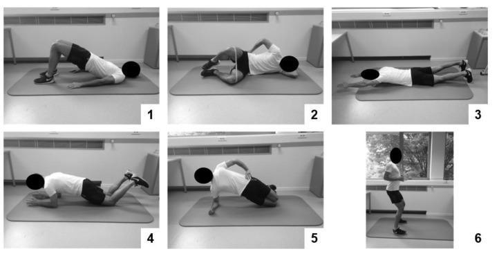
- Stage 1 — Activation: Draw-in manoeuvre; isolated transversus abdominis/multifidus co-contraction in supine
- Stage 2 — Static stability: Dead bug, supine bridge (glute bridge), side plank
- Stage 3 — Dynamic stability: Bird-dog (quadruped alternate arm/leg), Swiss ball exercises
- Stage 4 — Functional: Squats, lunges, functional task-specific training
3. Aerobic Exercise
- Walking, swimming, aquatic exercise, cycling (low-impact)
- Minimum 30 min moderate intensity, 5 days/week
- Consistent evidence for pain reduction and prevention of recurrence
4. Pilates
- 2024 meta-analysis (PMID 37632387) confirms effectiveness for pain reduction and functional improvement in LBP
- Focuses on body awareness, alignment, controlled movement, and breathing
5. Yoga / Tai Chi
- Modest improvement in chronic LBP; evidence Grade B
- Mindfulness + movement combination especially beneficial for psychosocial components
6. Aquatic Therapy (Hydrotherapy)
- Buoyancy reduces loading; warmth reduces spasm
- Ideal for obese, elderly, or those with severe pain limiting land-based exercise
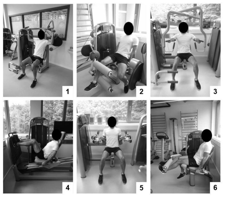
7. General Resistance Training
D. Specific Physiotherapy Approaches by Diagnosis
| Condition | Preferred PT Approach |
|---|
| Acute muscle strain | Heat, TENS, gentle mobilisation, early walking |
| Disc herniation + radiculopathy | McKenzie extension; neural mobilisation (nerve flossing); traction |
| Spinal stenosis | Flexion-based exercises (Williams'); aquatic therapy; stationary cycling |
| Facet joint pain | Passive lumbar extension mobilisation; manipulation; deep heat |
| Spondylolisthesis | Neutral spine stabilisation; avoid end-range extension |
| Chronic non-specific LBP | Graded activity; CBT + exercise; interdisciplinary rehabilitation |
| SI joint dysfunction | SI joint mobilisation; pelvic stability exercises; muscle energy technique |
E. Spinal Traction
- Mechanical or motorised lumbar traction (intermittent or sustained)
- May benefit acute disc herniation with radiculopathy
- Not recommended as sole treatment; limited long-term evidence
F. Postural and Ergonomic Rehabilitation
- Lumbar support/bracing (short-term only)
- Ergonomic workstation assessment
- Correct lifting technique: bend the knees (squat), keep load close to body, neutral spine, no twisting
- Driving posture, sleep position (mattress firmness: medium-firm preferred for chronic LBP)
G. Integrative Approaches
| Therapy | Evidence |
|---|
| Massage | Beneficial for acute/subacute LBP with prominent soft tissue involvement; short-lived benefit |
| Acupuncture | Modest, short-lived benefit for pain in both acute and chronic LBP; sham acupuncture also > no treatment |
| Spinal manipulation | Small benefit for acute LBP; non-inferior to other physical therapies |
| TENS | Short-term pain relief; no long-term benefit demonstrated |
H. Psychological Rehabilitation
Co-prevalence with psychopathology in chronic LBP:
- Depression: 33–67%
- Anxiety: 10–30%
- Substance misuse: 13–40%
Interventions:
- CBT: Fear-avoidance behaviour modification; activity pacing; pain coping skills
- Operant therapy: Reinforcing healthy behaviour
- Biofeedback: EMG biofeedback for paraspinal muscle retraining
- Pain neuroscience education (PNE): Explaining central sensitisation to patients
I. Rehabilitation Staging Protocol
| Phase | Timing | Focus |
|---|
| Phase 1 — Acute | Week 0–2 | Pain control, reduce spasm, protect |
| Phase 2 — Subacute | Week 2–6 | Restore ROM, begin core activation, education |
| Phase 3 — Functional | Week 6–12 | Strengthening, neuromuscular control, endurance |
| Phase 4 — Return to Activity | 3–6 months | Sport/work-specific training, relapse prevention |
| Phase 5 — Maintenance | Ongoing | Home exercise programme, lifestyle modification |
11. PROGNOSIS
- Acute mechanical LBP: Resolves in 30 days (most patients); 90% within 3 months
- Recurrence: ~50% within 1 year
- Chronic LBP risk factors: obesity, low education, tobacco, high initial pain, depression, job dissatisfaction, heavy lifting work, somatisation
- Exercise + education is proven to prevent recurrence
12. PREVENTION
- Primary: Core strengthening, correct lifting technique, workplace ergonomics, healthy weight, smoking cessation
- Secondary: Early active rehabilitation after acute LBP, avoid prolonged bed rest, early return to work
- Tertiary: Interdisciplinary rehabilitation for chronic LBP, psychosocial intervention, graded activity
📚 Sources
- Goldman-Cecil Medicine International Edition, Chapter 369: Neck and Low Back Pain — pp. 3837–3842
- Harrison's Principles of Internal Medicine 22E, Multimodal and Interdisciplinary Strategies — pp. 170+
- Bailey and Love's Short Practice of Surgery, 28th Ed. — Table 37.5 Red Flags
- Li Y et al. (2023). Exercise intervention for patients with chronic low back pain: a systematic review and network meta-analysis. Front Public Health. [PMID 38035307]
- Patti A et al. (2024). Effectiveness of Pilates exercise on low back pain. Disabil Rehabil. [PMID 37632387]
- Zhang SK et al. (2023). Effects of exercise therapy on disability in elderly with chronic LBP. J Orthop Surg Res. [PMID 37468931]
- Arslan S et al. (2025). Exercise in treatment of lumbar disc herniation: a systematic review. Acta Neurol Belg. [PMID 40128486]This is a comprehensive, evidence-based review of low back pain covering every domain requested. Here's a quick navigation guide:
What's covered:
- Definition & Epidemiology — prevalence, risk factors, burden
- Anatomy & Biomechanics — lumbar spine structures, disc mechanics, pain-generating mechanisms
- Etiology & Classification — mechanical (97%) vs. serious/non-mechanical causes with differential diagnosis flowchart
- Pathobiology — spondylosis cascade, disc degeneration, genetic factors
- Signs & Symptoms — acute vs. chronic LBP, radiculopathy patterns, neurogenic claudication, root-level neurology table
- Red Flags — cauda equina syndrome, fracture, malignancy, infection — with a full table from Bailey & Love
- Clinical Examination — inspection, palpation, ROM, neurology, special tests (SLR/Lasègue, FABER, FADIR, Femoral stretch, Slump, Waddell's signs) with images
- Investigations — imaging indications, MRI vs. CT, labs, EMG/NCS
- Treatment — pharmacologic (NSAIDs, muscle relaxants, duloxetine, opioids), interventional (epidural injections, RFA), surgery criteria
- Physiotherapy & Rehabilitation — McKenzie method, core stabilisation programme (staged), aerobic exercise, Pilates, aquatic therapy, resistance training, ergonomics, integrative therapies, psychosocial rehabilitation, and a 5-phase rehabilitation staging protocol
All sections are supported by illustrative diagrams from peer-reviewed medical imaging databases and authoritative textbooks.