
Urinary bladder position and external features
urinary bladder anatomy external features position
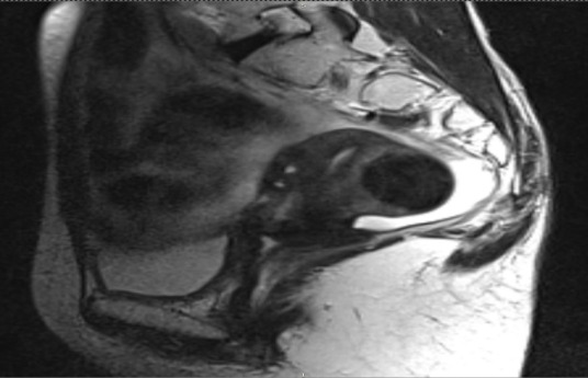
**Imaging Modality:** Magnetic Resonance Imaging (MRI), T2-weighted sequence. **Anatomical Region:** Sagittal view of the female pelvis. **Key Landmarks:** Visible structures include the sacrum and coccyx posteriorly, the pubic symphysis anteriorly, the urinary bladder, and the rectum. **Observed Findings:** - **Uterine Orientation:** The uterus is clearly visualized in a retroverted and retroflexed position, with the fundus directed posteriorly toward the sacrum. - **Uterine Morphology:** The zonal anatomy of the uterus (endometrium, junctional zone, and myometrium) is discernible. The cervical region and vaginal vault are visible inferior to the uterine body. - **Adjacent Organs:** The urinary bladder is situated anteriorly, appearing partially decompressed. The rectum is located posterior to the retroverted uterus. - **Pathology/Context:** The image displays the pelvic anatomy post-external beam radiotherapy (EBRT). There is no gross evidence of large exophytic masses, though the retroverted positioning is a key anatomical variant relevant for brachytherapy planning and treatment monitoring. **Diagnostic Features:** The sagittal plane highlights the posterior angulation of the uterine long axis relative to the vaginal axis, confirming retroversion. Signal intensities are consistent with post-treatment tissue characteristics in a T2-weighted acquisition.
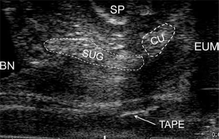
**Imaging Modality:** Transvaginal ultrasound (biplane electronic probe, linear array). **Anatomical Region:** Female pelvic floor, specifically the urethral complex and mid-urethral region. **Clinical Context:** Post-operative evaluation of a suburethral sling procedure for stress urinary incontinence. **Key Landmarks and Observed Structures:** * **Bladder Neck (BN):** Visible at the proximal aspect of the urethral tract. * **Symphysis Pubis (SP):** Hyperechoic landmark located anterior to the urethra. * **Urethral Complex:** Differentiation of the **suburethral gland (SUG)** and **compressor urethrae (CU)** indicated by dashed outlines, showing the anatomical relationship between glandular tissue and muscular components. * **External Urethral Meatus (EUM):** Distal terminus of the urethra. * **Sling/Tape:** A distinct hyperechoic, linear structure (labeled "TAPE") is visible in the mid-urethral position, representing the synthetic suburethral mesh. **Visual Features:** The image demonstrates the spatial orientation of a mid-urethral sling relative to the urethral anatomy. The use of high-frequency linear arrays allows for the visualization of subtle soft-tissue contrasts within the urethral wall and surrounding supportive structures. This is a characteristic view for assessing sling placement, tension, and proximity to the urethral lumen.
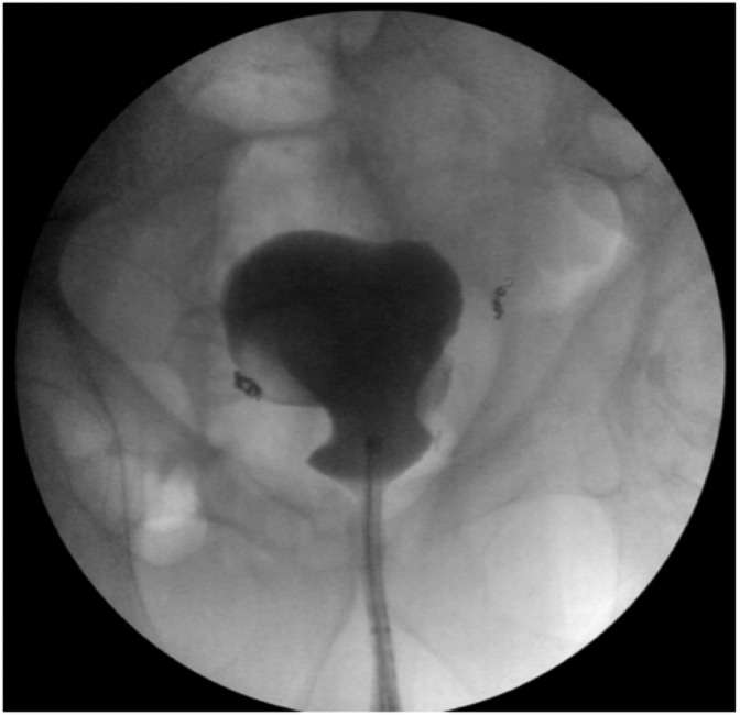
**Imaging Modality:** Intraoperative fluoroscopic cystogram (retrograde cystography). **Anatomical Region:** Lower abdomen and pelvis, specifically the lower urinary tract. **Observed Anatomy and Findings:** The image demonstrates a contrast-filled urinary bladder and urethra. The bladder exhibits a normal anatomical globular morphology and midline position within the bony pelvis. There is clear luminal continuity between the urethra and the bladder neck, indicating a patent urethral tract. Contrast opacification is uniform without evidence of extravasation, filling defects, or gross wall irregularities. **Characteristic Visual Features:** - **Contrast Distribution:** Dense opacification of the bladder and the narrow, linear passage of the urethra. - **Surgical Hardware:** Small, radiopaque surgical clips or coils are visible in the bilateral pelvic regions, lateral to the bladder, consistent with recent surgical intervention. - **Bony Landmarks:** The pelvic girdle, including the pubic symphysis and iliac wings, provides anatomical orientation. **Clinical Context:** The study confirms post-surgical integrity of the vesicourethral junction and the absence of obstructive uropathy or anastomotic leaks. This is a characteristic appearance of a successful urethral reconstruction or bladder repositioning procedure.
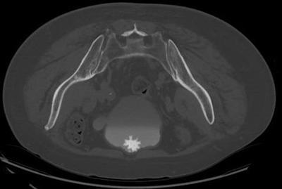
This diagnostic image is an axial CT scan of the pelvis, performed in the prone position. It demonstrates a classic 'jackstone' urinary bladder calculus, characterized by its highly hyperdense, spiculated, and star-shaped morphology with a dense central core. The calculus is located anteriorly within the bladder lumen, which also contains layered contrast material from a delayed phase study. Additionally, a distinct outpouching or diverticulum is visible on the left lateral wall of the bladder. The surrounding pelvic anatomy, including the hyperdense iliac bones and sacrum, as well as the pelvic musculature and soft tissues, are well-visualized. This case illustrates key radiological features of specialized bladder calculi often associated with urinary stasis or chronic infection. The image serves as a high-quality educational example of obstructive uropathy and vesical lithiasis for urology and radiology trainees.
urinary bladder parts apex body fundus neck diagram anatomy
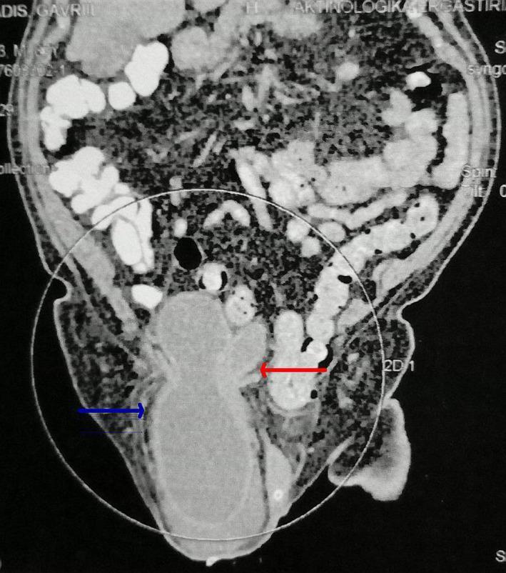
This diagnostic image is a coronal CT scan of the pelvis and lower abdomen demonstrating a large inguinal hernia. The image focuses on a massive herniation of the urinary bladder into the right inguinoscrotal sac. A blue arrow indicates the herniated bladder body, apex, and fundus, which appear as a homogeneous, fluid-filled structure within the scrotal sac. A red arrow points to the bladder trigone, illustrating its significant inferior displacement from the pelvic floor into the hernia neck. There is a notable absence of bowel loops within the hernia sac in this specific section. This clinical finding is characteristic of a massive inguinoscrotal cystocele, a rare condition that can lead to obstructive uropathy and acute kidney injury due to ureterovesical junction compression. The surrounding abdominal cavity shows contrast-filled intestinal loops superior to the hernia neck. The image serves as a high-level educational example of atypical hernia contents and their potential complications in geriatric urology.
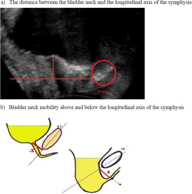
This composite educational graphic illustrates the assessment of bladder neck mobility using transperineal ultrasound. Panel (a) is a diagnostic grayscale ultrasound image in the sagittal plane, showing the pelvic floor anatomy. A red circle highlights the symphysis pubis, which serves as a fixed bony landmark. Red overlaid lines demonstrate the measurement of the vertical distance between the bladder neck and the longitudinal axis of the symphysis. Panel (b) features two comparative schematic diagrams showing different states of bladder neck position relative to the symphysis axis. The diagram on the left shows the bladder neck (yellow) at a higher position relative to the diagonal reference line, indicated as a negative value (-X). The diagram on the right shows the bladder neck descended below the reference line, indicated as a positive value (X). This comparison is clinically relevant for evaluating pelvic organ prolapse and urinary incontinence by measuring bladder neck descent during rest versus a Valsalva maneuver. The visual focus is on the dynamic relationship between the bladder, urethra, and the longitudinal symphyseal axis.
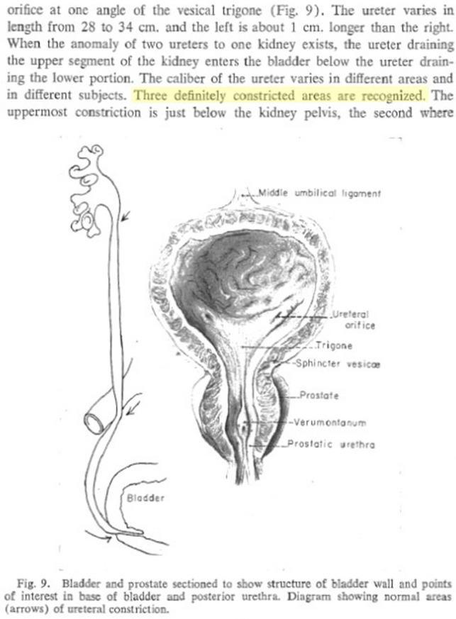
This medical illustration depicts the gross anatomy of the male lower urinary tract, focusing on the kidneys, ureters, bladder, and prostate. The diagram specifically highlights the historical anatomical concept of three physiological ureteral constrictions (indicated by arrows): the ureteropelvic junction (UPJ), the crossover of the iliac vessels (pelvic inlet), and the ureterovesical junction (UVJ) where the ureter enters the bladder. A detailed coronal section of the bladder and prostate is shown, labeling internal landmarks including the ureteral orifice, vesical trigone, sphincter vesicae, verumontanum, and prostatic urethra. The image demonstrates the spatial relationship where the bladder is positioned superior to the prostate, with the ureters descending retroperitoneally to insert into the posterior-inferior bladder wall. This diagram serves as a primary educational resource for urology and anatomy, illustrating common sites where urinary calculi may lodge and the internal structure of the bladder neck and posterior urethra.
Urinary Bladder - Position and External Features
Position
- When empty: The bladder is entirely pelvic, lying behind (posterior to) the pubic symphysis. It is bowl-shaped, with the apex and upper wall descending.
- When full: The apex and anterior wall are drawn forward and upward between the peritoneum and the anterior abdominal wall, extending into the lower abdomen. It can rise as high as the superior border of the pubic symphysis and assumes an ovoid shape.
External Features (Parts of the Bladder)
| Part | Description |
|---|---|
| Apex (Vertex) | Anterosuperior tip; connected to the median umbilical ligament (obliterated urachus) running to the umbilicus |
| Body | The largest part, making up most of the organ |
| Fundus (Base) | Posteroinferior surface; the ureters enter here on its lateral and posterior aspects |
| Neck | Inferiormost part; continuous anteriorly with the urethra; the most fixed part of the bladder |
Detailed Notes on Each Part
- Points anterosuperiorly
- Gives attachment to the obliterated urachus, which becomes the median umbilical ligament running in the midline to the umbilicus
- Moves up and down with filling/emptying
- Constitutes the bulk of the organ
- Has anterosuperior, posterior, and two inferolateral surfaces
- Covered superiorly by peritoneum (the serosa covers the superior surface and the posterior surface above the trigone)
- The muscle coat (detrusor) has three interlacing layers
- Faces posteriorly and inferiorly
- Receives the two ureters on its lateral and posterior aspects
- The internal surface of the fundus contains the trigone of the bladder - a smooth, triangular, mucosa-covered region bounded by the two ureteric orifices and the internal urethral orifice
- Continuous with the urethra below
- The most fixed and immobile part of the bladder
- Lies on the superior surface of the urogenital diaphragm/pelvic floor
- In males: rests on the prostate (which produces the uvula vesicae - a conical projection into the internal urethral orifice)
Shape Changes with Filling
| State | Shape | Apex position | Wall folds |
|---|---|---|---|
| Empty | Bowl-shaped | Descended, pelvic | Prominent mucosal folds |
| Full | Ovoid / pear-shaped | Elevated, abdominal | Folds smooth out |
Peritoneal Relations
- Superiorly: peritoneum covers the superior surface and upper posterior surface
- Inferiorly/anteriorly: no peritoneum - separated from the pubic symphysis by the retropubic (prevesical) space of Retzius
- In males: peritoneum reflects off the posterior bladder to form the rectovesical pouch
- In females: peritoneum reflects to form the vesicouterine pouch anteriorly and the rectouterine pouch (of Douglas) posteriorly
Anatomical Illustration (Color Atlas of Human Anatomy)
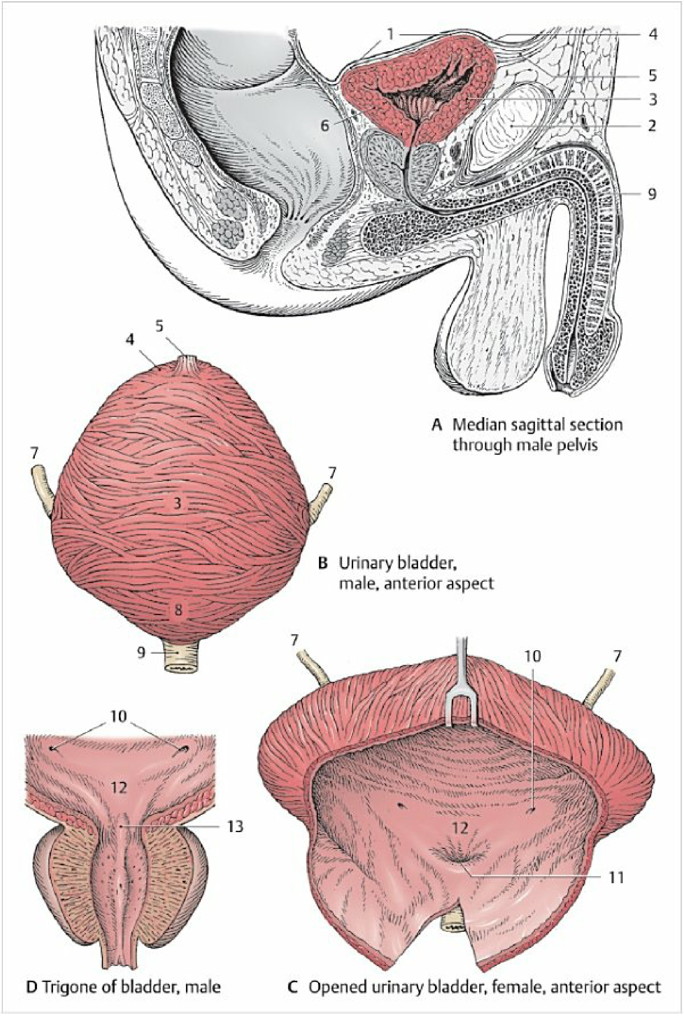
- Color Atlas of Human Anatomy Vol. 2, Internal Organs, p. 405