
For my dermatology practical exam please give me a proper case description of “becker’s nevus” and also prepare me extensively for the questions that examiner can ask from medium to hard to very hard
Reading File
Reading File
Finding Sources
Finding Sources
Searching PubMed
"Becker Nevus"
Searching the Web
Becker nevus DermNet clinical features differential diagnosis
Searching Images
Becker nevus clinical photograph unilateral hyperpigmented hairy patch shoulder chest
Reading File
Reading File
Finding Sources
Finding Sources
Finding Sources
query_images_db
Reading Web Page
https://dermnetnz.org/topics/becker-naevus
Becker's nevus: practical case description
One-line diagnosis
Becker's nevus (Becker melanosis) is a benign, usually acquired, unilateral hyperpigmented and often hypertrichotic cutaneous hamartoma that typically appears around puberty in males, most often over the shoulder, scapular area, or upper chest.
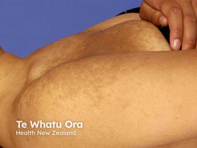
Ready-to-say long case presentation
This is a [age]-year-old male presenting with an asymptomatic dark patch over the [right/left] upper chest/shoulder/scapular region, noticed since [early adolescence / around puberty]. It initially began as a light-brown patch and gradually became darker, with development of increased coarse terminal hair over the lesion. There is no history of itching, pain, scaling, ulceration, bleeding, or rapid recent change. There is no preceding trauma, dermatitis, drug application, or similar family history.On cutaneous examination, there is a single, unilateral, well-demarcated but irregularly bordered, geographic hyperpigmented patch/plaque, measuring approximately [x × y] cm, over the [site]. It is tan to dark brown in colour, with a mildly thickened or corrugated surface and hypertrichosis, consisting of coarse dark terminal hairs. There is no scale, atrophy, ulceration, induration, satellite lesion, or sensory loss. The rest of the mucocutaneous examination is normal.On systemic examination, I would specifically look for ipsilateral breast or pectoral muscle hypoplasia, limb asymmetry, chest wall or vertebral abnormalities, scoliosis, and other soft-tissue anomalies, to exclude Becker nevus syndrome.My provisional diagnosis is Becker's nevus / Becker melanosis. Differentials include café-au-lait macule, congenital melanocytic nevus, congenital smooth muscle hamartoma, and plexiform neurofibroma.The diagnosis is primarily clinical. A biopsy is usually unnecessary, but if done, it may show acanthosis, hyperkeratosis, elongation of rete ridges, basal-layer hyperpigmentation without a major increase in melanocyte number, and variable smooth-muscle hyperplasia. Management is reassurance, screening for associated developmental abnormalities, sun protection and camouflage. Hair-removal methods or laser hair reduction may be offered for cosmetic concern; pigment laser results are variable and recurrence or incomplete clearing may occur.
The usual course is a light-brown patch in late childhood or adolescence that darkens, becomes hairier and may mildly thicken after puberty. It may enlarge for 1 to 2 years and then usually stabilizes. Dermatology, 2-Volume Set, 5e, p. 2357. The practical clinical features and management points are also consistent with DermNet's Becker naevus reference.
Examination description: high-scoring morphology
Use this sequence whenever the examiner asks, “Describe the lesion.”
| Feature | What to say |
|---|---|
| Number | Solitary |
| Distribution | Usually unilateral, segmental or block-like |
| Site | Upper trunk, shoulder, pectoral area, scapular region, proximal arm |
| Primary lesion | Patch or slightly elevated plaque |
| Colour | Tan, light brown, or dark brown |
| Margin | Well-defined but irregular, geographic, sometimes broken-up |
| Surface | Usually smooth, sometimes mildly thickened or corrugated |
| Hair | Hypertrichosis with coarse, dark terminal hairs |
| Other signs | Acneiform papules may occur within the lesion |
| Symptoms | Usually asymptomatic |
| Evolution | Appears around puberty, increases in pigmentation and hair, then becomes stable |
Exam pearl: Hyperpigmentation generally appears first. Hypertrichosis appears later and may be subtle or may not precisely overlap the pigmented region. Dermatology, 2-Volume Set, 5e, p. 2357.
Viva preparation
A. Medium-level questions
1. What is Becker's nevus?
It is a benign acquired cutaneous hamartoma characterized by a unilateral hyperpigmented patch, often with hypertrichosis, usually arising around puberty.
2. What are its synonyms?
- Becker melanosis
- Becker pigmentary hamartoma
- Pigmented hairy epidermal nevus
- Becker's pigmented hairy nevus
3. Who first described it?
S. William Becker, in 1949.
4. What is the usual age of onset?
Usually during late childhood or, more commonly, adolescence, around puberty.
5. What is the sex distribution?
It is recognized more often in males, traditionally reported with a marked male predominance. Females can be affected, and their lesions may be less conspicuous because androgen-dependent hypertrichosis is less prominent.
6. What are the common sites?
Shoulder, upper chest, scapular area, upper back, and proximal upper limb. Less commonly it can occur on the face, neck, lower trunk, buttocks, or limbs.
7. What is the typical morphology?
A large unilateral tan-to-brown patch with irregular but defined borders, often later becoming hypertrichotic and slightly thickened.
8. Is Becker's nevus congenital?
Usually no. It is classically acquired around puberty, although rare congenital or childhood-onset cases are reported.
9. Is it hereditary?
Usually it is sporadic and nonhereditary. It is best understood as a mosaic disorder, although occasional familial occurrence has been reported.
10. Is it symptomatic?
Usually asymptomatic. The concern is commonly cosmetic or psychosocial.
11. Can acne occur in Becker's nevus?
Yes. Acneiform lesions can be localized within the nevus, supporting androgen responsiveness.
12. Does Becker's nevus turn malignant?
It is a benign lesion and does not routinely require excision or cancer surveillance solely due to its presence. Any new ulceration, rapid asymmetrical change, or atypical lesion should be evaluated on its own merits.
B. Hard questions
13. Why does Becker's nevus usually become obvious at puberty?
Lesional skin has increased androgen receptor expression and androgen sensitivity. Pubertal androgen exposure can increase pigmentation, terminal hair growth, sebaceous activity, acne, and dermal thickening.
14. What is the current genetic basis?
Postzygotic mosaic mutations in ACTB, the gene encoding beta-actin, have been identified in many Becker nevi and in Becker nevus syndrome. This explains its mosaic, unilateral, patchy distribution. Dermatology, 2-Volume Set, 5e, p. 2357.
15. Why is it called a hamartoma?
Because it represents a localized, disorganized overgrowth of mature cutaneous components normally present at that site, including epidermal structures, pigmentary change, hair follicles, and often arrector pili smooth muscle.
16. Is it an epidermal nevus or melanocytic nevus?
It is generally classified as a cutaneous hamartoma or late-onset epidermal nevus, not a conventional melanocytic nevus. The pigmentation is mainly from increased basal melanin, not necessarily an increased number of melanocytes.
17. What are the histopathological features?
Histology may show:
- Mild acanthosis and hyperkeratosis
- Elongated rete ridges
- Increased basal layer melanin
- Variable pigment incontinence and dermal melanophages
- Hyperplasia of arrector pili smooth muscle bundles
- Sometimes enlarged sebaceous glands
The melanocyte count is generally normal or only minimally altered. The major pathology is hyperpigmentation and hamartomatous structural change rather than a melanocytic proliferation.
18. Why is smooth muscle important in Becker's nevus?
Many lesions demonstrate smooth-muscle hamartomatous proliferation, particularly arrector pili muscle. Becker nevus and congenital smooth muscle hamartoma may be considered part of a clinical and pathological spectrum.
19. What is Becker nevus syndrome?
It refers to Becker's nevus associated with ipsilateral developmental abnormalities, especially hypoplasia of breast, pectoral muscle, subcutaneous tissue, chest wall, or limb, along with skeletal and other soft-tissue abnormalities.
20. What associations should you actively look for?
Look for:
- Ipsilateral breast hypoplasia, particularly important in females
- Pectoralis major hypoplasia
- Chest wall asymmetry
- Limb hypoplasia or asymmetry
- Scoliosis or vertebral abnormalities
- Scapular asymmetry
- Smooth muscle hamartoma
- Occasionally supernumerary nipples, urogenital anomalies, or other soft-tissue anomalies
The most testable association is ipsilateral breast hypoplasia.
21. Why should a female patient with Becker's nevus be examined carefully?
Because Becker nevus syndrome may present with ipsilateral breast hypoplasia. It may be more cosmetically and functionally significant in female patients.
22. What investigations are required?
Usually none. Diagnosis is clinical. Consider:
- Dermoscopy or clinical photography when uncertain
- Skin biopsy if the diagnosis is unclear, especially to distinguish it from café-au-lait macule or melanocytic nevus
- Targeted musculoskeletal, breast, or imaging assessment only when clinical examination suggests Becker nevus syndrome
23. What would dermoscopy show?
There is no single pathognomonic dermoscopic sign. Common reported findings include a pigment network, areas of darker pigmentation, perifollicular hypopigmentation, and prominent hair follicles. Dermoscopy is supportive, not diagnostic.
24. How does it differ from a café-au-lait macule?
| Becker's nevus | Café-au-lait macule |
|---|---|
| Usually appears around puberty | Often congenital or appears in early infancy |
| Often male predominant | No sex preference |
| Upper trunk and shoulder common | Any site |
| Irregular geographic border | Usually smooth, sharply defined border |
| Hypertrichosis may develop | Hypertrichosis absent |
| May be slightly thickened/corrugated | Flat, smooth macule |
| Androgen-responsive | Not androgen-responsive |
| May be associated with ipsilateral hypoplasia | Multiple lesions may indicate NF1, McCune-Albright syndrome, or other syndromes |
25. How does it differ from congenital melanocytic nevus?
| Becker's nevus | Congenital melanocytic nevus |
|---|---|
| Usually acquired around puberty | Present at birth or becomes apparent early in life |
| Commonly upper trunk | Any site |
| Hypertrichosis develops later | Hair may be present from early life |
| No true melanocytic proliferation required | Proliferation of nevomelanocytes |
| Often associated with smooth muscle hyperplasia | May extend deeply around adnexa and neurovascular structures |
| Very low concern for malignancy | Risk depends on size and phenotype |
26. How does it differ from congenital smooth muscle hamartoma?
Congenital smooth muscle hamartoma is typically present at birth or infancy, usually smaller, may have hypertrichosis and hyperpigmentation, and can show a pseudo-Darier sign on rubbing. Becker's nevus more often arises around puberty and becomes more pigmented and hairy with androgen influence.
27. What is a pseudo-Darier sign?
Transient piloerection, induration, or wrinkling of a lesion after rubbing due to contraction of smooth muscle fibers. It may occur in smooth muscle hamartoma and occasionally in Becker's nevus with prominent smooth muscle.
28. How does it differ from plexiform neurofibroma?
Plexiform neurofibroma is often soft, tortuous, “bag of worms” in consistency, may cause neurological symptoms or be associated with NF1 features such as café-au-lait macules, axillary freckling, Lisch nodules, and other neurofibromas. Becker's nevus is primarily a pigmented, hairy patch without the characteristic soft neural tumor texture.
C. Very hard questions
29. Explain the pathogenesis in a structured way.
A strong answer:
Becker's nevus is a mosaic cutaneous hamartoma. A postzygotic mutation, often involving ACTB, produces a clone of altered skin cells. This creates a localized lesion in a mosaic distribution. Increased androgen receptor activity in lesional skin explains why the lesion typically becomes more pigmented, thicker, hairier, and sometimes acneiform around puberty. The variable smooth muscle hyperplasia explains the overlap with smooth muscle hamartoma.
30. Why does a mutation occurring after fertilization produce a unilateral lesion?
A postzygotic mutation occurs in only one cell lineage after the zygote has formed. Descendants of that cell populate only a portion of skin, producing cutaneous mosaicism. Therefore, the lesion is typically patchy, unilateral, segmental, or block-like rather than generalized.
31. Why is ACTB mutation biologically plausible?
ACTB encodes beta-actin, a cytoskeletal protein important in cell structure, migration, and tissue organization. Mosaic ACTB mutations may alter signaling and the development of multiple cutaneous components. A possible relationship with Hedgehog-pathway activation has been proposed in these organoid hamartomas. Dermatology, 2-Volume Set, 5e, p. 2357.
32. Why may pigmentation persist despite laser therapy?
Pigment may be located not only superficially in the epidermis but also in follicular structures and may coexist with dermal or hamartomatous changes. Lesion heterogeneity, hair follicles, deeper pigment, and continued biologic behavior contribute to inconsistent lightening and recurrence.
33. Which component responds better to treatment: pigment or hair?
Hair is usually easier to improve than pigmentation. Laser hair reduction, electrolysis, shaving, waxing, threading, and depilatories can address hypertrichosis. Pigment-targeting laser outcomes are variable and often incomplete. Dermatology, 2-Volume Set, 5e, p. 2357.
34. Which lasers may be used?
For pigmentation, pigment-targeting lasers such as Q-switched ruby, alexandrite, or Nd:YAG lasers have been tried. Fractional ablative lasers have also been used. For hair, laser hair reduction can be useful. However, set realistic expectations because results vary and relapse can occur.
35. Is surgical excision appropriate?
Not usually, because lesions are commonly large and benign. It may be considered only for a small lesion if cosmetic benefit clearly outweighs scarring risk. Routine surgery is not indicated.
36. What counseling would you provide?
- It is benign and typically stable after an initial period of enlargement.
- It is not contagious and usually not inherited.
- No treatment is medically necessary.
- Sun protection and camouflage can reduce contrast.
- Hair-removal techniques are safe and do not worsen the nevus.
- Laser treatment may help hair more reliably than pigmentation.
- Examine for associated ipsilateral breast, chest wall, muscle, limb, or skeletal abnormalities.
- Return for review if there is a separate suspicious evolving lesion, ulceration, bleeding, or a rapid atypical change.
37. Can Becker's nevus be bilateral or occur outside the upper trunk?
Yes, but this is atypical. It may occur on the face, neck, lower trunk, buttocks, or extremities; multiple and bilateral lesions are rare.
38. Is hypertrichosis mandatory for diagnosis?
No. Hypertrichosis is characteristic but may appear later, be subtle, or be absent. Early lesions may look like a hyperpigmented patch alone.
39. How can you distinguish an early Becker's nevus from a café-au-lait macule?
History is central. Becker's nevus tends to begin around puberty, gradually darkens and becomes hairy or mildly thickened. A café-au-lait macule is often earlier in onset, uniformly flat, nonhairy, and lacks pubertal progression. When uncertain, follow-up or biopsy may be considered.
40. How would you answer if the examiner asks, “Why is it more recognized in males?”
The lesion's visible phenotype is amplified by androgen action. Males tend to develop more obvious terminal hair, acneiform changes, and pigmentation after puberty. This may contribute to apparent male predominance and underrecognition in females.
Rapid-fire examiner traps
-
Is it a malignant lesion?
No, it is benign. -
Does it require biopsy in every case?
No. It is usually a clinical diagnosis. -
Is hypertrichosis present from birth?
Usually no. It often develops after pigmentation, around or after puberty. -
What is the most important syndrome association?
Ipsilateral breast hypoplasia, especially in females. -
What mutation is associated with it?
Postzygotic mosaic ACTB mutation. -
What is the main hormonal explanation?
Increased androgen receptor expression and androgen sensitivity in lesional skin. -
What is the key differential in a child?
Café-au-lait macule and congenital smooth muscle hamartoma. -
What sign suggests smooth-muscle involvement?
Pseudo-Darier sign. -
Does laser cure it?
No. Results, especially for pigmentation, are variable. -
What treatment works better: pigment laser or hair reduction?
Hair reduction generally has better cosmetic results.
Final 30-second answer
Becker's nevus, or Becker melanosis, is a benign mosaic cutaneous hamartoma that usually develops around puberty, predominantly in males. It presents as a unilateral, irregular tan-to-dark-brown patch, usually over the upper trunk or shoulder, which later develops hypertrichosis, mild thickening, or acneiform lesions. It is associated with increased androgen sensitivity and postzygotic ACTB mutations. Diagnosis is mainly clinical. Important differentials are café-au-lait macule, congenital melanocytic nevus, congenital smooth muscle hamartoma, and plexiform neurofibroma. I would examine for Becker nevus syndrome, particularly ipsilateral breast or pectoral hypoplasia and skeletal abnormalities. Management is reassurance and cosmetic treatment if needed, with hair reduction usually responding better than pigmentation.
Primary textbook points: Andrews' Diseases of the Skin: Clinical Dermatology, “Becker Nevus” section; Dermatology, 2-Volume Set, 5e, p. 2357.
This is a shared conversation. Sign in to Orris to start your own chat.