
Osteoology - mainly Pelvis Hip bone Normal basalis Mandible
Reading File
Now let me search the library for all four topics simultaneously.
Finding Sources
Finding Sources
Finding Sources
Good. Now let me read the key anatomy sections from Gray's for pelvis/hip bone, mandible, and also search for "norma basalis" (the skull base view):
Reading File
Reading File
Finding Sources
Finding Sources
Let me search for "norma" (skull base norma) and also read more content on hip bone and the pelvis bony anatomy:
Finding Sources
Finding Sources
Reading File
Good. Let me now read: the inferior skull base (norma basalis) section from Gray's block5, the hip bone / pelvis bony anatomy from the relevant block, and Mandible details:
Reading File
Reading File
Reading File
Now let me read the key detailed pelvis bony anatomy and norma basalis content:
Finding Sources
Reading File
Now let me read the "Inferior view" of skull (norma basalis) section from Gray's:
Reading File
Reading File
I now have enough content from Gray's Anatomy for Students and the THIEME Atlas to compile a comprehensive answer on all four topics. Let me fetch a couple of key images and then write the full response:
Loading Image
Loading Image
Loading Image
Now I have comprehensive content to write a detailed, well-illustrated osteology note. Let me fetch the inferior skull view and mandible images too:
Loading Image
I now have all the content and images needed. Here is the comprehensive osteology study note:
Osteology - Study Notes
1. PELVIS
Gray's Anatomy for Students
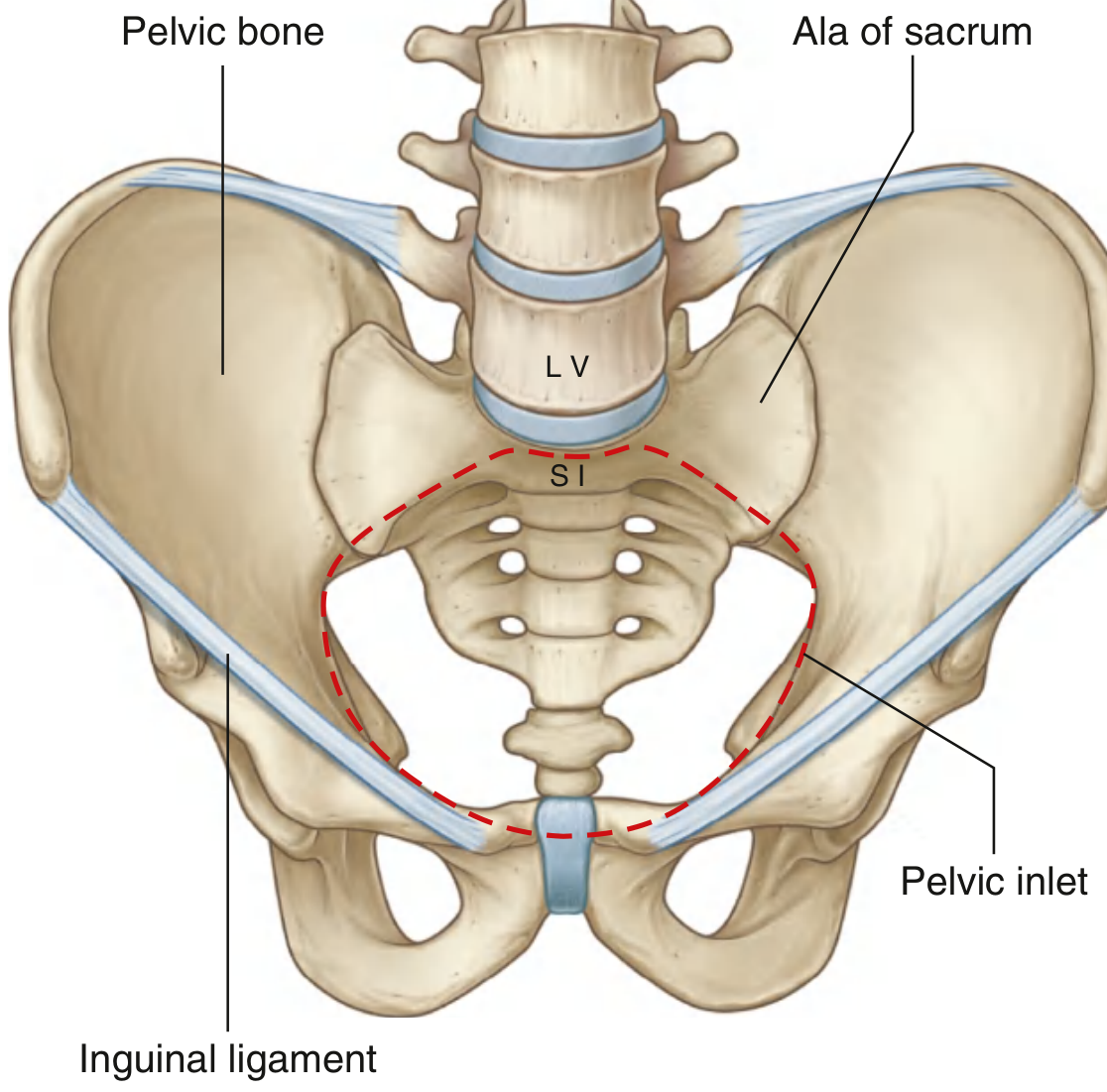
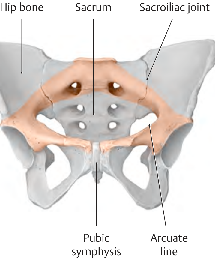
The pelvis is a bony ring that links the vertebral column above to the lower limbs below. It is formed by three bones: the two hip bones (one on each side) and the sacrum posteriorly. The two hip bones articulate with each other anteriorly at the pubic symphysis (a cartilaginous joint) and with the sacrum at the paired sacroiliac joints (SI joints). Together these form a stable, near-rigid ring.
Pelvic Inlet (Superior Aperture)
The pelvic inlet (pelvic brim) separates the pelvis into:
- Greater (false) pelvis - above the brim, part of the abdominal cavity
- Lesser (true) pelvis - below the brim; the actual pelvic cavity
The pelvic inlet is bounded by:
- Posteriorly: sacral promontory
- Laterally: arcuate line of ilium + pectineal line of pubis
- Anteriorly: upper border of pubic symphysis
Pelvic Outlet (Inferior Aperture)
Bounded by:
- Posteriorly: tip of coccyx
- Laterally: ischial tuberosities and sacrotuberous ligaments
- Anteriorly: inferior border of pubic symphysis, pubic arch (subpubic angle)
Communication with Lower Limb
Structures pass between pelvis and lower limb through two major routes:
- Greater sciatic foramen (posterior) - transmits piriformis, sciatic nerve, superior/inferior gluteal nerves & vessels, pudendal nerve, internal pudendal artery
- Obturator canal (anteriorly, through obturator foramen) - transmits obturator nerve and vessels
- Lesser sciatic foramen - transmits obturator internus tendon; pudendal nerve and internal pudendal artery re-enter perineum here
Abdominal-Pelvic Continuity
The peritoneum of the abdominal cavity is continuous with that of the pelvic cavity. The pelvic inlet opens directly into the abdomen, so infections can spread freely between the two regions. The bladder expands superiorly from the pelvis into the abdomen, as does the uterus during pregnancy.
2. HIP BONE (Os Coxae / Innominate Bone)
Gray's Anatomy for Students; THIEME Atlas of Anatomy; Campbell-Walsh Urology
The hip bone (innominate bone) is formed by the fusion of three primary bones:
- Ilium (upper fan-shaped part)
- Ischium (posteroinferior)
- Pubis (anteromedial)
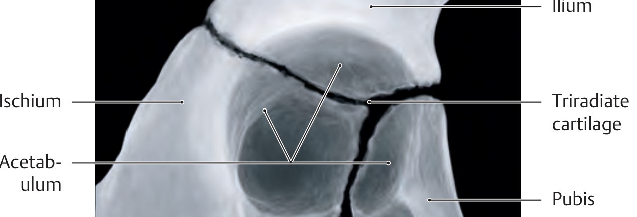
All three meet at the acetabulum (hip socket):
- Ilium: 2/5 of the acetabulum
- Ischium: 2/5 of the acetabulum
- Pubis: 1/5 of the acetabulum
The junction of all three bones in the acetabulum is marked in childhood by the triradiate (Y-shaped) cartilage, which fuses between ages 14-16 years.
Ilium
- Large fan-shaped bone forming the upper part
- Iliac crest - the superior curved border; extends from ASIS to PSIS
- Anterosuperior iliac spine (ASIS) - attachment for inguinal ligament and sartorius
- Anteroinferior iliac spine (AIIS) - attachment for rectus femoris
- Posterosuperior iliac spine (PSIS) and posteroinferior iliac spine (PIIS)
- Iliac fossa - medial concave surface; floor for iliacus muscle
- Auricular surface - posteriorly; articulates with sacrum (SI joint)
- Arcuate line - continues as pectineal line to form the pelvic brim
- Greater sciatic notch - between PIIS and ischial spine
Ischium
- Posteroinferior part of hip bone
- Body of ischium - forms posteroinferior acetabulum
- Ischial spine - projects medially; separates greater from lesser sciatic notch; attachment for sacrospinous ligament
- Lesser sciatic notch - below ischial spine
- Ischial tuberosity - weight-bearing prominence when sitting; attachment for sacrotuberous ligament and hamstrings (biceps femoris, semimembranosus, semitendinosus)
- Ramus of ischium - joins inferior pubic ramus to complete the obturator foramen
Pubis
- Anteromedial part of hip bone
- Body - medial, bears pubic symphysis
- Superior pubic ramus - projects laterally from body to acetabulum; bears the pectineal line (pecten pubis) and pubic tubercle
- Inferior pubic ramus - descends and meets ischial ramus
- The two pubic bones articulate at the pubic symphysis (secondary cartilaginous joint)
Obturator Foramen
Large opening bounded by the body and rami of the pubis and ischium. Closed in life by the obturator membrane, which leaves a small superior gap - the obturator canal - for the obturator nerve and vessels.
Key Ossification Points (Clinically Important)
- Iliac crest fuses with ilium: by ~20 years
- Ischial tuberosity fuses with ischium: by ~20 years
- Triradiate cartilage of acetabulum: fuses between 14-16 years
3. NORMA BASALIS (Inferior View of Skull / External Cranial Base)
Gray's Anatomy for Students
The norma basalis is the view of the skull from below. It extends from the middle incisor teeth anteriorly to the superior nuchal lines posteriorly.
For descriptive purposes it is divided into three parts:
Anterior Part
Main features: teeth and hard palate
- Teeth are anchored in the alveolar processes of the two maxillae, arranged in a U-shaped alveolar arch
- Hard palate is formed by:
- Palatine processes of maxillae (anterior 2/3)
- Horizontal plates of palatine bones (posterior 1/3)
- Sutures of hard palate:
- Intermaxillary suture - midline, between the two maxillae
- Palatomaxillary suture - between maxillae and palatine bones
- Interpalatine suture - midline, between the two palatine bones
- Incisive fossa - anterior midline, behind the incisor teeth; contains incisive foramina (nasopalatine nerve and vessels)
- Greater palatine foramina - near posterolateral border of hard palate (greater palatine nerve and vessels)
- Lesser palatine foramina - just posterior to greater palatine foramina (lesser palatine nerves)
- Posterior nasal spine - pointed projection at free posterior border of hard palate (palatoglossus and uvular muscles)
Middle Part (behind hard palate to anterior edge of foramen magnum)
- Anterior half: vomer and sphenoid bones
- Posterior half: occipital bone and temporal bones
Key features:
- Choanae (posterior nasal apertures) - between vomer and medial pterygoid plates of sphenoid
- Medial and lateral pterygoid plates of sphenoid - medial plate ends in pterygoid hamulus (around which tensor veli palatini wraps)
- Scaphoid fossa - at root of medial pterygoid plate (origin of tensor veli palatini)
- Pterygoid canal (Vidian canal) - opens just above scaphoid fossa (transmits Vidian nerve = greater petrosal + deep petrosal)
- Foramen lacerum - irregular gap at junction of sphenoid, temporal, and occipital; filled with fibrocartilage in life (only internal carotid artery traverses its superior part)
- Foramen ovale - transmits mandibular nerve (V3), accessory meningeal artery, lesser petrosal nerve
- Foramen spinosum - transmits middle meningeal artery and meningeal branch of V3
- Foramen rotundum - (opens into the middle cranial fossa, not visible from below)
- Carotid canal - entrance for internal carotid artery
- Jugular foramen - between temporal and occipital bones; transmits CN IX (glossopharyngeal), CN X (vagus), CN XI (accessory), and internal jugular vein
- Styloid process + stylomastoid foramen (CN VII exits here) medial to mastoid process
- Mastoid process - with mastoid notch (posterior belly of digastric)
- Occipital condyles - articulate with atlas (C1); flanked by hypoglossal canal (CN XII)
Posterior Part (foramen magnum to superior nuchal lines)
- Foramen magnum - largest foramen of skull; transmits medulla oblongata, vertebral arteries, anterior and posterior spinal arteries, spinal roots of CN XI
- Pharyngeal tubercle - midline anteriorly on occipital bone; attachment of pharyngeal raphe (fibrous pharynx)
- External occipital crest - midline ridge descending from foramen magnum to external occipital protuberance
- External occipital protuberance (inion) - most prominent midline point
- Superior nuchal lines - extend laterally from inion (trapezius, sternocleidomastoid)
- Inferior nuchal lines - about 1 inch (2.5 cm) below superior nuchal lines
4. MANDIBLE
Gray's Anatomy for Students
The mandible is the bone of the lower jaw - the only moveable bone of the skull.
General Parts
- Body - the horseshoe-shaped horizontal portion; formed by fusion of right and left halves at the mandibular symphysis (visible externally as a vertical midline ridge)
- Rami - two vertical plates rising from the posterior body; one on each side
- Angle of mandible - where inferior border of body meets posterior margin of ramus
Body of Mandible
Divided into:
- Base (lower part) - contains the mental protuberance at the anterior midline, flanked by mental tubercles on each side
- Alveolar part (upper part) - contains the alveolar arch with the teeth; resorbed when teeth are lost
External surface features:
- Mental foramen - on lateral surface halfway between upper alveolar border and lower base; transmits mental nerve (terminal branch of inferior alveolar nerve) and mental vessels
- Oblique line - continues posteriorly from the front of the ramus; attachment for muscles depressing the lower lip
Internal surface features:
- Mental spines (genial spines) - 4 small spines in 2 pairs at midline, posterior to mandibular symphysis
- Superior pair: origin of genioglossus (into tongue)
- Inferior pair: origin of geniohyoid
- Mylohyoid line - oblique ridge running posterosuperiorly from below the mental spines to below the last molar; attachment for mylohyoid muscle (floor of mouth)
- Sublingual fossa - above the anterior 1/3 of mylohyoid line; houses sublingual gland
- Submandibular fossa - below the posterior 2/3 of mylohyoid line; houses submandibular gland
- Lingual nerve groove - shallow groove between last molar and mylohyoid line
- Retromolar triangle - small triangular depression just posterior to last molar on medial upper surface; attachment for pterygomandibular raphe (which connects to buccinator/superior pharyngeal constrictor)
Ramus of Mandible
- Coronoid process - anterior superior projection; attachment for temporalis muscle
- Condylar process - posterior superior projection; participates in temporomandibular joint (TMJ) with the temporal bone
- Mandibular notch (sigmoid notch) - between coronoid and condylar processes
- Mandibular foramen - on medial surface of ramus; transmits inferior alveolar nerve and vessels into the mandibular canal; guarded anteriorly by the lingula (spine of Spix)
- Pterygoid fovea - small depression anterior to condylar neck; attachment for lateral pterygoid muscle
- Masseteric groove/tuberosity - on lateral surface at angle; for masseter
- Pterygoid tuberosity - on medial surface at angle; for medial pterygoid
Ossification of Mandible
- The mandible ossifies in membrane (intramembranous ossification) in association with Meckel's cartilage
- The two halves fuse at the mandibular symphysis by the end of year 1 (early childhood)
Quick Reference Summary Table
| Topic | Key Feature | Clinical Relevance |
|---|---|---|
| Pelvis - Inlet | Sacral promontory → arcuate line → pectineal line → pubis | Obstetric measurements |
| Hip bone | Triradiate cartilage fuses age 14-16 | Paediatric hip X-ray interpretation |
| Hip bone - Ischium | Ischial tuberosity bears weight in sitting | Avulsion fractures in athletes |
| Hip bone - Obturator foramen | Closed by obturator membrane; obturator canal at top | Obturator hernia |
| Norma basalis - Middle | Foramen ovale (V3), spinosum (middle meningeal a.) | Trigeminal neurotomy, meningeal haemorrhage |
| Norma basalis - Posterior | Foramen magnum (medulla, vertebral arteries) | Tonsillar herniation |
| Norma basalis - Hypoglossal canal | CN XII | Hypoglossal nerve palsy |
| Mandible - Lingula | Guards mandibular foramen | Inferior alveolar nerve block landmark |
| Mandible - Mental foramen | Level of 2nd premolar, halfway up body | Dental anaesthesia |
| Mandible - Symphysis fusion | End of year 1 | Age estimation in forensics |
Sources: Gray's Anatomy for Students (9780323934237); THIEME Atlas of Anatomy - General Anatomy and Musculoskeletal System (9781626237186); Campbell-Walsh-Wein Urology (9780323546423)
This is a shared conversation. Sign in to Orris to start your own chat.