
Calcium homeostasis
Reading File
Now let me search the library for calcium homeostasis content in parallel:
Finding Sources
Finding Sources
Searching PubMed
"calcium homeostasis" AND regulation
Excellent sources found. Let me now read the most comprehensive sections in parallel:
Reading File
Reading File
Reading File
Now let me fetch the key diagram and read more sections on hormonal regulation:
Loading Image
Reading File
Reading File
I now have comprehensive content. Let me also fetch the PTH secretion graph and read the Vitamin D section:
Loading Image
Reading File
I now have comprehensive material. Let me compile the full answer:
Calcium Homeostasis
Calcium homeostasis is the set of integrated physiological mechanisms that maintain the extracellular fluid (ECF) Ca2+ concentration within a tight normal range (~8.5-10.5 mg/dL or ~2.1-2.6 mmol/L). Three organ systems (bone, intestine, kidney) and three principal hormones (PTH, vitamin D, calcitonin) work in concert to achieve this.
1. Body Calcium Distribution
- Total body calcium: ~1000-1300 g
- 99.3% in bone and teeth as hydroxyapatite crystal [Ca₁₀(PO₄)₆(OH)₂]
- 0.6% in soft tissues (intracellular)
- 0.1% in ECF, including 0.03% in plasma
- Intracellular free Ca2+ is ~100 nM vs. extracellular ~1 mM - a 10,000-fold gradient is maintained
Fractions of Plasma Calcium
| Fraction | % of Total | Notes |
|---|---|---|
| Protein-bound (mainly albumin) | ~40% | Not ultrafilterable |
| Complexed (citrate, phosphate) | ~10% | Not ionized |
| Ionized (free) Ca2+ | ~50% | Biologically active; regulated |
The ionized fraction is what the parathyroid glands sense and regulate.
2. The Overall Calcium Balance (Daily Fluxes)
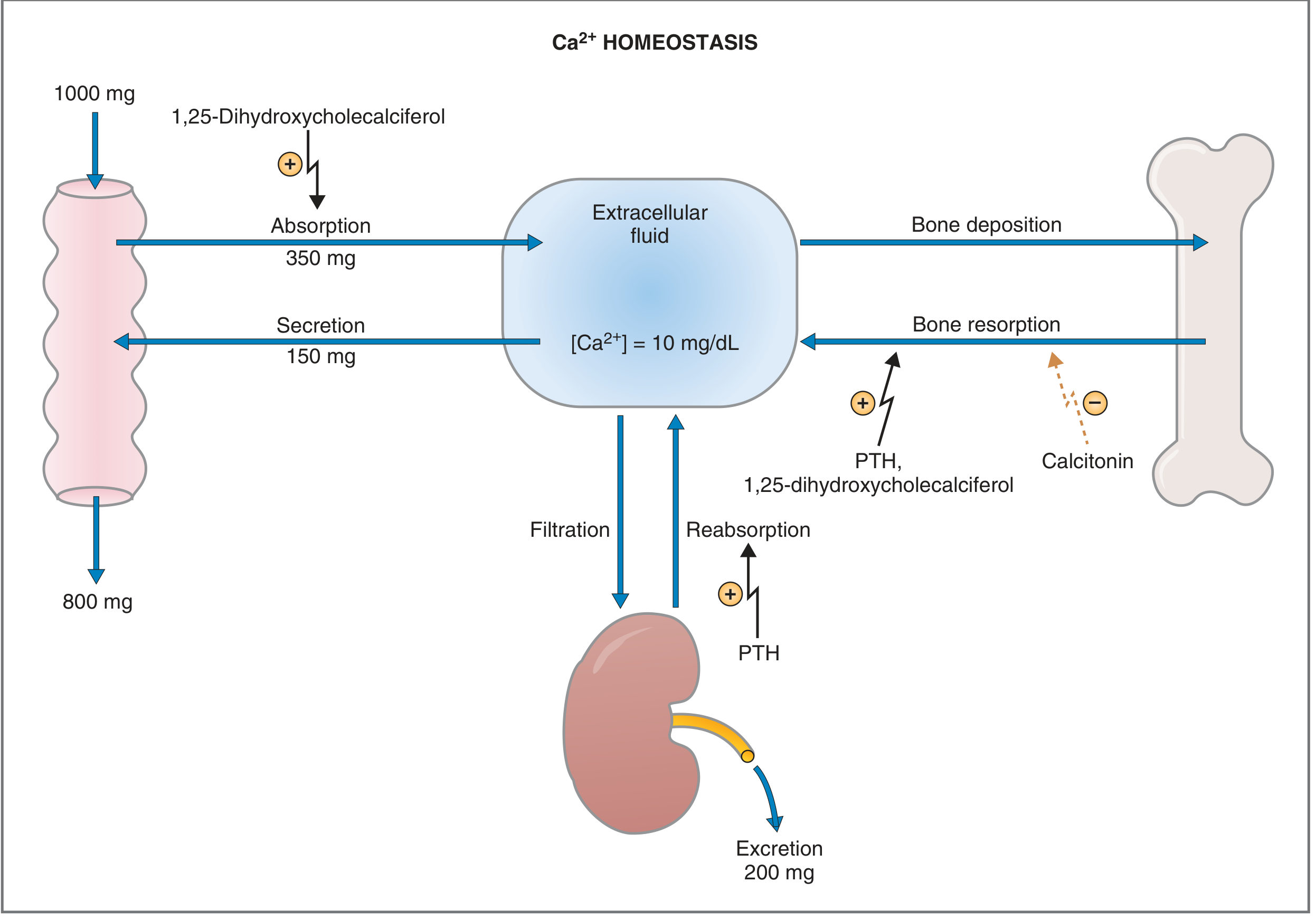
Fig. Ca2+ homeostasis in an adult eating 1000 mg/day. - Costanzo Physiology, 7th ed., p. 453
In a healthy adult eating 1000 mg/day of calcium:
- Intestine: 350 mg absorbed (stimulated by 1,25-dihydroxycholecalciferol), 150 mg secreted back in digestive juices. Net absorption = 200 mg/day; 800 mg excreted in feces.
- Bone: Continuous remodeling with ~500 mg/day released (resorption) and ~500 mg/day deposited - net zero in healthy adults.
- Kidney: ~10 g filtered daily; ~9.8 g reabsorbed; ~200 mg excreted in urine (equal to net intestinal absorption, maintaining balance).
3. Parathyroid Hormone (PTH) - The Primary Regulator
Structure
- Synthesized by chief cells of the 4 parathyroid glands
- 84-amino acid single-chain polypeptide; biologic activity in N-terminal 34 amino acids
- Precursors: preproPTH (115 aa) → proPTH (90 aa) → PTH (84 aa)
Regulation of PTH Secretion - The Calcium-Sensing Receptor (CaSR)
PTH secretion is inversely related to plasma ionized Ca2+:
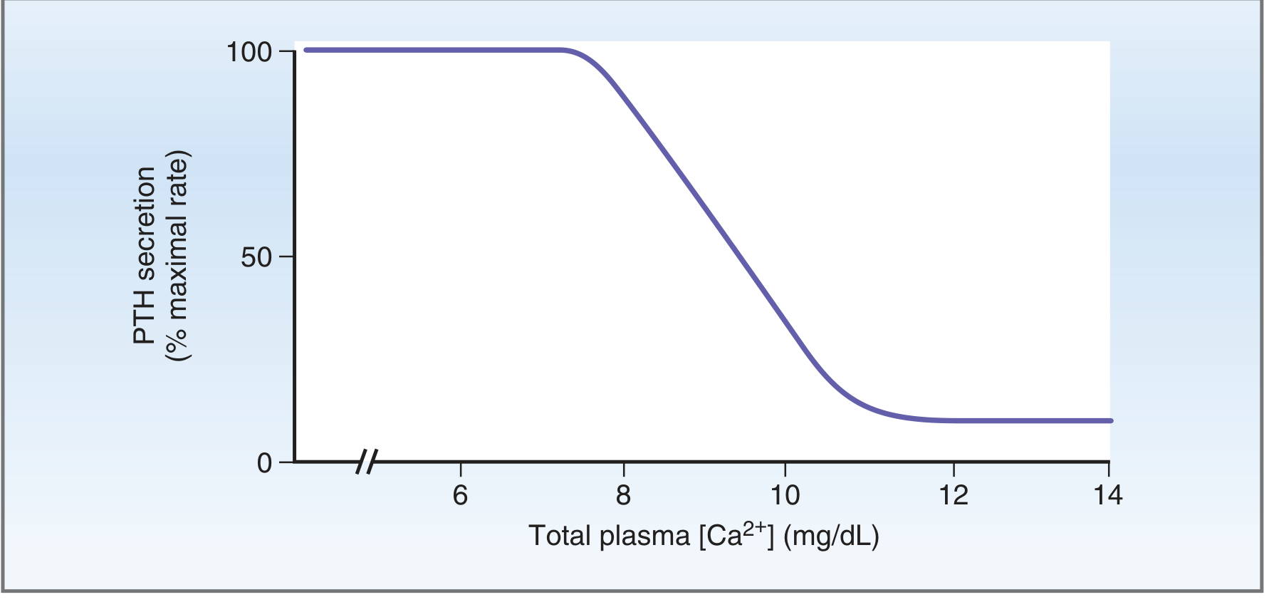
Fig. PTH secretion vs. plasma Ca2+ concentration. - Costanzo Physiology, 7th ed., p. 454
Mechanism:
- Parathyroid chief cells express the calcium-sensing receptor (CaSR), a Gq-coupled GPCR
- When ECF Ca2+ is high: Ca2+ binds CaSR → activates phospholipase C → ↑ IP₃/Ca2+ → inhibits PTH secretion
- When ECF Ca2+ is low: reduced CaSR activation → stimulates PTH secretion within seconds
- PTH response to falling Ca2+ is remarkably fast; the faster the fall, the greater the PTH secretory response
Actions of PTH on Target Organs
Bone
PTH acts in two phases:
- Rapid phase (minutes-hours): Activates existing osteocytes and osteoblasts of the osteocytic membrane system - an interconnected network that separates bone fluid from ECF. PTH activates the osteocytic pump, pulling Ca2+ from bone fluid into ECF. Called osteolysis - occurs without matrix resorption.
- Slow phase (days-weeks): Proliferation of osteoclasts → increased bone resorption (matrix + mineral). Also mobilizes phosphate from bone.
Kidney
PTH has two renal effects:
- Proximal tubule: Inhibits Na⁺-phosphate cotransport → phosphaturia (urinary phosphate excretion). This is functionally critical: the phosphate released from bone would otherwise complex ECF Ca2+ and blunt the calcium rise. By excreting phosphate, PTH "allows" ionized Ca2+ to increase.
- Distal convoluted tubule: Stimulates Ca2+ reabsorption, directly reducing urinary calcium loss.
Intestine (Indirect)
PTH has no direct effect on the intestine. Instead, PTH stimulates renal 1α-hydroxylase, converting 25-hydroxycholecalciferol → 1,25-dihydroxycholecalciferol (calcitriol), which then directly stimulates intestinal Ca2+ absorption.
PTH's Net Effect
↓ Ca2+ → ↑ PTH → ↑ bone resorption + ↑ renal Ca2+ reabsorption + ↓ phosphaturia + ↑ vitamin D activation → ↑ plasma Ca2+
4. Vitamin D (Calcitriol)
Synthesis and Activation
- Skin: UV-B converts 7-dehydrocholesterol → cholecalciferol (D3)
- Liver: 25-hydroxylase converts D3 → 25-hydroxycholecalciferol [25(OH)D3] (storage form; measured for vitamin D status)
- Kidney: 1α-hydroxylase converts 25(OH)D3 → 1,25-dihydroxycholecalciferol [1,25(OH)₂D3] = calcitriol (active form)
- This step is stimulated by PTH, hypocalcemia, and hypophosphatemia
- Inhibited by calcitriol itself (negative feedback) and by FGF-23
Actions of Calcitriol
- Intestine: Major target. Stimulates synthesis of calcium-binding proteins (e.g., calbindin) → markedly increases Ca2+ and phosphate absorption (both active and passive transport)
- Bone: At physiologic levels, promotes bone mineralization. At pharmacologic/excess levels, enhances PTH-mediated bone resorption
- Kidney: Mildly increases Ca2+ and phosphate reabsorption
- Parathyroid: Directly suppresses PTH gene transcription (negative feedback loop)
5. Calcitonin
- Secreted by parafollicular C cells of the thyroid gland
- 32-amino acid peptide
- Released in response to elevated plasma Ca2+ (opposite trigger to PTH)
Actions
- Bone: Directly inhibits osteoclast activity → decreases bone resorption → lowers plasma Ca2+
- Rapid effect: reduces osteoclastic absorption
- Prolonged effect: decreases formation of new osteoclasts
- Kidney: Minor effects opposing PTH (↑ Ca2+ excretion)
Physiologic Role - Limited in Adults
Calcitonin has a weak and transient effect on plasma Ca2+ in adult humans for two reasons:
- Any drop in Ca2+ from calcitonin powerfully stimulates PTH, which overrides the calcitonin effect
- Daily calcium flux rates are small relative to the 1 g total ECF calcium
Calcitonin is more relevant in children (rapid bone remodeling) and in conditions like Paget disease (accelerated osteoclast activity).
6. The Calcium-Sensing Receptor (CaSR) - Central Sensor
The CaSR is a class C GPCR expressed on:
- Parathyroid chief cells (primary regulator of PTH secretion)
- Thyroid C cells (controls calcitonin secretion)
- Renal tubular cells (directly regulates tubular Ca2+ handling)
- Bone cells and intestinal epithelium
Loss-of-function mutations → Familial Hypocalciuric Hypercalcemia (FHH) - CaSR set-point shifted, PTH not suppressed at normal Ca2+ levels
Gain-of-function mutations → Autosomal Dominant Hypocalcemia (ADH) - CaSR oversensitive, excess PTH suppression
7. FGF-23 and Phosphatonins (Indirect Role)
Fibroblast growth factor 23 (FGF-23), secreted by osteocytes, primarily regulates phosphate and vitamin D homeostasis:
- Inhibits renal 1α-hydroxylase → ↓ calcitriol production → indirectly lowers intestinal Ca2+ absorption
- Stimulates renal phosphate excretion (phosphaturia)
- FGF-23 does not directly modify ECF calcium
8. Three Lines of Defense Against Calcium Perturbation
| Line | Mechanism | Speed |
|---|---|---|
| 1st | Exchange of calcium from amorphous bone salts (CaHPO4) - the "exchangeable pool" | Seconds to minutes |
| 2nd | PTH feedback via CaSR - bone resorption, renal Ca2+ retention, ↑ calcitriol | Minutes to hours |
| 3rd | Changes in vitamin D synthesis and intestinal absorption | Hours to days |
9. Pathophysiology Summary
| Disorder | Mechanism | Ca2+ | PO4 | PTH |
|---|---|---|---|---|
| Primary hyperparathyroidism | PTH-secreting adenoma | ↑ | ↓ | ↑ |
| Hypoparathyroidism | PTH deficiency (post-surgical, autoimmune) | ↓ | ↑ | ↓ |
| Pseudohypoparathyroidism | Target organ resistance to PTH (Gs mutation) | ↓ | ↑ | ↑↑ |
| Secondary hyperparathyroidism | Chronic kidney disease → ↓ calcitriol → ↓ Ca2+ → ↑ PTH | ↓ | ↑ | ↑ |
| Hypercalcemia of malignancy | PTHrP from tumor mimics PTH; bone metastases | ↑ | ↓ | ↓ |
| Vitamin D toxicity | Excess calcitriol → ↑ intestinal absorption + bone resorption | ↑ | ↑ | ↓ |
The classic mnemonic for primary hyperparathyroidism is "stones, bones, and groans": kidney stones (hypercalciuria), bone resorption, and constipation/abdominal pain.
Sources:
- Costanzo Physiology, 7th Edition, pp. 453-456 (Overall Ca2+ homeostasis, PTH, Vitamin D)
- Guyton and Hall Textbook of Medical Physiology, pp. 989-992 (PTH bone effects, calcitonin, Ca2+ control summary)
- Brenner and Rector's The Kidney, pp. 796-798 (Whole-body Ca2+ homeostasis, PTH-Vitamin D system)
This is a shared conversation. Sign in to Orris to start your own chat.