
Ascending tracts of CNS
ascending tracts spinal cord diagram
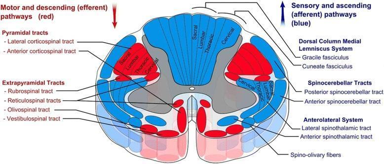
This medical anatomical diagram illustrates a transverse section of the human spinal cord, specifically detailing the organization of white matter tracts. The visual uses a color-coded schema to differentiate between motor and sensory pathways: descending (efferent) motor tracts are highlighted in red, while ascending (afferent) sensory tracts are shown in blue. Key anatomical structures identified include the Pyramidal tracts (lateral and anterior corticospinal tracts), Extrapyramidal tracts (rubrospinal, reticulospinal, olivospinal, and vestibulospinal), the Dorsal Column-Medial Lemniscus system (gracile and cuneate fasciculi), Spinocerebellar tracts, and the Anterolateral system (lateral and anterior spinothalamic tracts). A significant educational feature of the diagram is the depiction of somatotopic organization, showing the concentric arrangement of sacral, lumbar, thoracic, and cervical fibers within the lateral corticospinal and dorsal column tracts. The image provides a comprehensive overview of spinal cord neuroanatomy, useful for understanding clinical localization of spinal cord lesions and the functional topography of the central nervous system.
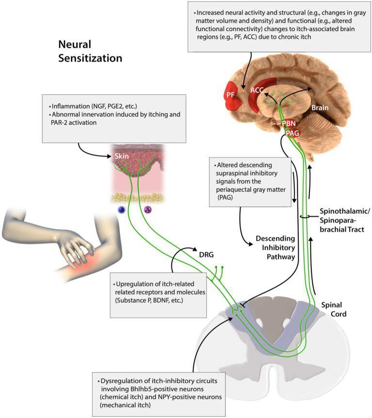
An educational pathophysiology diagram illustrating the pathways of peripheral and central neural sensitization in chronic itch. The diagram traces signals from the skin, through the spinal cord, to the brain. Primary cutaneous stimuli include inflammation (NGF, PGE2) and abnormal innervation via PAR-2 activation. Pruritic signals travel through the dorsal root ganglion (DRG), where upregulation of Substance P and BDNF occurs, and enter the spinal cord. The spinal level highlights dysregulation of itch-inhibitory circuits involving Bhlhb5-positive (chemical itch) and NPY-positive (mechanical itch) neurons. Ascending transmission via the spinothalamic and spinoparabrachial tracts carries signals to the brain, specifically the periaqueductal gray (PAG) and parabrachial nucleus (PBN). The diagram depicts increased neural activity and structural/functional changes (gray matter volume, connectivity) in cortical regions including the prefrontal cortex (PF) and anterior cingulate cortex (ACC). A descending inhibitory pathway from the PAG to the spinal cord is also shown, noting altered supraspinal inhibitory signals that contribute to the chronic itch cycle.
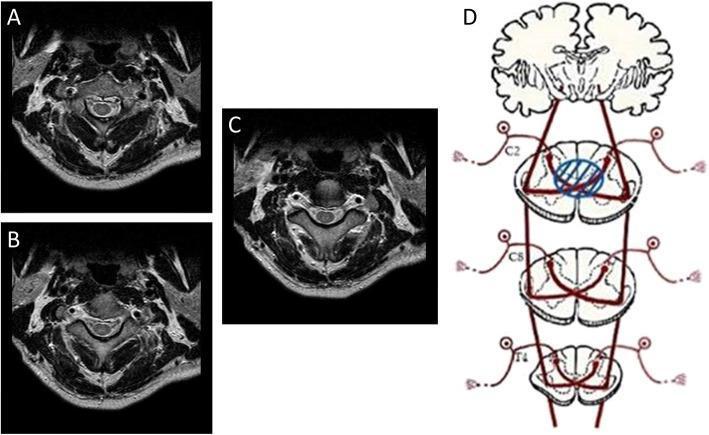
This composite educational image illustrates the radiological and pathophysiological features of Central Cord Syndrome. Panels A, B, and C present three sequential cranial-to-caudal axial T2-weighted MRI scans of the cervical spine at the C2-3 level. These images reveal a focal area of hyperintensity (high signal) localized in the central portion of the spinal cord, corresponding to the central canal and surrounding gray matter. Panel D is a neuroanatomical schematic diagram illustrating the lateral spinothalamic tracts across C2, C8, and T4 spinal levels. A blue circle highlights a central lesion at the C2 level, specifically demonstrating how the injury interrupts the decussating fibers of the spinothalamic tract as they cross the anterior white commissure. This visual correlation explains the mechanism of bilateral neuropathic pain and sensory deficits localized to the dermatome level of the injury (C2) while sparing more laterally located ascending tracts. The content serves to teach the clinical-radiological correlation of traumatic spinal cord injuries and the functional anatomy of sensory pathways within the spinal cord.
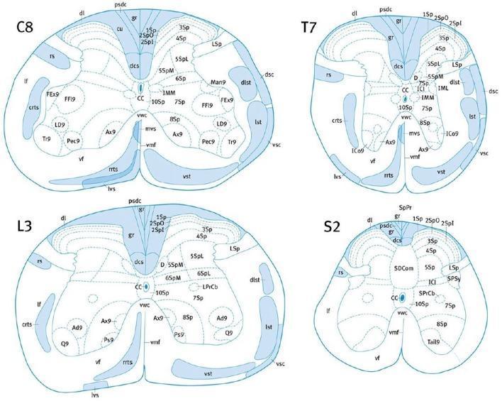
This medical anatomical diagram illustrates the cross-sectional neuroanatomy and white matter tract localization within the mouse spinal cord at four distinct levels: Cervical (C8), Thoracic (T7), Lumbar (L3), and Sacral (S2). Each level depicts the characteristic morphology of the spinal cord, showing the transition from a rounded C8/T7 shape to a more laterally expanded L3 and a flattened, compressed S2. Key ascending (sensory) and descending (motor) pathways are highlighted in blue. Notable structures include the dorsal corticospinal tract (dcs) located centrally along the dorsal midline, flanked by the gracile fasciculus (gr) and cuneate fasciculus (cu, prominent in higher levels). Lateral tracts shown include the rubrospinal (rs), dorsal spinocerebellar (dsc), and lateral spinothalamic (lst) tracts. Ventral regions display the vestibulospinal (lvs, mvs) and reticulospinal (crts, rrts) tracts. The diagrams also delineate Rexed laminae (1Sp-10Sp) within the gray matter and the central canal (CC). This educational resource is designed for neuroanatomical mapping and comparative spinal pathology research.
dorsal column medial lemniscus pathway diagram
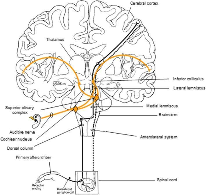
A pathophysiology diagram illustrating the ascending sensory pathways from the periphery to the cerebral cortex. The schematic uses a coronal section of the human brain, brainstem, and spinal cord to trace three major systems: the auditory-lateral lemniscal system (solid orange line), the dorsal column-medial lemniscus system (solid black line), and the anterolateral system (dashed black line). The auditory pathway is traced from the ear through the auditive nerve, cochlear nucleus, superior olivary complex, lateral lemniscus, and inferior colliculus, relaying through the medial geniculate nucleus of the thalamus to the auditory cortex. Somatosensory pathways are traced from receptor endings in the finger through the dorsal root ganglion to the spinal cord. The dorsal column system ascends ipsilaterally before decussating in the medulla (medial lemniscus) and relaying via the thalamus to the somatosensory cortex. The anterolateral system decussates at the spinal level before ascending. This visual summarizes the functional neuroanatomy required for sensory reactivity, commonly assessed in electroencephalography (EEG) clinical evaluations.
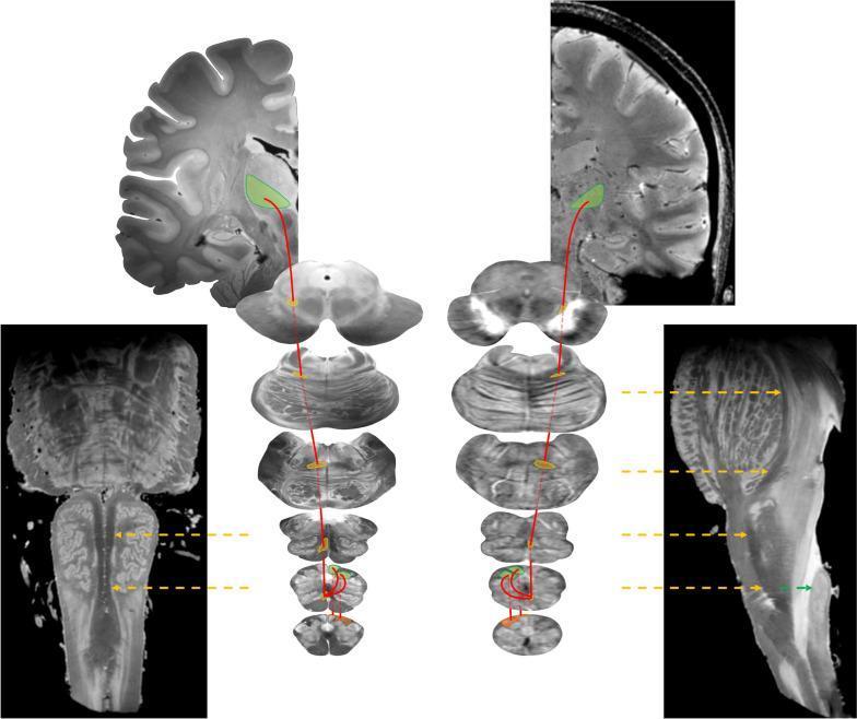
Educational neuroanatomical diagram and MRI composite illustrating the dorsal column-medial lemniscus (DCML) system. The visual uses a side-by-side comparison of ex vivo (left) and in vivo (right) high-resolution 7-T MR images to trace the somatosensory pathway. Red lines represent the trajectory of second-order neurons from the brainstem to the thalamus. Key structures are color-coded: the gracile and cuneate fasciculi are highlighted in orange at the caudal medulla; the gracile and cuneate nuclei are in green, marking the site of first-order synapse. The internal arcuate fibers are shown decussating before forming the medial lemniscus, which is highlighted in yellow across axial slices of the medulla, pons, and midbrain. Yellow arrows in the oblique sagittal and coronal brainstem views indicate the ascending trajectory of the medial lemniscus. The pathway terminates at the ventral posterolateral (VPL) nucleus of the thalamus, highlighted in green on coronal sections. This infographic serves as a diagnostic and anatomical reference for tracing epicritic sensation and conscious proprioception pathways.
Ascending Tracts of the CNS
Overview Diagram
1. Posterior Column - Medial Lemniscal Pathway
Three-neuron chain:
| Neuron | Location | Details |
|---|---|---|
| 1st order | Dorsal root ganglion | Enters spinal cord via medial bundle of dorsal root; heavily myelinated fibers ascend ipsilaterally in the dorsal funiculus |
| 2nd order | Nucleus gracilis / Nucleus cuneatus (caudal medulla) | Axons cross as internal arcuate fibers and ascend as the medial lemniscus |
| 3rd order | VPL nucleus of thalamus | Projects via posterior limb of internal capsule to primary somatosensory cortex (S1) |
Two subdivisions:
- Fasciculus gracilis (Goll's column) - carries fibers from sacral, lumbar, and lower 6 thoracic levels (lower limb/trunk); located medially
- Fasciculus cuneatus (Burdach's column) - carries fibers from upper 6 thoracic and all cervical levels (upper limb/neck); located laterally
Fibers cross in the lower medulla - a lesion above the decussation gives contralateral sensory loss; a lesion below (in the spinal cord itself) gives ipsilateral loss.
2. Anterolateral System (Spinothalamic Tracts)
A. Lateral Spinothalamic Tract (Neospinothalamic)
- Carries: pain and temperature (sharp, well-localized pain)
- 1st order neuron: Dorsal root ganglion → enters via lateral bundle → ascends/descends 1-2 segments in Lissauer's tract → synapses in laminae I and V of the dorsal horn
- 2nd order neuron: Crosses obliquely over 2-3 segments via the anterior white commissure → ascends in the ventrolateral quadrant → reaches VPL nucleus of thalamus
- 3rd order neuron: VPL → posterior limb of internal capsule → primary somatosensory cortex
- Sacral fibers: ventrolateral (outermost)
- Cervical fibers: dorsomedial (innermost)
- Temperature fibers: dorsolateral to pain fibers
B. Spinoreticulothalamic Tract (Paleospinothalamic)
- Carries: poorly localized, diffuse pain (aching, visceral pain) and the affective/emotional aspects of pain
- 2nd order axons project to brainstem reticular formation (nucleus gigantocellularis in medulla, parabrachial region, periaqueductal gray) → intralaminar/medial nuclei of thalamus → limbic and frontal lobes
- This pathway continues to transmit pain even after lateral spinothalamic tractotomy - explaining why cordotomy for pain often fails long-term
C. Spinomesencephalic (Spinoreticular) Tract
- Projects to: periaqueductal gray (PAG) and superior colliculus of the midbrain
- Function: central modulation of pain (descending pain inhibition via PAG)
D. Spinohypothalamic Tract
- A direct pathway to the hypothalamus in the medial anterolateral fasciculus
- Role in autonomic and neuroendocrine responses to pain
Ventral Spinothalamic Tract
- Carries: light touch (crude tactile sensation)
- Fibers cross via the anterior white commissure and ascend in the ventral funiculus
- Light touch is also transmitted via the dorsal columns, explaining why isolated dorsal column lesions do not abolish touch completely
3. Spinocerebellar Tracts (Unconscious Proprioception)
| Tract | Side of Ascent | Source | Termination |
|---|---|---|---|
| Posterior (dorsal) spinocerebellar tract | Ipsilateral (uncrossed) | Lower limb, inferior trunk (T1 and below) | Cerebellum via inferior cerebellar peduncle |
| Anterior (ventral) spinocerebellar tract | Contralateral then recrosses | Lower limb (L2 and below) | Cerebellum via superior cerebellar peduncle |
| Cuneocerebellar tract | Ipsilateral | Upper limb, rostral trunk (C2-T1) | Cerebellum via inferior cerebellar peduncle |
| Rostral spinocerebellar tract | Ipsilateral | Upper limb (equivalent of anterior spinocerebellar) | Cerebellum via superior or inferior cerebellar peduncle |
4. Summary Table
| Tract | Modality | 1st Neuron | Decussation | 2nd Neuron Destination | Fiber Location in Cord |
|---|---|---|---|---|---|
| Fasciculus gracilis | Fine touch, vibration, proprioception (lower body) | DRG | Lower medulla | Nucleus gracilis | Dorsal funiculus (medial) |
| Fasciculus cuneatus | Fine touch, vibration, proprioception (upper body) | DRG | Lower medulla | Nucleus cuneatus | Dorsal funiculus (lateral) |
| Lateral spinothalamic | Pain, temperature | DRG | 2-3 segments above entry (anterior commissure) | VPL thalamus | Ventrolateral funiculus |
| Ventral spinothalamic | Crude/light touch | DRG | Anterior commissure | VPL thalamus | Ventral funiculus |
| Spinoreticulothalamic | Diffuse/visceral pain, affect | DRG | Anterior commissure | Intralaminar thalamus | Medial anterolateral |
| Spinomesencephalic | Pain modulation | DRG | Anterior commissure | PAG, superior colliculus | Anterolateral |
| Posterior spinocerebellar | Unconscious proprioception (lower) | Nucleus dorsalis (Clarke's column, T1-L2) | Does NOT cross | Cerebellum (inf. peduncle) | Dorsolateral funiculus |
| Anterior spinocerebellar | Unconscious proprioception (lower) | Spinal border cells (L2-S3) | Crosses (then recrosses in pons) | Cerebellum (sup. peduncle) | Ventrolateral funiculus |
Key Clinical Points
- Brown-Séquard syndrome (hemisection of cord): Ipsilateral loss of fine touch/proprioception (dorsal columns) + contralateral loss of pain/temperature (spinothalamic, already crossed)
- Syringomyelia: Cavity in the center of cord disrupts crossing spinothalamic fibers in the anterior commissure → bilateral suspended "cape-like" loss of pain/temperature at the level of the lesion, with preserved dorsal column function
- Tabes dorsalis (syphilis): Destruction of dorsal columns → loss of vibration, proprioception, two-point discrimination; positive Romberg sign
- Anterior cord syndrome: Loss of pain/temperature (spinothalamic) and motor function, with preserved posterior column modalities
- Localization in Clinical Neurology, 8e, pp. 245-246
- Gray's Anatomy for Students - ascending tracts section
- Adams and Victor's Principles of Neurology, 12th Edition, pp. 149-150