
What is tubercular otitis media and what are the stages of it and why there is perforation in TM
Reading File
Finding Sources
Finding Sources
Searching PubMed
"tuberculosis"[MeSH Terms] AND "otitis media"[MeSH Terms]
Reading File
Reading File
Searching Images
tuberculous otitis media tympanic membrane perforation otoscopy
Reading File
Tubercular (tuberculous) otitis media
Tuberculous otitis media (TOM) is infection of the middle ear cleft and mastoid, usually by Mycobacterium tuberculosis. Atypical mycobacteria such as M. avium and M. fortuitum occasionally cause it. It is rare. One African series of more than 11,000 TB patients found it in only 0.37% (Scott-Brown's Otorhinolaryngology, Vol 2, p. 1100). It is more common with HIV and drug-resistant TB. Think of it in any chronic ear discharge that does not respond to usual treatment.
Routes of infection (Scott-Brown, p. 1101; Cummings):
- Through the Eustachian tube, for example by swallowed or aspirated sputum.
- Directly through an existing tympanic membrane (TM) perforation, as in pre-existing chronic otitis media.
- By blood or lymphatic spread from a distant focus, usually pulmonary. Up to 50% of childhood cases are secondary to another focus.
Classical features:
- Painless otorrhoea.
- Multiple TM perforations.
- Pale granulation tissue.
- Early, severe hearing loss, often out of proportion to the otoscopic findings.
- Bone necrosis.
- Facial palsy in 10 to 20% of patients, appearing early. Complications are commoner and more severe in children.
- Non-tender cervical or high jugular lymphadenopathy.
Stages (pathological phases)
Scott-Brown describes three phases:
- Acute (exudative) phase.
- The TM and mucosa become congested, then thickened, with loss of landmarks.
- Tubercles appear, followed by perforations.
- There is middle ear effusion, and the hearing loss is conductive.
- Chronic (granulation) phase.
- The course is slow and painless.
- Perforations persist, and pale pink granulation tissue fills the middle ear.
- Polyps may appear in the canal.
- Necrotic or caseous phase.
- Caseation destroys bone and forms sequestra.
- Ossicles, mastoid cortex and otic capsule are destroyed.
- Complications include facial nerve palsy, labyrinthitis with sensorineural loss, and a "cold" Bezold abscess from mastoid tip destruction (Cummings).
Many exam notes describe the same sequence in other words: tubercle formation, then multiple perforations, then coalescence into a total or large perforation, then granulations, then bone sequestration and complications. This is the same progression, not a different staging system.
Why does the TM perforate?
The textbooks describe what happens but not the mechanism in detail. The mechanism below is standard pathology that I am supplying from my own knowledge.
- Granuloma formation in the TM. Bacilli reach the middle ear mucosa and the inner mucosal and fibrous layers of the TM. Tubercles form there. The TM first becomes thickened and congested, and Cummings shows it greatly thickened by granulation tissue containing epithelioid and multinucleated giant cells.
- Caseous necrosis and ischaemia. Caseation destroys the TM tissue. Endarteritis of the small vessels cuts off its blood supply. The TM is thin and has a limited blood supply, so it breaks down easily.
- Multiple small perforations that coalesce. Necrosis happens at several foci at once, so many small perforations appear, often with seropurulent discharge. Cummings notes they "quickly coalesce to cause loss of the tympanic membrane". Scott-Brown adds that multiple perforations are now reported less often than once thought, and a single large perforation is common.
- Painless course. Pain is minimal because the process is chronic, granulomatous and non-suppurative. It is not the acute pyogenic inflammation of ordinary acute otitis media.
- Perforations enlarge, not heal. Ongoing necrosis keeps enlarging them. A myringotomy site in an intact TM can also enlarge rapidly (Cummings).
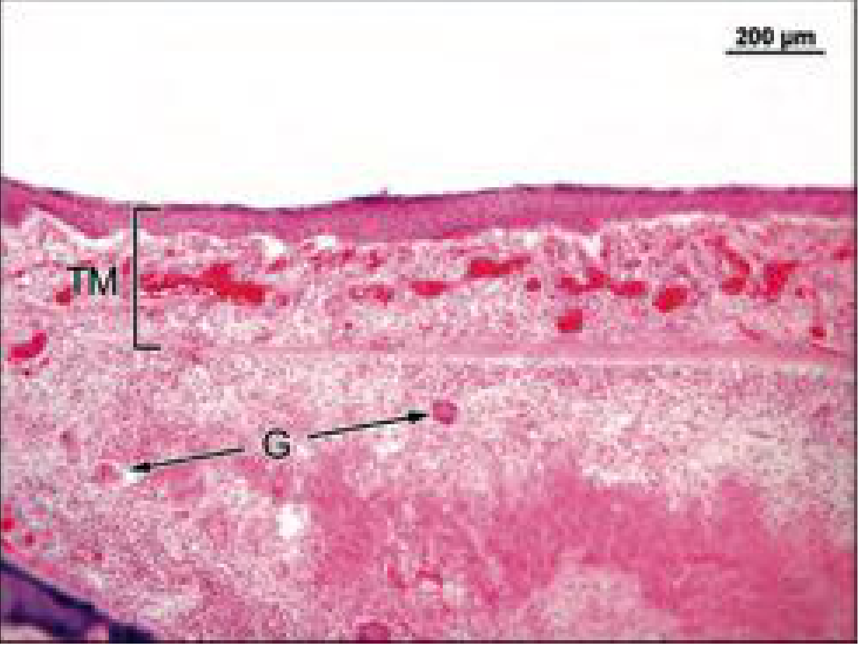
Histology of the thickened TM with tuberculous granulation tissue and giant cells (Cummings Otolaryngology, Fig. 151.7).
Diagnosis and treatment
- Culture of ear discharge is positive in under 20% of cases.
- Histology of a biopsy of granulation tissue or polyp is the most definitive test. It shows caseating granulomas with epithelioid and Langerhans cells. PCR can also be used.
- Chest X-ray is needed, because pulmonary TB is present in 14 to 93% of patients.
- HRCT temporal bone is the imaging of choice.
- Treatment is standard anti-tubercular therapy. Surgery is reserved for complications or for obtaining tissue.
A PubMed search of recent meta-analyses and systematic reviews found nothing on TOM that updates or contradicts the textbook account.
Why there is perforation in post superior quadrant most common in csom
Finding Sources
Finding Sources
Searching PubMed
"otitis media, suppurative"[MeSH Terms] AND "tympanic membrane perforation"[MeSH Terms]
Reading File
Reading File
Posterosuperior quadrant disease in CSOM
The textbooks I searched don't give one definitive reason for this. The explanation below combines what they say with standard teaching, and I've marked which is which.
First, a correction to the premise. The most common perforation in CSOM overall is the central perforation of the pars tensa. It is seen in the safe (tubotympanic, mucosal) type, and it can sit in any quadrant, though it is often anteroinferior or large and kidney-shaped.
The posterosuperior quadrant is the typical site of retraction and marginal or attic-type perforation in the unsafe (atticoantral, squamous) type, which is linked to cholesteatoma. Scott-Brown says cholesteatoma arises from retraction of Shrapnell's membrane or of the posterior superior quadrant of the pars tensa (Scott-Brown's Otorhinolaryngology, Vol 2). Shambaugh also notes retraction pockets most often involve the attic, the posterior quadrant, or both together as a posterior-superior retraction pocket (Shambaugh Surgery of the Ear).
Why this quadrant is affected (retraction first, then perforation)
- Eustachian tube dysfunction causes negative pressure. Retraction pockets form where negative pressure, over time, pulls the TM into the middle ear and attic. The membrane is drawn in and then thinned (Cummings Otolaryngology, Ch. 137).
- The posterosuperior area is mechanically weaker or less supported. This is standard teaching rather than something stated in the passages I found:
- The attic and posterior mesotympanum are ventilated through the narrow tympanic isthmus. Folds, the ossicles and the tensor tympani tendon can block it, so pressure equalisation fails here first.
- The TM near the pars flaccida and its adjoining region is less rigid. Cummings notes the pars flaccida is less rigid than the rest of the TM, which is held to the bony canal by the tympanic annular ligament.
- The posterior-superior part is relatively thin and poorly supported, so it is the first to invaginate under persistent negative pressure.
- Retraction turns into perforation. The thinned, atelectatic membrane can adhere to the incus or stapes and erode. Infection and keratin debris accumulating in the pocket also break it down, so the pocket becomes a marginal perforation. These perforations involve the annulus, so they have no rim of TM on one side. Cummings notes that perforations and pockets involving the annulus are more likely to trap debris and lead to cholesteatoma.
- Bone erosion follows. Squamous epithelium trapped in the pocket produces keratin and spreads. It erodes the scutum, ossicles and posterior canal wall by enzymatic and pressure effects. This is why this site gives unsafe disease with complications.
Contrast with the central perforation
| Feature | Central (tubotympanic) | Posterosuperior/marginal (atticoantral) |
|---|---|---|
| Location | Pars tensa, rim of TM all round | Posterosuperior or attic, involves annulus |
| Cause | Past acute otitis media or necrosis from infection | Retraction pocket from Eustachian tube dysfunction |
| Discharge | Mucoid, odourless | Scanty, foul, with cholesteatoma |
| Risk | Safe | Unsafe, with erosion and complications |
I found nothing in the last two years of meta-analyses or systematic reviews on this topic that would change the above.
This is a shared conversation. Sign in to Orris to start your own chat.