I now have comprehensive information from multiple ENT textbooks. Let me compile the complete, detailed answer.
Cochlear Implant (CI) - Complete ENT Textbook Overview
Sources: KJ Lee's Essential Otolaryngology, Cummings Otolaryngology Head & Neck Surgery, Scott-Brown's Otorhinolaryngology Head & Neck Surgery
1. WHAT IS A COCHLEAR IMPLANT?
A cochlear implant is an electronic auditory prosthesis that bypasses the damaged hair cells of the cochlea and directly stimulates the auditory (spiral ganglion) nerve fibers with electrical impulses. Unlike a hearing aid (which amplifies sound), a CI converts sound into electrical signals and delivers them to the cochlear nerve.
A normal cochlea has approximately 35,000 auditory nerve fibers. At least 10,000 functioning nerve fibers are needed for speech recognition using a CI. The success of an implant depends on:
- Transmission of the electrical signal to the auditory cortex
- Appropriate central processing for understanding speech
2. DEVICE COMPONENTS
External Components
| Component | Function |
|---|
| Microphone | Placed near the ear hook; picks up natural acoustic sound, converts to analog electrical signal |
| Sound Processor | Behind-the-ear (BTE) unit; amplifies, compresses, and filters the signal; digitizes it via Fourier analysis to convey timing, frequency, and intensity information |
| RF Transmitter Antenna | Sends the processed digital signal transcutaneously to the internal receiver |
| External Magnet | Aligns with the internal magnet to ensure accurate transcutaneous signal transmission |
| Battery | Powers the external unit |
Internal Components (surgically implanted)
| Component | Function |
|---|
| Receiver-Stimulator | Placed in a bony well behind the ear; receives the RF signal and generates electrical pulses |
| Internal Magnet | Aligns with the external magnet |
| Electrode Array | Flexible multi-channel array inserted into the scala tympani of the cochlea; each electrode corresponds to a different frequency region (tonotopic organization) |
3. PRINCIPLES OF HEARING WITH CI
The cochlea has a tonotopic organization:
- High-pitched sounds → spiral ganglion cells at the basal turn of the cochlea
- Low-pitched sounds → spiral ganglion cells at the apex
The electrode array mimics this arrangement by stimulating different electrodes for different frequency bands, allowing the brain to interpret sounds as speech.
4. INDICATIONS
Adults (FDA/Standard Criteria)
- Bilateral moderate-to-profound sensorineural hearing loss (SNHL)
- Aided sentence recognition scores ≤50% in the ear to be implanted and ≤60% in the best-aided condition (binaural hearing aids)
- Failure to benefit from hearing aids after adequate trial
- No upper age limit - patients aged 65-80 years show significant improvement in speech perception and quality of life
Children
| Age Group | Criteria |
|---|
| 12-24 months | Profound SNHL (>90 dB), limited benefit from hearing aids on MAIS scale |
| >24 months (2-17 years) | Severe-to-profound SNHL (>70 dB), aided open-set word recognition ≤30%, limited benefit from hearing aid trial over 3-6 months |
Special note for children: The hearing aid trial is waived if meningitis occurred with radiographic evidence of cochlear ossification (to prevent total ossification from blocking electrode insertion).
Special/Non-Traditional Indications
- Auditory Neuropathy Spectrum Disorder (ANSD): Normal OAEs but absent ABR - reliable open-set speech recognition can be achieved with CI
- Single-Sided Deafness (SSD): CI can reduce tinnitus and improve sound localization (not FDA-approved but widely practiced)
- Far-Advanced Otosclerosis: CI outcomes equivalent to or better than other causes of deafness
- Neurofibromatosis Type II with bilateral vestibular schwannomas: CI can achieve open-set speech recognition when cochlear nerve is intact
- Elderly patients: No upper age limit; good evidence for improvement even in patients aged 70-80
- Pre-lingual deafness: Early implantation gives language development rates comparable to normal hearing children
NICE Criteria (England & Wales)
- Severe to profound deafness defined as hearing only sounds louder than 90 dBHL at 2 kHz and 4 kHz without hearing aids
- Hearing aids used for at least 3 months unless contraindicated
- Adequate benefit with hearing aids = score of ≥50% on BKB sentence testing at 70 dB SPL (adults) or appropriate speech/language milestones (children)
- Simultaneous bilateral CI recommended for children and blind adults
5. CONTRAINDICATIONS
Absolute Contraindications
- Cochlear aplasia (absent cochlea) - no structure for electrode insertion
- Cochlear nerve aplasia - no nerve to stimulate; MRI is the preferred investigation to identify this preoperatively
- Active middle ear infection or cholesteatoma - must be fully eradicated before implantation due to increased risk of meningitis
Relative Contraindications
- Cochlear ossification (post-meningitis, cochlear otosclerosis) - may complicate electrode insertion; requires specialized surgical approaches
- Unfavorable anatomy: Low-lying dura, anterior sigmoid sinus, anomalous facial nerve - may require alternative surgical approaches
- Previous canal wall-down mastoid surgery - mastoid and middle ear must be obliterated before implantation
- Tympanic membrane perforation - must be repaired before CI to prevent infection pathway
- Patients unfit for general anesthesia - though CI under local anesthesia is possible in exceptional cases
- Severe cognitive impairment or autism - psychologist assessment required; may impact device usage and rehabilitation outcomes
6. PRE-OPERATIVE ASSESSMENT (Multidisciplinary)
The CI team includes: surgeon, audiologist, speech & language therapist, psychologist, educational specialist, radiologist, and in children: teachers of the deaf, early interventionists.
Audiological Assessment
- Pure tone audiometry (PTA)
- Speech discrimination testing (CNC word scores, AzBio sentences in noise)
- Hearing aid trial (3-6 months minimum)
Imaging
- MRI (preferred first): Identifies cochlear nerve aplasia, cochlear aplasia, acoustic neuromas, inner ear anomalies - most important in infants
- CT scan: Identifies cochlear lumen obliteration (post-meningitis ossification), otosclerosis, anatomical variants (low dura, anterior sigmoid sinus), middle ear pathology; used selectively in infants to limit radiation exposure
Psychological Assessment
- Counseling to ensure realistic expectations
- Assessment of cognitive ability, developmental disorders
- Family support evaluation
Pre-operative Vaccinations
- Children 2-5 years: At least 2 previous doses of PCV13 and 1 dose of PPSV23 (8 weeks after last PCV13)
- Children 6-19 years: At least 1 previous dose of PCV13 and 1 dose of PPSV23 (at least 8 weeks apart)
- (To reduce risk of post-implantation meningitis)
7. SURGICAL PROCEDURE - STEP BY STEP
Approach
The standard approach is the transmastoid-posterior tympanotomy (facial recess) approach. Alternative approaches include the suprameatal approach and transcanalicular approach.
Anesthesia
General anesthesia without muscle relaxant to allow continuous facial nerve monitoring (FNM) throughout the procedure.
STEP 1 - INCISION
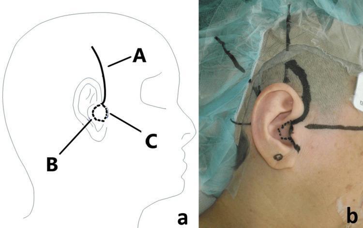
- A postauricular (retroauricular) skin incision is made, sufficiently long to allow introduction of the receiver-stimulator
- The incision is deliberately not placed directly over the receiver-stimulator to minimize wound complications
- The soft tissue is dissected down to the periosteum
- An anteriorly or posteriorly based periosteal flap is created
STEP 2 - CREATING THE RECEIVER-STIMULATOR BED
- A subperiosteal pocket is created under the temporalis muscle to house the receiver-stimulator
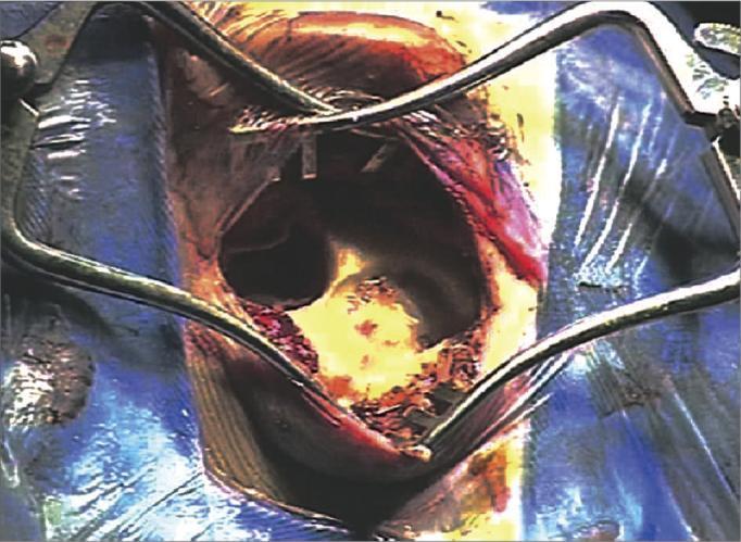
- Alternatively, a shallow bony well (trough) may be drilled into the outer cortex of the skull for secure implant placement
- A gutter/trough is drilled in the cortical bone for the electrode lead as it passes toward the mastoid cavity
- Some surgeons use tie-down sutures to secure the device
STEP 3 - CORTICAL MASTOIDECTOMY
- A simple (cortical) mastoidectomy is performed with a drill to open the mastoid air cells
- The posterior EAC wall is carefully thinned for better visualization through the facial recess
- The posterior and inferior mastoid cavities are NOT saucerized - bony overhangs are preserved to retain the electrode array coiled within the mastoid
- The incus and ossicular chain are carefully preserved, especially in patients with residual hearing
STEP 4 - POSTERIOR TYMPANOTOMY (FACIAL RECESS APPROACH)
- The facial recess (triangular space bounded by the facial nerve posteriorly, chorda tympani anteriorly, incus buttress superiorly) is opened
- This gives access to the round window niche through the middle ear without entering the EAC
- Care is taken to preserve the chorda tympani (taste nerve) - dividing it risks dysgeusia
- The round window niche is visualized; removal of bone around it exposes the round window membrane
STEP 5 - COCHLEAR ENTRY (COCHLEOSTOMY or ROUND WINDOW)
Two approaches:
-
Round Window Insertion (preferred for hearing preservation):
- The round window membrane is opened in an anteroinferior direction
- Minimally traumatic entry into the scala tympani
-
Cochleostomy:
- A small opening is drilled anteroinferior to the round window
- Directly enters the scala tympani
- Used when round window is not accessible due to poor visualization or ossification
STEP 6 - ELECTRODE INSERTION
- The flexible electrode array is inserted slowly and gently into the scala tympani in a controlled atraumatic motion ("soft surgery" principles)
- Most conventional-length electrodes are inserted to a minimum angular depth of 360°-400° (just over one full cochlear turn)
- Residual perilymph is NOT suctioned (to preserve inner ear environment)
- The electrode is inserted very slowly to minimize intracochlear pressure changes
Soft Surgery Principles (for Hearing Preservation)
- Use of flexible, thin electrodes requiring less insertion force
- Preference for round window insertion over cochleostomy
- Slow electrode insertion
- No suctioning of perilymph
- Preserving ossicular chain integrity
- Perioperative steroids (e.g., dexamethasone at induction; triamcinolone soaked onto spongistan placed on round window membrane)
STEP 7 - INTRAOPERATIVE TESTING
- Electrocochleography (ECochG) may be used during insertion to monitor for cochlear damage in hearing preservation cases
- Neural response telemetry (NRT) / Electrically evoked compound action potentials (ECAP) confirm electrode function
- Impedance testing to verify electrode integrity
- Intraoperative imaging (X-ray or fluoroscopy) may confirm electrode position
STEP 8 - SEALING AND WOUND CLOSURE
- After electrode insertion, the cochlear opening is sealed with small pieces of fascia or muscle to prevent perilymph leak
- The residual electrode wire is coiled within the mastoid cavity
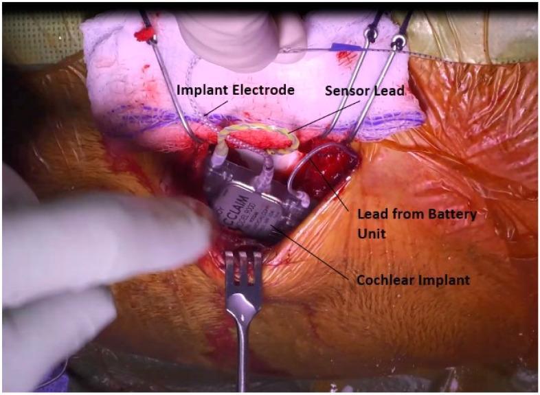
- The receiver-stimulator is seated in its subperiosteal pocket or bony well
- The wound is closed in layers (periosteum, subcutaneous tissue, skin)
- A mastoid dressing is applied
8. CHALLENGING SURGICAL SITUATIONS
| Situation | Approach |
|---|
| Common cavity deformity | Double posterior labyrinthotomy technique |
| Underdeveloped mastoid, narrow facial recess, anomalous facial nerve, very anterior sigmoid sinus | Suprameatal approach (tympanomeatal flap elevated to visualize round window) |
| Unfavorable anatomy, uncontrollable CSF leak, recurrent meningitis | Subtotal petrosectomy (canal wall-down with EAC overclosure) |
| Post-meningitis ossification, otosclerosis | Intentional scala vestibuli insertion |
| Previous canal wall-down mastoid surgery | Obliterate mastoid + blind sac closure of EAC first, then CI (simultaneous or staged 3-6 months later) |
9. POST-OPERATIVE CARE
- The external processor is NOT activated immediately after surgery
- Device activation ("switch-on") and mapping occurs approximately 4-6 weeks after surgery once wound healing is complete
- Mapping/Programming: The audiologist programs each electrode's stimulation levels (T-levels = threshold, C-levels = comfort levels)
- Intensive auditory rehabilitation: Speech therapy, auditory training, listening exercises
- More than 85% of implant recipients can use telephone speech understanding after implantation
10. COMPLICATIONS
Intraoperative Complications
| Complication | Details | Management |
|---|
| Facial nerve injury | Rare (<1%); direct transection or heat from drill | Continuous FNM; decompression/primary repair if identified |
| Injury to tegmen ± dural tear | Risk of CSF leak and meningitis | Fascia underlay graft + bone pate; may still proceed with implantation |
| EAC violation | Risk of electrode extrusion through EAC skin | Bone pate alone if bony; cartilage/fascia graft if skin violated |
| Tympanic membrane tear | Risk of infection | Concurrent cartilage/fascia tympanoplasty |
| Malpositioned/damaged electrode | Kinking, electrode shorts | Remove and reinsert; confirmed on intraoperative imaging/electrophysiology |
Early Post-operative Complications
| Complication | Details |
|---|
| Taste disturbance (dysgeusia) | Chorda tympani stretch/division; usually temporary, permanent in ~3% |
| Tinnitus | May be new or exacerbated; usually short-lived; CIs actually suppress tinnitus in most patients |
| Vestibular dysfunction/vertigo | Reported in ~30% of adults; usually transient; BPPV common - responds to Epley maneuver |
| Wound hematoma/seroma | Minor complication; may need aspiration |
Late Complications
| Complication | Rate/Details | Management |
|---|
| Wound infection / skin flap breakdown | Historically most common; reduced with smaller devices; delayed prosthetic infection possible | IV antibiotics; device removal and reimplantation after 3-6 months if unresponsive |
| Device failure | ~4% over device lifetime; spontaneous electrical malfunction or trauma | Explantation and reimplantation with new electrode (safe, no performance decrement) |
| CSF leak | Rare; from dural tear, unsealed cochleostomy, or oval/round window fistula | Revision surgery, lumbar drain, Eustachian tube obliteration |
| Meningitis | Children with CI have 30x higher incidence than normal-hearing children | Pre-op pneumococcal vaccination; prompt antibiotic treatment |
| Facial nerve stimulation | 7-25%; more common in otosclerosis; non-modiolar electrodes in severe disease | Device reprogramming (resolves in nearly all cases) |
| Magnet displacement | After head trauma in devices with removable magnets | Repositioning under local anesthesia |
11. SPECIAL TOPICS
Electric-Acoustic Stimulation (EAS) / Hybrid Cochlear Implant
- For patients with "ski-slope" hearing loss: normal/moderate low-frequency hearing + severe-to-profound high-frequency hearing loss
- A shorter electrode array covers only the basal (high-frequency) cochlea
- A hearing aid provides low-frequency acoustic hearing in the same implanted ear
- Requires meticulous hearing preservation surgery with soft surgery principles
Bilateral Cochlear Implants
- Advantages over unilateral: improved sound localization, spatial acuity, better speech understanding in noise
- Recommended for children and visually impaired adults
- Sequential implantation: relative increase in quality of life is higher after the first implant
Auditory Outcomes
- More than 85% of CI recipients can use telephone for speech understanding
- Music perception remains generally poor despite advances (limited melody/pitch/timbre identification, but normal rhythm recognition)
- Postlingual deafened adults perform better than prelingual deafened recipients
- Children implanted earlier have language development rates comparable to normal-hearing peers
Summary Flowchart
Referral for CI
       ↓
Audiological evaluation (PTA, speech discrimination, hearing aid trial)
       ↓
Imaging (MRI first, then CT if needed)
       ↓
Multidisciplinary team assessment (psychologist, speech therapist, audiologist, surgeon)
       ↓
Pre-op vaccinations (pneumococcal)
       ↓
SURGERY (GA, no muscle relaxant, facial nerve monitoring throughout)
  → Postauricular incision
  → Receiver-stimulator bed creation (subperiosteal pocket or bony well)
  → Cortical mastoidectomy
  → Posterior tympanotomy (facial recess approach)
  → Round window visualization
  → Cochlear entry (round window membrane or cochleostomy)
  → Electrode insertion into scala tympani (slow, atraumatic "soft surgery")
  → Intraoperative testing (ECAP, impedance, imaging)
  → Fascia seal at cochlear opening
  → Layered wound closure + mastoid dressing
       ↓
Post-op wound healing (4-6 weeks)
       ↓
Device activation + audiological mapping
       ↓
Intensive auditory rehabilitation (speech therapy, listening training)
Sources: KJ Lee's Essential Otolaryngology (9th Ed.) | Cummings Otolaryngology Head & Neck Surgery (7th Ed.) | Scott-Brown's Otorhinolaryngology Head & Neck Surgery (8th Ed.)
Relevant surgical images from the literature:
Intraoperative view showing the bony well drilled into the temporal bone to house the receiver-stimulator of a cochlear implant system
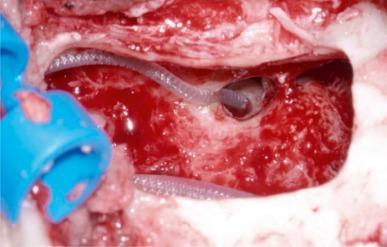
High-magnification intraoperative view showing the flexible cochlear electrode traversing the mastoid and being secured at the posterior tympanotomy, with the facial nerve canal visible