
Blood transfusion in pathology
blood transfusion reaction hemolysis pathology
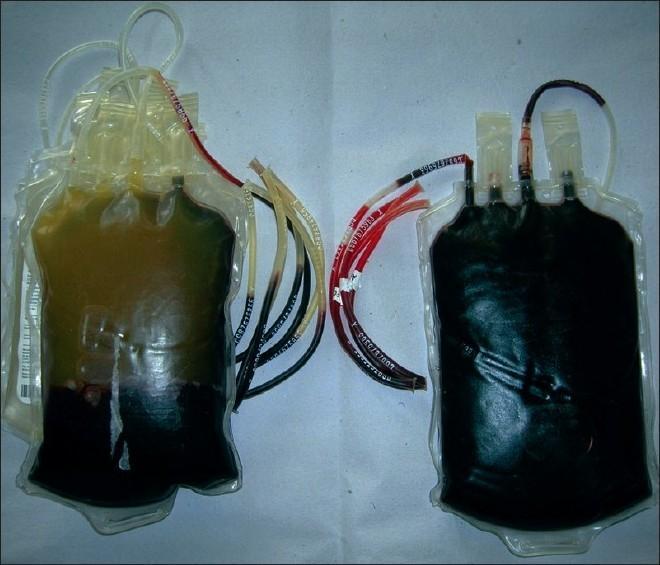
A clinical photograph displaying a side-by-side comparison of two whole blood units, illustrating the visual differences between normal and hemolyzed blood. The unit on the left represents a normal blood bag where distinct layering is visible: the upper portion contains clear, straw-colored (yellowish) plasma separated from the dark red sedimented red blood cells. The attached tubing segments on this bag also demonstrate clear, transparent plasma. In contrast, the unit on the right displays gross hemolysis, characterized by a uniform, dark reddish-black appearance throughout the entire bag. Crucially, the attached segments for the right-hand bag are filled with dark, opaque fluid instead of clear plasma, indicating the breakdown of red blood cells. This comparison serves as an educational tool for blood bank technicians and medical professionals to identify transfusion product contamination, thermal injury, or storage-related hemolysis during pre-issue inspections.
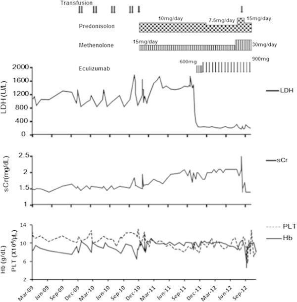
A multi-panel line graph illustrating the clinical course and treatment response of a patient with Paroxysmal Nocturnal Hemoglobinuria (PNH). The timeline spans from March 2009 to September 2012. The top panel monitors lactate dehydrogenase (LDH) levels (U/L), showing significant baseline hemolysis (fluctuating between 800-1600 U/L) that drops precipitously and stabilizes within normal limits following the initiation of Eculizumab in late 2011. The middle panel tracks serum creatinine (sCr) (mg/dL), indicating a gradual rise in renal dysfunction over time. The bottom panel displays hemoglobin (Hb) (g/dL) and platelet (PLT) (x10^4/μL) counts, highlighting periods of anemia and the frequency of blood transfusions (marked by vertical arrows). The figure also overlays pharmaceutical interventions, including dosages for prednisolone, methenolone, and eculizumab. This chart demonstrates the efficacy of terminal complement inhibition in controlling intravascular hemolysis and alleviating transfusion dependence in PNH patients.
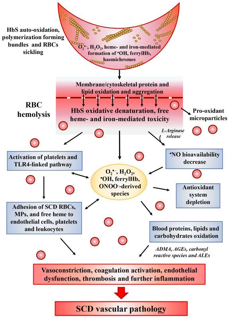
A medical pathophysiology diagram illustrating the oxidative stress cascade in Sickle Cell Disease (SCD) and its progression to vascular pathology. The diagram follows a vertical flow starting with a sickled red blood cell (RBC) undergoing HbS auto-oxidation and polymerization, leading to the generation of reactive oxygen species (ROS) such as H2O2, ferrylHb, and hemichromes. This triggers a central event of RBC hemolysis, characterized by membrane lipid oxidation, protein aggregation, and the release of pro-oxidant microparticles and L-arginase. Downstream pathways depict the systemic consequences, including the activation of platelets and the TLR4-linked inflammatory pathway, adhesion of SCD RBCs to endothelial cells and leukocytes, and a significant decrease in nitric oxide (NO) bioavailability. The lower section highlights the depletion of the antioxidant system and the oxidative modification of blood components into ADMA, AGEs, and ALEs. The cascade culminates in clinical manifestations such as vasoconstriction, endothelial dysfunction, and thrombosis, collectively summarized as SCD vascular pathology. This illustration is designed for advanced medical education regarding the molecular mechanisms of hematologic disorders.

Summary : This image is a logo for "Transfusion Medicine," featuring stylized red blood cells and bold text. logo: TRANSFUSION MEDICINE Logo Elements : • Text: "TRANSFUSION MEDICINE" in uppercase, white font on a dark blue rectangular background. • Graphic: Multiple stylized red blood cells depicted to the left of the text, suggesting a medical or hematology theme. • Layout: The text is right-aligned with the blood cell graphics on the left, creating a balanced composition. • Colour palette: Dominant colours are dark blue (background for text) and red (blood cells), with white text for contrast. Dimensions & Placement : • Horizontal orientation. • Text box occupies the right half, blood cell graphics on the left. • No additional taglines or company names visible. Analysis : The logo visually communicates a focus on blood and transfusion medicine through the use of red blood cell imagery and clear, bold text. The colour scheme reinforces the medical theme and ensures high visibility and readability.
Blood Transfusion in Pathology
1. Blood Groups and Compatibility
- Full crossmatch: ABO + Rh typing of donor and recipient, then mixing recipient serum with donor cells. Takes ~45 minutes.
- Type-specific blood: ABO/Rh matched only, available in 10-15 minutes.
- Emergency blood: Group O-negative (universal donor) to females, O-positive to males.
2. Blood Components Used in Transfusion
| Component | Indication |
|---|---|
| Packed Red Blood Cells (PRBCs) | Anemia, active hemorrhage (threshold Hb <7-8 g/dL in stable patients; <9 g/dL with CAD) |
| Platelets | Thrombocytopenia (<50,000/μL in active bleeding); not recommended empirically for patients on antiplatelets without thrombocytopenia |
| Fresh Frozen Plasma (FFP) | Coagulopathy, elevated INR, liver disease, massive transfusion |
| Cryoprecipitate | Low fibrinogen; fibrinogen is essential to clot formation and stabilization |
| Prothrombin Complex Concentrate (PCC) | Alternative to FFP in anticoagulant reversal |
| Whole Blood | Being re-explored in massive transfusion protocols |
3. Hazards and Complications of Blood Transfusion
Immunological Reactions
a) Acute Haemolytic Transfusion Reaction (AHTR)
- Incidence: ~1:110,000 transfusions
- Cause: ABO incompatibility - almost always due to human error (misidentification of patient, wrong specimen labelling, failure to check compatibility)
- Mechanism: Recipient antibodies activate complement via the classical pathway → intravascular haemolysis → free haemoglobin release → renal tubular damage, DIC, shock
- Clinical features: Fever, chills/rigors, anxiety, chest and back/flank pain, haemoglobinuria (dark urine), diffuse bleeding, oliguria/anuria, hypotension
- Pathology at autopsy: Haemoglobinuric nephrosis - acute tubular necrosis with haemoglobin casts in renal tubules
- Outcome: ~50% of ABO-incompatible transfusions have no adverse effect; 5% are fatal; death from anaphylaxis, renal failure, electrolyte imbalance, or DIC
- Serum: ↑ haemoglobin, ↑ methemalbumin, ↓ haptoglobin (intravascular)
- Serum: ↑ unconjugated bilirubin (extravascular)
- Urine: haemoglobinuria, urobilinogen, red cell casts
- Serology: positive direct Coombs (DAT) test, incompatible crossmatch, ↑ LDH
- 2% red cell suspension may show agglutinates
b) Febrile Non-Haemolytic Transfusion Reaction (FNHTR)
- Incidence: ~1:1,100 transfusions (most common reaction type)
- Cause: In platelets - leukocyte-derived cytokines accumulate during storage; in RBCs - donor leukocytes interact with recipient white cell antibodies (graft-vs-host-type response)
- Clinical features: Temperature rise ≥1°C or >38°C + chills/rigors within 4 hours of transfusion cessation
- Management: Stop transfusion; supportive care; exclude more serious reaction
- Prevention: Leukoreduction (leukodepletion) markedly reduces incidence
c) Allergic Transfusion Reaction
- Incidence: ~1:1,200 transfusions
- Cause: Sensitivity to donor plasma proteins (leukocytes, platelets, plasma components)
- Features: Urticaria to anaphylaxis; eosinophilia may be present; IgA-specific antibodies found in IgA-deficient recipients reacting to donor IgA
- Severe anaphylaxis: Rare; seen in IgA-deficient patients with anti-IgA antibodies
d) Transfusion-Related Acute Lung Injury (TRALI)
- Definition: Non-cardiogenic pulmonary oedema (ARDS-like picture) within minutes to hours of transfusion of blood products (FFP > platelets > RBCs)
- Incidence: 1:1,000 to 1:100,000; leading cause of transfusion-related mortality
- Pathobiology (two mechanisms):
- Antibody-mediated: Donor HLA class I/II or neutrophil-specific antigen (NSA) antibodies bind recipient leukocytes → release of oxidative/non-oxidative products → pulmonary endothelial damage. HLA antibodies develop in multiparous female donors.
- Two-hit model: Pre-existing neutrophil priming (trauma, sepsis, surgery) + activation by bioactive lipids/cytokines from stored blood products → lung injury
- Pathology: Alveolar filling with protein-rich fluid; increased microvascular permeability from pulmonary endothelial damage
- Clinical: Hypoxia, bilateral pulmonary infiltrates on CXR, fever, hypotension; indistinguishable from ARDS/cardiogenic oedema
- Prevention: Use of male or never-pregnant female donors for plasma-containing products; pathogen-reduction technologies
Non-Immunological Reactions
| Complication | Mechanism / Notes |
|---|---|
| Circulatory overload (TACO) | Volume excess → pulmonary oedema; distinguish from TRALI by raised BNP and response to diuretics |
| Coagulation defects | Dilutional coagulopathy in massive transfusion; DIC in haemolytic reactions |
| Hyperkalaemia | Stored RBCs leak K⁺; risk with rapid/massive transfusion |
| Hypocalcaemia | Citrate (anticoagulant in stored blood) chelates Ca²⁺; seen in massive/rapid transfusion |
| Hypothermia | Rapid transfusion of cold blood; lungs oedematous on autopsy if circulatory overload |
| Air embolism | Air entering transfusion line; autopsy shows air in right ventricle |
| Thrombophlebitis | At venous access site |
| Bacterial infection | Faulty storage; more common with platelets (stored at room temperature) |
| Viral infections | Hepatitis B, Hepatitis C, HIV, CMV; markedly reduced by modern screening |
| Parasitic infections | Malaria, toxoplasmosis (rare) |
| Iron overload | Repeated transfusions (e.g., thalassaemia); each unit of RBCs contains ~250 mg elemental iron → haemosiderosis |
| Hyperfibrinolysis | Common in trauma/massive haemorrhage → treat with tranexamic acid |
4. Classification of Blood Transfusion Hazards (Forensic/Pathology)
| Immunological | Non-Immunological |
|---|---|
| Intravascular haemolysis | Circulatory overload |
| Extravascular haemolysis | Coagulation defects |
| Sensitivity to WBCs, platelets, plasma components | Hyperkalaemia |
| Citrate toxicity | |
| Infections (syphilis, hepatitis, toxoplasmosis, AIDS) | |
| Air embolism | |
| Hypothermia | |
| Rigors |
- Clerical/labelling error (most common overall cause of ABO incompatibility)
- Confusing terminology ("group A serum" vs "anti-B serum")
- Failure to check bottle reference against lab compatibility report
- Similarly-named patients in the same ward
5. Forensic/Medicolegal Aspects
- Sample of blood transfused
- Sample of recipient blood before and after transfusion
- At autopsy: kidneys, blood, urine
- Acute intravascular haemolysis: Haemoglobinuric nephrosis - acute tubular necrosis, haemoglobin casts in tubules
- Circulatory overload: Pulmonary oedema (oedematous lungs)
- Air embolism: Air in right ventricle
6. Transfusion Thresholds (Pathophysiology Context)
- Stable patients without CAD: Transfuse when Hb <7-8 g/dL (restrictive strategy reduces mortality vs liberal strategy - supported by RCTs in GI bleeding)
- Known/symptomatic CAD: Target Hb >9 g/dL
- Portal hypertension bleeding: Monitor closely; overtransfusion worsens portal pressure and can increase bleeding
- Platelets: Maintain >50,000/μL in active GI bleeding (ACG guidelines)
- Massive transfusion protocol: RBCs:FFP:Platelets = 1:1:1 + cryoprecipitate for fibrinogen + tranexamic acid