
Mbbs saq answer including data from standard books to score maximum marks: Cervical pap smear diagram
cervical Pap smear diagram transformation zone squamocolumnar junction
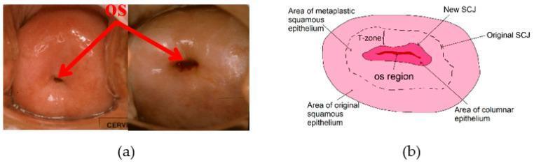
This composite educational graphic focuses on cervical anatomy and visual screening for gynecological health. Section (a) features two clinical cervigrams (endoscopic cervical photographs) demonstrating morphological variations of the external os. The first photograph shows a small, circular nulliparous os, while the second shows a larger, more irregular opening. Red arrows highlight the os in both clinical examples. Section (b) is an anatomical diagram of the cervix illustrating key histological regions and zones. It identifies the central 'os region' surrounded by the 'area of columnar epithelium.' The diagram further delineates the 'squamocolumnar junction (SCJ),' showcasing its physiological migration with age from an 'original SCJ' to a 'new SCJ' position. The 'transformation zone (T-zone)' is labeled between these two junctions, within the 'area of metaplastic squamous epithelium.' The peripheral region is identified as the 'area of original squamous epithelium.' This visual aid is designed to teach cervical topography and the clinical significance of the T-zone as the primary site for neoplastic development and colposcopic evaluation.
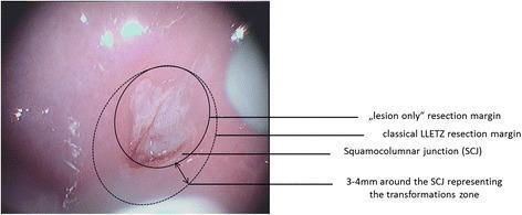
A colposcopic clinical photograph of a cervix showing a comparison of two surgical resection methods for cervical intraepithelial neoplasia (CIN). The image displays an acetowhite dysplastic lesion centered on the squamocolumnar junction (SCJ). Two concentric circular overlays illustrate different surgical margins: a solid line indicates the 'lesion only' resection margin, which closely contours the visible pathology; a larger dashed line represents the 'classical LLETZ' (Large Loop Excision of the Transformation Zone) resection margin, which encompasses the entire transformation zone. The transformation zone is visually defined as extending 3 to 4 mm peripheral to the SCJ. Labels and arrows specify the SCJ, the transformation zone dimensions, and the comparative extent of both excision techniques. This comparison chart serves as an educational tool for gynecological oncology and dysplasia management, highlighting the difference between targeted lesion excision and traditional excision of the whole transformation zone.
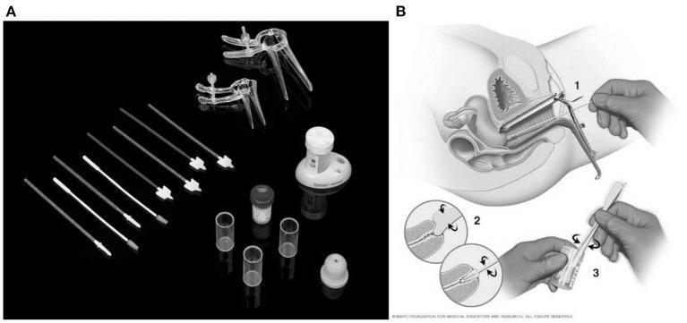
This composite educational graphic illustrates the tools and clinical technique for a Pap smear, a common screening procedure for cervical cancer. Figure A is a photograph of a Pap smear test kit containing various instruments on a black background: clear plastic vaginal speculums of different sizes, multiple cervical sampling devices including spatulas and endocervical brushes (cytobrushes), and liquid-based cytology collection vials. Figure B is a multi-step anatomical diagram showing the clinical procedure: (1) Cross-sectional view of a speculum inserted into the vagina to visualize the cervix, with a collection device entering the cervical canal; (2) Close-up insets showing the rotation of the cervical brush and spatula at the transformation zone to collect ectocervical and endocervical cells; and (3) The final step of transferring the collected cellular material into a preservative vial (liquid-based cytology) or onto a glass slide. The content is designed for medical training, highlighting essential gynecological screening equipment and correct sampling methodology.
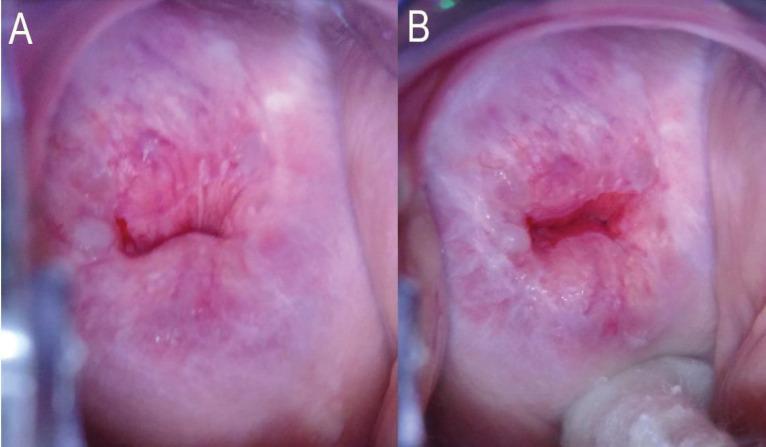
Two side-by-side colposcopic clinical photographs (A and B) demonstrating the assessment of the Transformation Zone (TZ) and Squamocolumnar Junction (SCJ) of the human cervix. In Image A, the cervix shows a pinkish, smooth ectocervix with a partially obscured SCJ, where the upper limit of the junction disappears into the endocervical canal without manipulation. This is categorized as TZ Type 3A because the entire circumference of the SCJ is not visible. In Image B, the same cervix is shown while a cotton-tipped applicator is used to exert pressure on the posterior fornix, successfully exposing the endocervical canal. This maneuver reveals the entire circumference of the SCJ, which is found to extend less than 5 mm into the canal, reclassifying the finding as TZ Type 2A. These images illustrate the importance of clinical manipulation techniques in cervical cancer screening to differentiate between patients eligible for ablative therapy (TZ 1 and 2) versus those requiring excisional procedures (TZ 3).
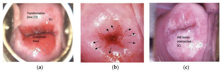
This set of clinical photographs depicts cervical anatomy and pathology as seen during colposcopy or cervicography after the application of acetic acid. Image (a) illustrates normal cervical landmarks, identifying the central red columnar area (CA) consisting of a single layer of columnar epithelium, the surrounding pink transformation zone (TZ) composed of stratified squamous epithelium, and the squamocolumnar junction (SCJ) representing the border between these two regions. Image (b) shows the progression of squamous metaplasia at the squamocolumnar junction. Image (c) demonstrates an abnormal clinical finding: an acetowhite (AW) lesion. This white, opaque area arises from the SCJ and extends into the transformation zone, a visual manifestation characteristic of cervical intraepithelial neoplasia (CIN) or precancerous lesions. The images serve as educational tools for identifying key anatomical structures and differentiating normal physiological changes from pathological acetowhite reactions during cervical cancer screening and visual inspection with acetic acid (VIA).
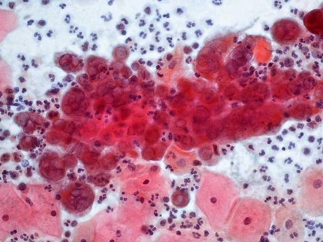
This cervical cytology image from a conventional Pap smear demonstrates the cytopathic effect of herpes simplex virus infection in squamous epithelium. Imaging modality is light microscopy with Pap staining; technique includes fixation, staining, and smear preparation, with magnification around 40x. Anatomical location corresponds to the cervix, specifically the transformation zone where squamous epithelium overlies glandular mucosa. Visual features include numerous large multinucleated squamous cells with nuclear molding, prominent ground-glass nuclei, and perinuclear halos; chromatin marginates and intranuclear eosinophilic inclusions (Cowdry type A) are variably present. A background inflammatory infiltrate contains lymphocytes and neutrophils; adjacent erythrocytes and epithelial cells provide a mixed cytology field. Pathology: characteristic HSV-induced cytopathic effect in cervical epithelium; findings reflect active viral replication and cell-cell fusion, leading to multinucleation and nuclear fragmentation. Diagnostic significance: supports diagnosis of HSV cervicitis in the appropriate clinical context; helps distinguish HSV infection from other cervicitis etiologies such as HPV-related koilocytosis or Chlamydia; may prompt confirmatory testing (HSV PCR, viral culture) and antiviral therapy. Differential considerations: CMV infection with owl’s eye inclusions; koilocytosis from HPV; nonspecific inflammatory changes. Clinical correlation: correlate with genital ulcers, dysuria, sexual history, and lesion distribution; documents viral shedding risk and informs patient counseling.
Cervical Pap Smear - SAQ Answer (MBBS)
Definition
Relevant Anatomy - The Transformation Zone
| Region | Epithelium |
|---|---|
| Ectocervix / exocervix | Stratified squamous epithelium (4 layers) |
| Endocervical canal | Single-layer columnar (mucus-secreting) epithelium |
- The region between the original SCJ and the new SCJ
- Where columnar epithelium undergoes squamous metaplasia - replacement by advancing squamous epithelium
- The T-zone is the primary sampling target of the Pap smear
- Immature metaplastic cells here are most susceptible to HPV infection, making this the site of origin of virtually all cervical precursor lesions and cancers
Diagram 1 - Cervical Anatomy: SCJ and Transformation Zone
Diagram 2 - Changes in SCJ Position Through Life
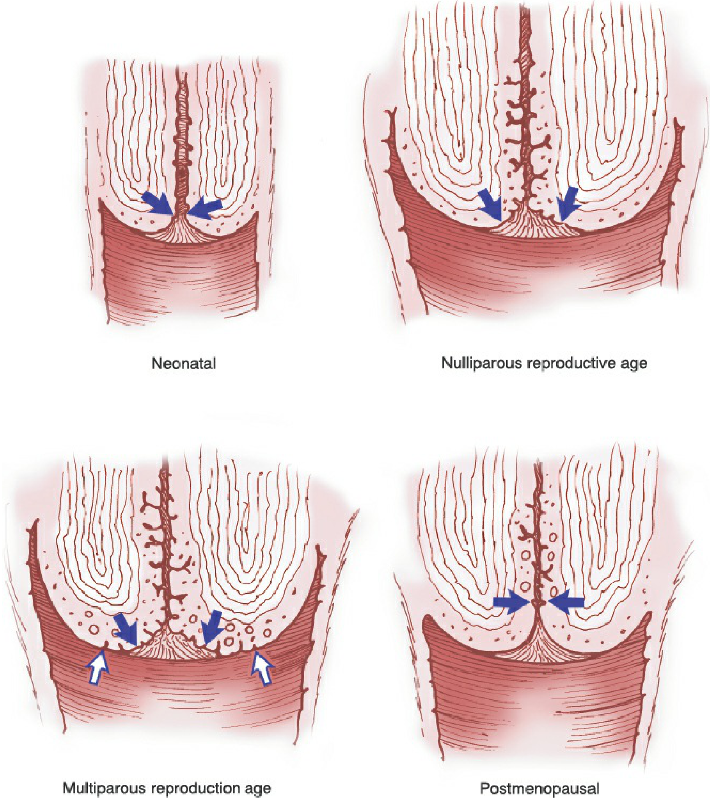
- Neonatal/Nulliparous: SCJ is at or just outside the external os
- Multiparous reproductive age: SCJ is maximally ectopic - the T-zone is wide and fully visible (ectropion); blue arrows = new (inner) SCJ; white arrows = original (outer) SCJ
- Postmenopausal: SCJ recedes into the endocervical canal - T-zone is no longer visible from outside, making sampling more difficult
Diagram 3 - Histology of the Squamocolumnar Junction
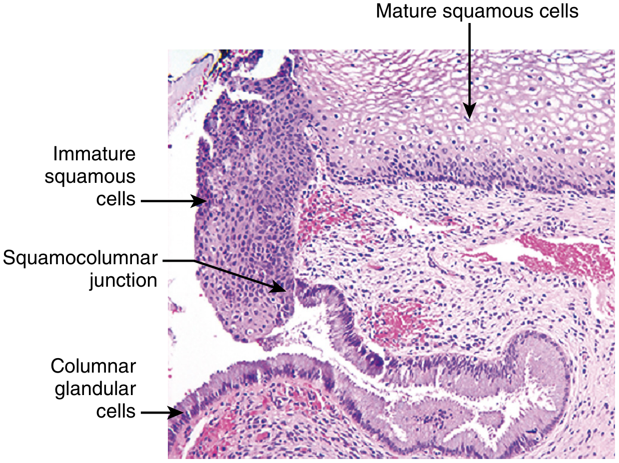
- Mature squamous cells (top right) - glycogenized, NOT susceptible to HPV
- Immature squamous cells / metaplastic cells (at junction) - HPV-susceptible
- Columnar glandular cells (bottom) - also HPV-susceptible
- Arrow marks the squamocolumnar junction itself
Diagram 4 - Pap Smear Collection Technique
- Speculum inserted to visualize the cervix
- Cervical spatula/brush rotated at the transformation zone - ectocervical and endocervical cells collected
- Cells transferred into liquid-based cytology vial (or smeared directly on a glass slide for conventional Pap)
Squamous Epithelium - 4 Layers (Berek & Novak's Gynecology)
| Layer | Features |
|---|---|
| Basal layer | Single row of immature cells, large nuclei, scant cytoplasm |
| Parabasal layer | 2-4 rows; mitotic figures; replacement cells for overlying epithelium |
| Intermediate layer | 4-6 rows; polyhedral cells; glycogen production begins; intercellular bridges visible |
| Superficial layer | 5-8 rows; flattened; small pyknotic nuclei; glycogen-filled cytoplasm; cells exfoliate - these form the basis of Pap smear sampling |
Techniques of Collection
| Method | Details |
|---|---|
| Conventional Pap smear | Cells spread on glass slide + fixative applied by examiner immediately |
| Liquid-based cytology (LBC) | Cells suspended in vial of liquid preservative; debris removed in lab; slide made in lab; equal sensitivity/specificity to conventional |
Staining - Papanicolaou (Pap) Stain
- Fixation: 95% ethanol (wet fixation) or spray fixative immediately after collection
- Stains used: Haematoxylin (nuclei - blue/purple), Orange G (keratin - orange), EA (eosin-azure - stains cytoplasm in shades of pink and green)
- Characteristic appearance: Superficial cells - orangeophilic/eosinophilic; Intermediate cells - cyanophilic (pale green/blue)
Bethesda Classification System (2001) - Reporting Pap Smear Results
1. Negative for Intraepithelial Lesion or Malignancy (NILM)
- Normal result
2. Epithelial Cell Abnormalities: Squamous
| Category | Meaning | Equivalent |
|---|---|---|
| ASC-US | Atypical Squamous Cells of Undetermined Significance | - |
| ASC-H | ASC - cannot exclude HSIL | - |
| LSIL | Low-grade Squamous Intraepithelial Lesion | HPV changes + CIN 1 (mild dysplasia) |
| HSIL | High-grade Squamous Intraepithelial Lesion | CIN 2, CIN 3, CIS (moderate/severe dysplasia, carcinoma in situ) |
| Squamous Cell Carcinoma | Frankly invasive | - |
3. Epithelial Cell Abnormalities: Glandular
| Category |
|---|
| Atypical glandular cells (endocervical, endometrial, or NOS) |
| Atypical glandular cells, favors neoplasm |
| Endocervical adenocarcinoma in situ (AIS) |
| Adenocarcinoma |
Sensitivity & Specificity
| Test | Sensitivity | Specificity |
|---|---|---|
| Conventional Pap - HSIL | ~56% | ~97% |
| Conventional Pap - LSIL | ~77% | ~80% |
| HPV testing for CIN 2/3 | Higher than Pap | Lower (more false positives, especially <35 yrs) |
Screening Guidelines (Current)
| Parameter | Recommendation |
|---|---|
| Start screening | Age 21, regardless of onset of sexual activity |
| Age 21-65 | Pap cytology every 3 years |
| Age 30-65 (preferred) | Co-testing (Pap + HPV testing) every 5 years |
| Stop screening | Age 65 with adequate prior normal results |
| Not indicated | Women who have had a total hysterectomy for benign disease |
Why the Transformation Zone is Critical
"The unique epithelial environment of the cervix renders it highly susceptible to infection with HPV, the main cause of cervical cancer. Immature squamous cells in the transformation zone are most susceptible to HPV infection, and as a result most cervical precursor lesions and cervical cancers develop in the transformation zone."
- HPV gains entry into immature basal epithelial cells at sites of squamous metaplasia
- HPV-16 accounts for ~60% of cervical cancers; HPV-18 ~10%
- E6 protein of high-risk HPV binds and degrades p53 (via proteasome)
- E7 protein promotes degradation of RB and inhibits p21, p27
- Result: uncontrolled cell cycle progression + impaired DNA repair + telomerase upregulation = neoplastic transformation
Adequacy of a Pap Smear (for exam marks)
- Sufficient squamous cells are present
- Endocervical/transformation zone component is present (endocervical cells or metaplastic squamous cells must be seen)
- An unsatisfactory smear lacks these cells - usually due to: poor technique, failure to sample the T-zone, obscuring blood/inflammation, or improper fixation
Quick Summary Box (Write in SAQ)
Pap Smear = Papanicolaou cytology for cervical cancer screening

Site: Transformation zone (between original & new SCJ)
Stain: Papanicolaou stain (wet-fixed)
Techniques: Conventional smear OR Liquid-based cytology (LBC)
Reporting: Bethesda System 2001
  - NILM / ASC-US / ASC-H / LSIL (CIN 1) / HSIL (CIN 2, 3) / SCC
  - Glandular: AGC / AIS / Adenocarcinoma
Screening: Every 3 years (cytology alone) from age 21
           Every 5 years (co-testing with HPV) from age 30-65
Key role: Detects precancer lesions (over years) before invasion