
CAN YOU CREATE A STEP BY STEP GUIDE FOR THE SHOULDER EXAMINATION WITH PICTURES FOR EACH STEPS
stepwise clinical shoulder examination inspection palpation range of motion strength special tests diagram
"shoulder" AND "physical examination"
shoulder examination clinical inspection palpation range of motion special tests guidance
https://www.aafp.org/afp/2000/0515/p3079
An educational eight-panel clinical shoulder examination picture guide. Show a fully clothed patient with shoulder and upper arm exposed and a clinician performing: 1 observation from front and back, 2 palpation of clavicle and acromioclavicular joint, 3 active shoulder flexion and abduction, 4 internal and external rotation with elbow at side, 5 resisted empty-can supraspinatus strength test, 6 Hawkins impingement test at 90 degrees forward flexion with internal rotation, 7 cross-body adduction test for acromioclavicular joint, 8 anterior apprehension test. Clear medically accurate anatomy and hand positioning, no text labels.
Step-by-step shoulder examination guide
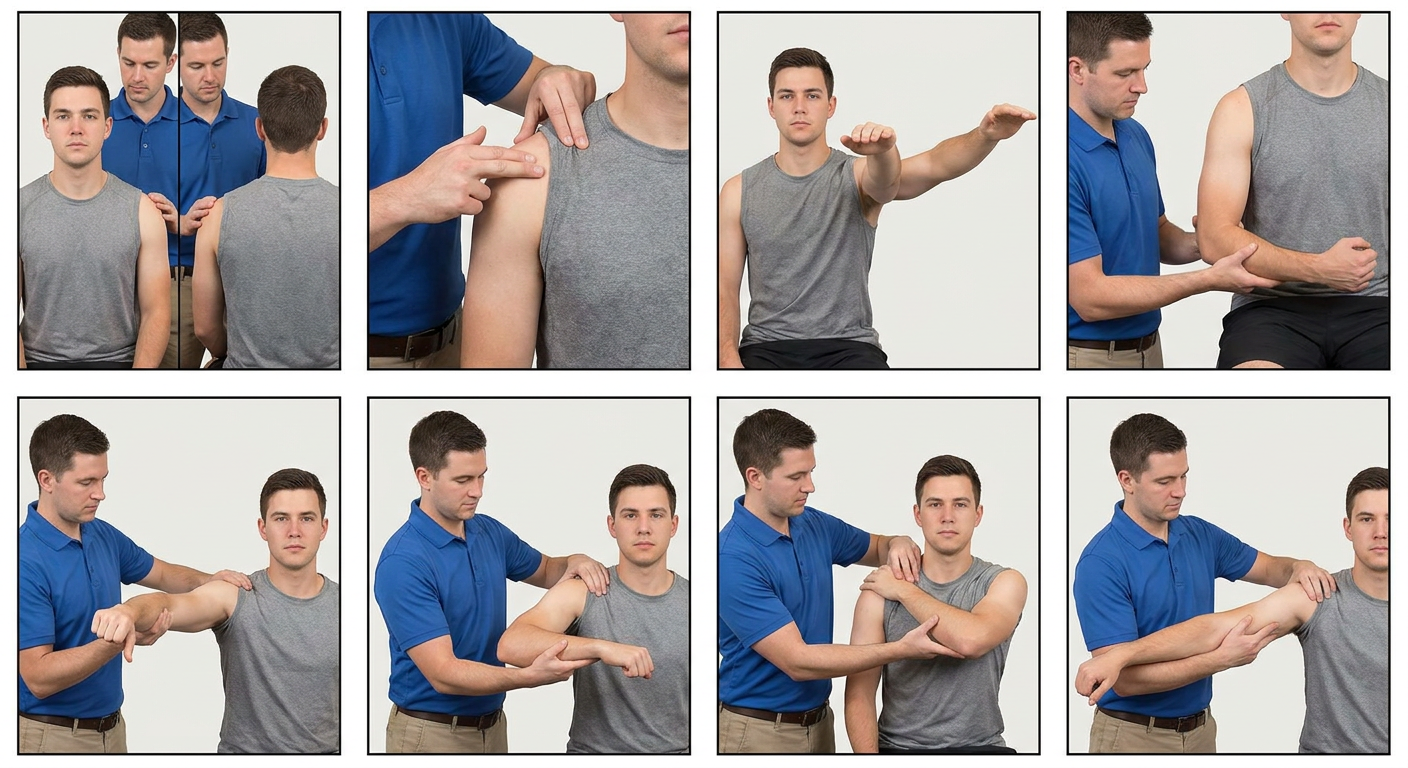
Picture key
| Panel | Examination step |
|---|---|
| 1 | Inspection |
| 2 | Palpation |
| 3 | Flexion and abduction |
| 4 | Internal and external rotation |
| 5 | Empty-can strength test |
| 6 | Hawkins impingement test |
| 7 | Cross-body adduction test |
| 8 | Apprehension test |
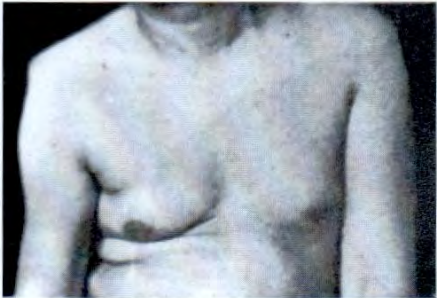
1. Inspect - look from the front, side, and back
- Shoulder level and contour symmetry
- Swelling, bruising, redness, skin lesions, or surgical scars
- Prominence or step deformity at the clavicle or AC joint
- Muscle wasting: deltoid, supraspinatus, infraspinatus
- Scapular winging or abnormal scapular movement
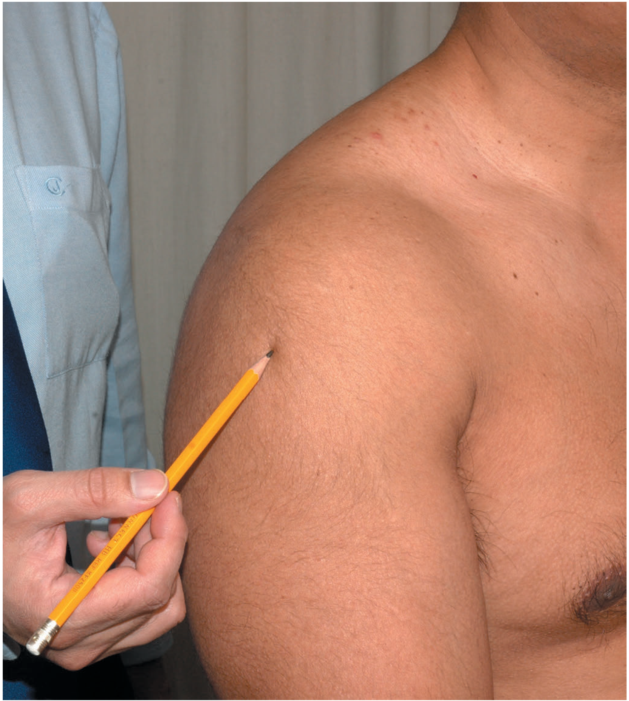
2. Palpate - compare both sides
- Sternoclavicular joint
- Clavicle along its entire length
- Acromioclavicular (AC) joint
- Acromion and coracoid process
- Bicipital groove, just medial to the anterior deltoid
- Greater tuberosity and subacromial region
- Scapular spine, medial border, and posterior joint line
3. Assess active range of motion
| Movement | Instruction | Typical reference range |
|---|---|---|
| Flexion | Raise straight arms forward and overhead | 0-180° |
| Extension | Move arms backwards | 0-60° |
| Abduction | Raise arms sideways overhead | 0-180° |
| Adduction | Bring arm across body | Variable |
| External rotation | Elbows by sides, rotate forearms outward | About 0-60° |
| Internal rotation | Reach thumb up the spine behind the back | Record vertebral level reached |
4. Assess passive range of motion
- Active restriction with relatively preserved passive motion: consider pain inhibition, weakness, or rotator cuff pathology.
- Both active and passive motion restricted: consider capsular stiffness, adhesive capsulitis, or glenohumeral osteoarthritis.
- Marked loss of external rotation is particularly helpful for identifying glenohumeral stiffness.
5. Test rotator cuff and shoulder strength
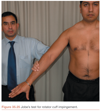
A. Supraspinatus - Empty-can / Jobe test
- Abduct arm to about 90° in the scapular plane, slightly forward of the body.
- Turn thumb downward, as if emptying a can.
- Apply gentle downward pressure while the patient resists.
- Positive: pain or weakness, which may indicate supraspinatus or rotator cuff pathology.
B. Infraspinatus and teres minor - External rotation resistance
- Elbows at 90° and tucked against the body.
- Ask the patient to rotate forearms outward against your resistance.
- Weakness or pain may suggest posterior rotator cuff involvement.
C. Subscapularis - Lift-off test
- Place the back of the hand on the lower back.
- Ask the patient to lift the hand away from the back against resistance.
- Inability or marked weakness suggests subscapularis dysfunction.
D. Deltoid and axillary nerve
- Resist abduction, particularly from roughly 15° to 90°.
- Combine with sensation over the lateral upper arm.
6. Test for subacromial pain or impingement
Hawkins test
- Flex shoulder and elbow to 90°.
- Stabilize the arm, then gently internally rotate the shoulder.
- Positive: familiar anterolateral shoulder pain can suggest subacromial pain or rotator cuff irritation.
Neer test
- Stabilize the scapula.
- Passively elevate the internally rotated arm in forward flexion.
- Positive: pain near end-range may indicate subacromial pain.
7. Test the acromioclavicular joint
Cross-body adduction test
- Flex shoulder to 90°.
- Move the arm across the chest toward the opposite shoulder.
- Positive: localized pain over the AC joint suggests AC-joint pathology.
8. Test for anterior instability
Apprehension test
- With the patient sitting or supine, abduct shoulder to about 90°.
- Slowly externally rotate the arm.
- Positive: a feeling that the shoulder will “come out,” not merely pain.
Relocation test
- In the same position, apply gentle posterior pressure to the humeral head.
- Relief of apprehension supports anterior instability.
9. Complete the examination
- Examine the neck: cervical motion and radicular symptoms can refer pain to the shoulder.
- Examine the elbow when relevant.
- Perform a brief distal neurovascular check: radial pulse, hand movement, and sensation.
- Summarize: side, tenderness, active/passive range, strength, provocative tests, and neurovascular status.
Quick documentation template
Right/left shoulder: no/positive deformity, swelling, scar, or muscle wasting. Tenderness at ____. Active ROM: ____. Passive ROM: ____. Strength: supraspinatus __/5, external rotation __/5, internal rotation __/5. Hawkins __, cross-body adduction __, apprehension __. Distal sensation and pulse intact/impaired.