
Adrenergic receptors in detail
Reading File
Now let me search the medical library for adrenergic receptors in detail.
Finding Sources
Searching Images
adrenergic receptor subtypes alpha beta pharmacology diagram
Excellent. Let me now read the most detailed sections from Goodman & Gilman and Braunwald's Heart Disease in parallel.
Finding Sources
Reading File
Reading File
Excellent content. Let me now read more from Braunwald (Alpha subtypes + beta desensitization) and Goodman & Gilman (beta receptors + antagonists section) in parallel.
Reading File
Reading File
Great content. Now let me get the Goodman & Gilman table on adrenergic receptor subtypes and the clinical pharmacology section, plus fetch the diagram from Braunwald.
Finding Sources
Loading Image
Now let me get the Goodman & Gilman table on adrenergic receptor subtypes and look for any diagrams of receptor structure/signaling.
Finding Sources
Reading File
Let me get the Goodman & Gilman table on receptor subtypes (Table 10-6 referenced) and the alpha1 receptor section.
Reading File
I now have extensive, authoritative content. Let me fetch the receptor structure diagram from Goodman & Gilman and compile the full answer.
Loading Image
Loading Image
I now have rich, authoritative content from two premier textbooks and three excellent diagrams. Let me compile the comprehensive answer.
Adrenergic Receptors - In Detail
Adrenergic receptors (ARs) are cell-surface proteins that mediate the physiological effects of the catecholamines norepinephrine (NE) and epinephrine (Epi). They belong to the superfamily of seven-transmembrane (hepta-spanning) G protein-coupled receptors (GPCRs) - their transmembrane domains are the site of agonist/antagonist binding, while the cytoplasmic domains interact with G proteins.
1. Overview and Classification
There are two main families - alpha (α) and beta (β) - each with three confirmed subtypes:
| Family | Subtypes |
|---|---|
| α1 | α1A, α1B, α1D |
| α2 | α2A, α2B, α2C |
| β | β1, β2, β3 |
Within each major type (~75% identity within the membrane-spanning domains), the α1 and α2 subtypes share only ~30-40% identity with each other, similar to the degree of difference between α and β types overall.
2. Structural Features
All adrenergic receptors share a hepta-spanning GPCR architecture:
- Seven transmembrane alpha-helical domains embedded in the plasma membrane
- Extracellular N-terminus - contains N-glycosylation sites (Ψ)
- Intracellular C-terminus and three cytoplasmic loops - interact with G proteins and GRKs (G protein-coupled receptor kinases)
- The coding genes differ: β receptor genes and α2 genes have their coding region in a single exon, while each α1 gene has a large intron separating the body of the receptor from the 7th TM domain and C-terminus
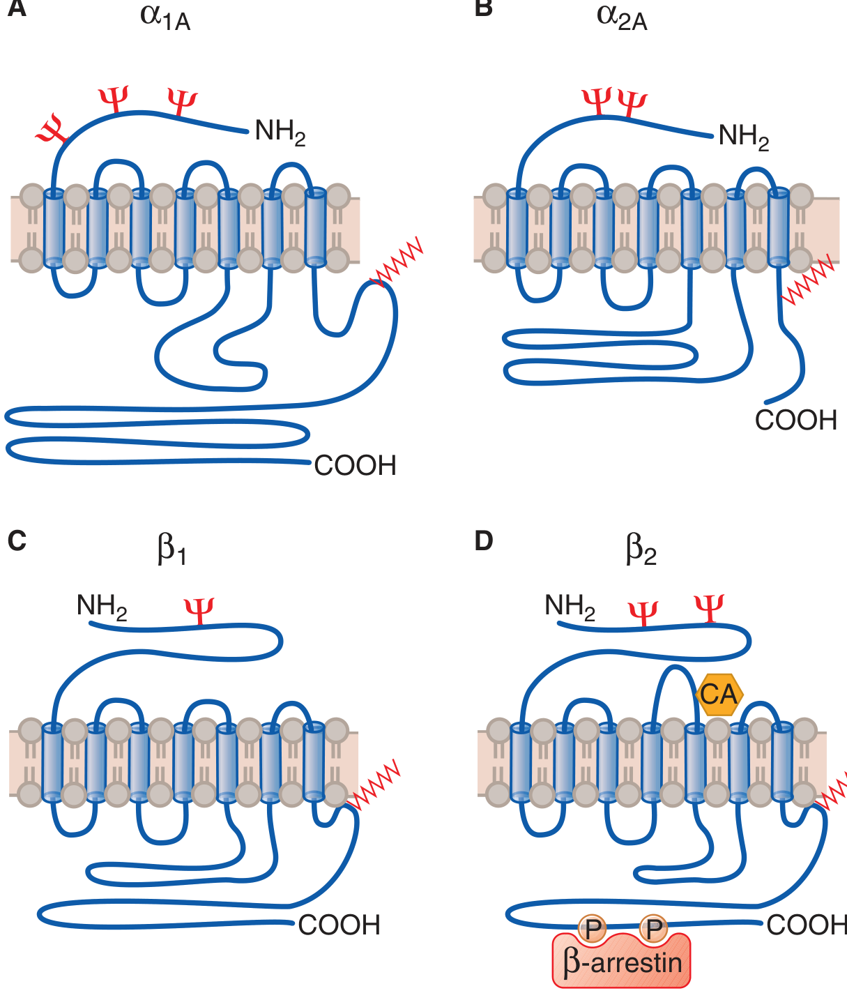
Structural features of adrenergic receptors: all are hepta-spanning GPCRs. Note the differences in intracellular loop sizes and the β-arrestin binding site on β2 after GRK-mediated phosphorylation. (Goodman & Gilman's)
3. Alpha-1 (α1) Receptors
Subtypes: α1A, α1B, α1D
G Protein Coupling
Gq → PLC-β → IP3 + DAG → Ca²+ + PKC
- Activation of Gq stimulates phospholipase C-β (PLC-β)
- PLC-β cleaves PIP2 into IP3 (increases intracellular Ca²+) and DAG (activates PKC)
- PKC phosphorylates multiple substrates: ion channels, pumps, Ca²+-transport ATPase
- Additional pathways: PLA2 stimulation releases arachidonic acid → prostaglandins (COX) and leukotrienes (LOX)
- Also activates p38/p42/p44 MAPKs, PI3K, JNK - regulates cell growth and proliferation
Subtype-Specific Roles
| Subtype | Key Roles |
|---|---|
| α1A | Predominant vasoconstrictor in mammary, mesenteric, splenic, hepatic, omental, renal, pulmonary, and epicardial coronary arteries; also in vena cava and pulmonary veins |
| α1B | Together with α1A promotes cardiac growth and structure; mediates novelty-seeking behavior; involved in addiction vulnerability |
| α1D | Most abundant in the heart; predominant vasoconstrictor in the aorta |
Notable Feature
α1 receptors (and β receptors) have been found on nuclear membranes of adult cardiac myocytes, where they activate intranuclear signaling with an apparent cardioprotective role.
Agonist Potency Order
NE > Epi > isoproterenol
Physiological Effects
- Vascular smooth muscle contraction (vasoconstriction) - major role of postsynaptic α1 in arterioles
- Cardiac myocyte effects: fine-tuning of Ca²+ transients, ionic currents, myofilament properties, and cardiac remodeling
- GI smooth muscle: α1 stimulation causes hyperpolarization and relaxation (via Ca²+-activated K+ channels - opposite to most smooth muscles)
4. Alpha-2 (α2) Receptors
Subtypes: α2A, α2B, α2C
G Protein Coupling
Gi → ↓cAMP; also activates K+ channels, inhibits Ca²+ channels
Primary effectors include:
- Inhibition of adenylyl cyclase (via Gi) → ↓cAMP (most systems)
- Activation of inwardly-rectifying K+ channels → membrane hyperpolarization (via Gβγ subunits)
- Inhibition of voltage-gated Ca²+ channels (via Gi)
- Activation of Na+/H+ exchange, stimulation of PLCβ2, arachidonic acid mobilization, increased intracellular Ca²+ (mediates smooth muscle contraction)
- Activation of MAPKs via Gi α and βγ components (growth factor receptor-like pathways)
Subtype-Specific Roles
| Subtype | Key Roles |
|---|---|
| α2A | Dominant CNS adrenergic receptor; mediates antinociception, sedation, hypothermia, hypotension; inhibits NE release from sympathetic nerve endings |
| α2B | Main receptor mediating α2-induced vasoconstriction |
| α2C | Predominant receptor inhibiting catecholamine release from adrenal medulla; modulates dopamine (DA) neurotransmission in brain (ventral/dorsal striatum, hippocampus) |
Key Functions (broadly)
- Presynaptic autoreceptors on sympathetic nerve terminals - the α2A and α2C subtypes inhibit NE release (feedback inhibition - see diagram below)
- Platelet aggregation promotion
- Insulin secretion inhibition and lipolysis regulation
- Suppression of sympathetic outflow from the CNS → clinically exploited by clonidine, dexmedetomidine
5. Beta (β) Receptors
Subtypes: β1, β2, β3
G Protein Coupling
All three → Gs → ↑adenylyl cyclase → ↑cAMP → PKA activation
- Gs-alpha subunit (Gαs) separates and stimulates adenylyl cyclase → ↑cAMP → activates protein kinase A (PKA)
- PKA phosphorylates numerous cellular targets (phospholamban, troponin I, L-type Ca²+ channels, etc.)
- Gβγ subunits can directly activate voltage-sensitive Ca²+ channels in skeletal and cardiac muscle
- β2 receptors also couple to Gi (in addition to Gs) - dual coupling that bifurcates signaling at the first postreceptor step
Subtype Details
β1 Receptors
- 80% of cardiac beta receptors (in left ventricle; 20% is β2)
- Linked exclusively to Gs
- Main mediator of the positive inotropic response to adrenergic activation in humans
- Agonist potency: isoproterenol > Epi = NE
- Located throughout the heart; activation increases HR, contractility (inotropy), relaxation speed (lusitropy), and conduction velocity (dromotropy)
β2 Receptors
- Predominant noncardiac beta receptor
- Dual coupling: Gs and Gi
- Gs path mediates bronchodilation, vasodilation, uterine relaxation, glycogenolysis
- Gi path causes negative chronotropy (can be blocked by pertussis toxin)
- Normally confined to caveolae in cardiac myocyte membranes - important for compartmentation of cAMP signaling
- Agonist potency: isoproterenol > Epi > NE
- β2 receptors are also present on presynaptic sympathetic nerve terminals - stimulation by circulating Epi promotes NE release
β3 Receptors
- Present in small numbers in cardiac myocytes
- Coupled to Gi (unlike β1/β2)
- Produces a negative inotropic effect, mediated partly through nitric oxide (NO)
- Also important in adipose tissue (lipolysis) and bladder
Agonist Potency Comparison
| Receptor | Order of Potency |
|---|---|
| β1 | Isoproterenol > Epi = NE |
| β2 | Isoproterenol > Epi > NE |
| α1 | NE > Epi > Isoproterenol |
| α2 | NE > Epi > Isoproterenol |
6. G Protein Signaling in Detail
G proteins are heterotrimers (Gα, Gβ, Gγ). On receptor activation:
- The receptor (activated by agonist) catalyzes GDP → GTP exchange on Gα
- Gα-GTP dissociates from Gβγ
- Both Gα-GTP and free Gβγ can independently regulate effectors:
- Adenylyl cyclase (AC)
- Phospholipase C (PLC)
- Ion channels
- Gα intrinsic GTPase activity hydrolyzes GTP → GDP, terminating the signal
| G Protein | Receptor Coupling | Effect on Adenylyl Cyclase |
|---|---|---|
| Gs | β1, β2, β3 | Stimulates AC → ↑cAMP |
| Gi | α2, β2 (partial), β3 | Inhibits AC → ↓cAMP |
| Gq | α1 | Stimulates PLC-β → IP3/DAG |
7. NE Release and Receptor Interaction at the Synapse
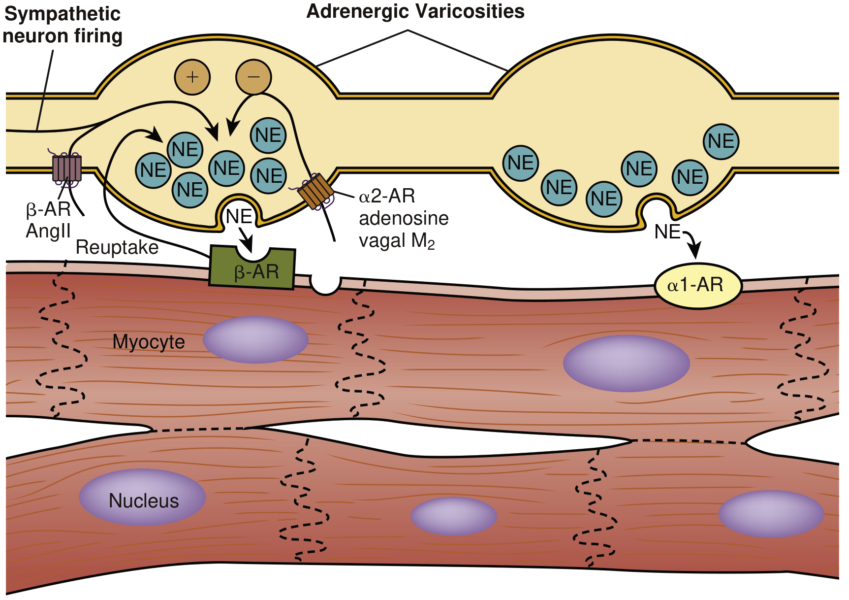
NE is released from storage granules into narrow synapse-like spaces. In cardiomyocytes, β-AR activation drives inotropy, chronotropy, lusitropy, and dromotropy. In arterioles, NE causes vasoconstriction via postsynaptic α1 receptors. Presynaptic α2 receptors limit NE release (feedback inhibition). Presynaptic β-AR and AngII receptors on the nerve terminal promote NE release. (Braunwald's Heart Disease)
8. Desensitization and Refractoriness
Prolonged or repeated exposure to agonists causes receptor desensitization (also called tachyphylaxis or refractoriness) - a critical regulatory mechanism:
- GRK phosphorylation: G protein-coupled receptor kinases (GRKs) phosphorylate the active (agonist-bound) receptor on intracellular serine/threonine residues
- β-arrestin recruitment: β-arrestin binds phosphorylated receptor, sterically blocks further G protein activation
- Two outcomes of β-arrestin binding:
- Receptor internalization/trafficking - receptor is removed from the surface
- Independent β-arrestin signaling - can itself initiate effector cell responses
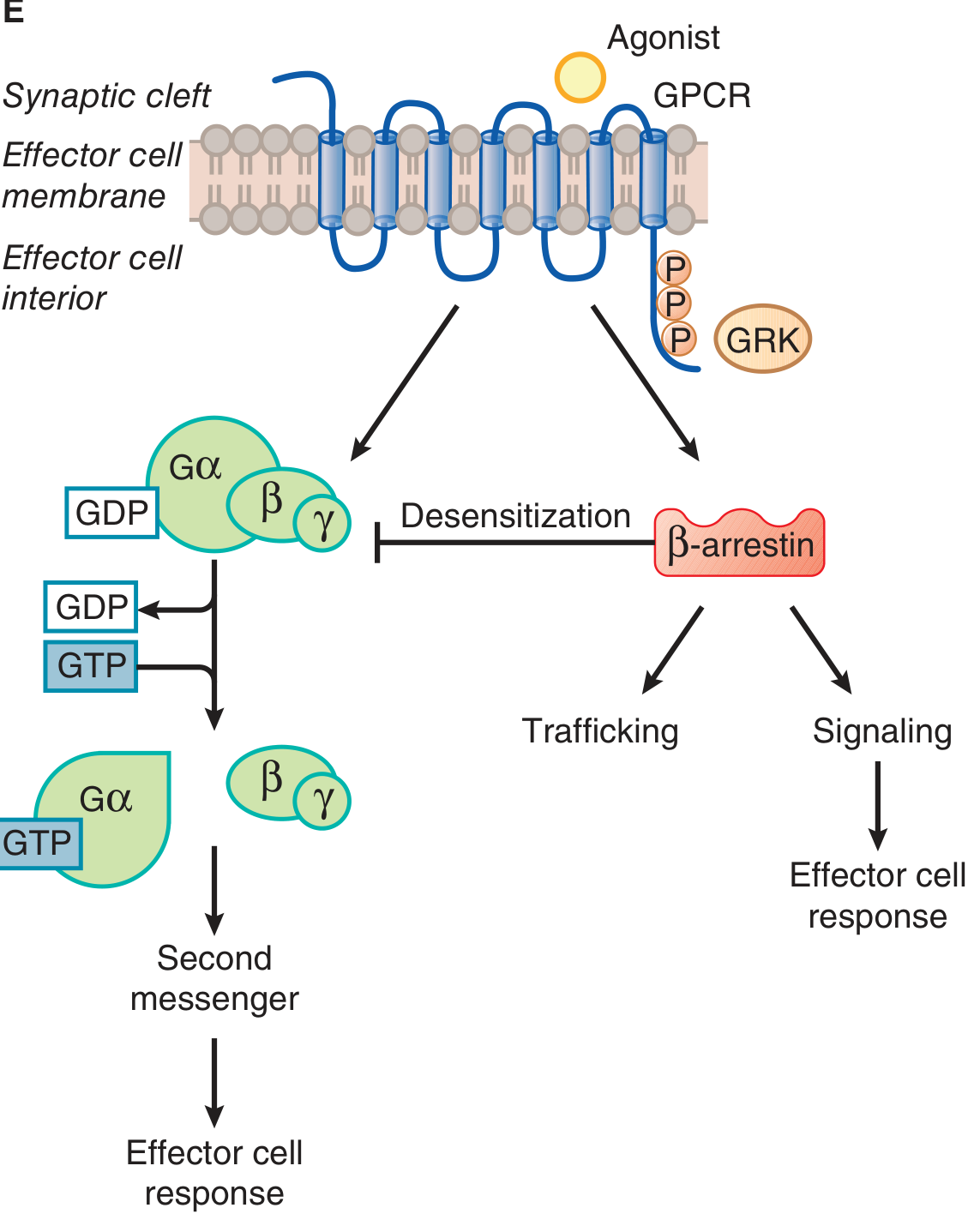
Two-armed signaling from adrenergic receptors: agonist activates both classical G protein signaling (left, via GDP/GTP exchange) and β-arrestin-mediated pathways (right, following GRK phosphorylation). β-arrestin causes desensitization and receptor trafficking but also initiates its own signaling arm. Biased agonists can preferentially activate one arm. (Goodman & Gilman's)
This is clinically relevant in heart failure, where chronic sympathetic activation leads to β1-receptor downregulation and Gs uncoupling, reducing inotropic responsiveness.
9. Cardiovascular Effects Summary
| Effect | Mediating Receptor |
|---|---|
| Increased HR (chronotropy) | β1 |
| Increased contractility (inotropy) | β1 > α1 |
| Increased conduction velocity (dromotropy) | β1 |
| Increased relaxation speed (lusitropy) | β1 |
| Arteriolar vasoconstriction | α1 (postsynaptic) |
| Arteriolar vasodilation | β2 |
| Presynaptic NE inhibition | α2 (presynaptic) |
| Negative inotropy (in heart failure context) | β3 (via NO/Gi) |
10. Receptor Polymorphisms and Clinical Significance
- Genetic polymorphisms in adrenergic receptors (studied extensively in Goodman & Gilman) can alter receptor function and clinical drug responses
- In heart failure, β1 receptors are downregulated; relative Gi signaling is augmented; β2-Gs coupling is diminished due to uncoupling
- Nuclear localization of α1 and β receptors in cardiac myocytes suggests additional intranuclear signaling roles beyond classical membrane signaling
11. Adrenergic Receptor Polymorphism (Pharmacogenomics)
Clinically important single-nucleotide polymorphisms (SNPs) have been identified in the genes encoding β1 and β2 receptors. These affect:
- Agonist-stimulated downregulation
- Coupling efficiency to Gs
- Response to beta-blocker therapy in heart failure and hypertension
Quick Reference Table
| Receptor | G Protein | 2nd Messenger | Key Locations | Key Effects | Prototype Agonist |
|---|---|---|---|---|---|
| α1A/B/D | Gq | ↑IP3/DAG/Ca²+ | Vascular SM, heart | Vasoconstriction, cardiac hypertrophy | Phenylephrine |
| α2A/B/C | Gi | ↓cAMP, ↑K+ curr | Nerve terminals, CNS, platelets | ↓NE release, sedation, ↓BP, platelet aggregation | Clonidine |
| β1 | Gs | ↑cAMP | Heart (80%) | ↑HR, ↑contractility, ↑conduction | Dobutamine |
| β2 | Gs (+Gi) | ↑cAMP | Lungs, vessels, uterus | Bronchodilation, vasodilation, tocolysis | Salbutamol |
| β3 | Gi | ↓cAMP/↑NO | Heart (small), adipose | ↓Inotropy, lipolysis | Mirabegron (bladder) |
Sources:
- Goodman & Gilman's The Pharmacological Basis of Therapeutics, Chapter 10 - Adrenergic Agonists and Antagonists
- Braunwald's Heart Disease: A Textbook of Cardiovascular Medicine, Chapter 46 - Adrenergic Receptor Subtypes and Signaling
This is a shared conversation. Sign in to Orris to start your own chat.