
Why femoral nerve does not pass through Femoral sheath
The femoral nerve is not enclosed in the femoral sheath because of its embryological origin and tissue compartment differences. The femoral sheath is derived from the transversalis fascia (anteriorly) and iliacus fascia (posteriorly) and encloses only structures of vascular origin (femoral artery, vein, and lymphatics). The femoral nerve, being a nerve of the lumbar plexus, travels within the iliacus fascia compartment - it lies lateral to the femoral sheath, separated from it by the iliopectineal arch (a thickening of the iliopsoas fascia).
Searching sources...
Reading File
Finding Sources
Finding Sources
Reading File
Reading File
Loading Image
Reading File
Finding Sources
Here is a thorough, textbook-grounded answer:
Why the Femoral Nerve Does NOT Pass Through the Femoral Sheath
1. What is the Femoral Sheath?
The femoral sheath is a funnel-shaped sleeve of fascia that forms as the transversalis fascia (anteriorly) and iliac fascia (posteriorly) extend downward beneath the inguinal ligament to wrap around the femoral vessels. It is, fundamentally, a vascular compartment - designed to enclose the femoral artery, femoral vein, and lymphatics (femoral canal) as they transition from the abdomen to the thigh.
- Gray's Anatomy for Students describes the sheath as "continuous superiorly with the transversalis fascia and iliac fascia of the abdomen."
2. The Iliopectineal Arch - the Key Dividing Structure
The critical anatomical reason the femoral nerve is excluded comes down to the iliopectineal arch. This is a condensation of the iliopsoas fascia (itself a part of transversalis fascia) that stretches from the inguinal ligament to the iliopectineal eminence of the hip bone.
This arch physically divides the space beneath the inguinal ligament into two distinct lacunae:
| Compartment | Contents |
|---|---|
| Lacuna vasorum (medial, vascular) | Femoral artery, femoral vein, femoral canal (lymphatics), femoral branch of genitofemoral nerve |
| Lacuna musculorum (lateral, muscular) | Iliopsoas muscle, femoral nerve, lateral femoral cutaneous nerve |
As Mulholland & Greenfield's Surgery states directly: "The iliopectineal arch divides the vascular compartment (lacuna vasorum), containing the femoral vessels, from the muscular portion (lacuna musculorum), which contains the iliopsoas muscle, femoral nerve, and lateral femoral cutaneous nerve."
3. The Femoral Nerve Travels Under the Iliac Fascia, NOT Inside the Sheath
The femoral nerve (L2-L4 of the lumbar plexus) descends through the psoas, then runs in the groove between the psoas and iliacus muscles - the entire time lying deep to the iliac fascia. This fascia covers the nerve as a continuous sheet. When the iliac fascia contributes to forming the femoral sheath, it does so only medially, around the vessels. The nerve stays lateral, wrapped by the iliac fascia but outside the sheath proper.
From Gray's Anatomy for Students: "The femoral nerve lies on the lateral side of the femoral artery and is outside the femoral sheath, which surrounds the vessels."
4. Summary Diagram from Textbook
Here is the femoral triangle layout showing the femoral nerve (yellow, lateral) sitting completely outside the sheath compartments:
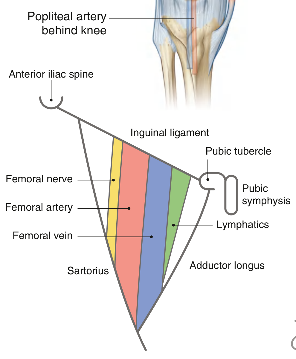
Notice the femoral nerve (yellow) is lateral to the femoral artery and is NOT enclosed within the same fascial compartment as the vessels.
5. The Simple Conceptual Reason
The femoral sheath exists to protect and contain vascular structures as they cross the inguinal ligament where pressure changes occur with body movement. Nerves do not require this same protection and, more importantly, nerves must not be compressed - enclosing a nerve in a tight vascular sheath would risk compression injury. The iliopectineal arch ensures permanent physical separation between the neural/muscular lane and the vascular lane.
Mnemonic - NAVL (lateral to medial at the femoral triangle):
Nerve - Artery - Vein - Lymphatics Only AVL are inside the femoral sheath; the Nerve stays outside.
- Gray's Anatomy for Students, femoral sheath section, p. 676
- Mulholland & Greenfield's Surgery (7e), Femoral Sheath, Canal, and Ring, p. 3652
This is a shared conversation. Sign in to Orris to start your own chat.