
I am going to study for my gynaecology block its my first time studying it I want you to use good references and resources like ten teacher irfan maasood first aid or other gynecology books out there andFor each disease, you'll explain it in a structured, exam-oriented way: Definition Etiology/Risk factors Pathogenesis (step-by-step) Clinical features Diagnosis Investigations Management Complications And other useful points or subheadings High-yield exam points Mnemonics Reference images
- Definition
- Etiology and risk factors
- Pathogenesis - step-by-step
- Clinical features
- Diagnosis and diagnostic criteria
- Investigations
- Management - acute, definitive, medical, and surgical where relevant
- Complications
- Differentials
- High-yield exam points and common traps
- Mnemonics
- Reference images or diagrams when they genuinely help
Gynaecology study map
-
Basics and symptoms
- Menstrual physiology and normal cycle
- Abnormal uterine bleeding (AUB)
- Pelvic pain, vaginal discharge, amenorrhea
-
Menstrual and endocrine disorders
- PCOS
- Primary and secondary amenorrhea
- Dysmenorrhea and PMS
- Hyperprolactinemia, thyroid-related menstrual disturbance
- Premature ovarian insufficiency
-
Benign uterine and ovarian disease
- Fibroids
- Adenomyosis
- Endometriosis
- Ovarian cysts and benign ovarian tumors
- Ovarian torsion
-
Infections
- Vaginitis and cervicitis
- Pelvic inflammatory disease
- Tuberculosis of the female genital tract
- Sexually transmitted infections
-
Infertility and reproductive medicine
- Female infertility work-up
- Ovulatory disorders
- Tubal, uterine, and endometriosis-related infertility
- Assisted reproduction basics
-
Gynecologic oncology
- Cervical intraepithelial neoplasia and cervical cancer
- Endometrial hyperplasia and cancer
- Ovarian cancer
- Vulvar and vaginal cancer
- Screening and HPV vaccination
-
Urogynecology
- Pelvic organ prolapse
- Urinary incontinence
- Urogenital fistulae
-
Contraception and menopause
- Combined hormonal contraception
- Progestin-only contraception
- IUDs and emergency contraception
- Menopausal symptoms and hormone therapy
-
Gynecologic emergencies
- Ectopic pregnancy is technically obstetric but commonly tested with gynecology
- Acute pelvic pain
- Hemorrhage and shock in gynecologic conditions
- Torsion, ruptured cyst, tubo-ovarian abscess
Pelvic organ prolapse
"pelvic organ prolapse" AND treatment
pelvic organ prolapse NICE guideline diagnosis management POP-Q pessary surgery
pelvic organ prolapse anatomical diagram anterior apical posterior compartments
https://www.nice.org.uk/guidance/ng123
PMID: 39666016
https://www.nice.org.uk/guidance/ng123/resources/urinary-inc…
Pelvic Organ Prolapse (POP)
1. Definition
| Compartment | Structure prolapsing | Common name |
|---|---|---|
| Anterior | Bladder, urethra | Cystocele, urethrocele |
| Apical | Uterus/cervix or post-hysterectomy vaginal vault | Uterine prolapse, vault prolapse |
| Posterior | Rectum or small bowel | Rectocele, enterocele |
2. Etiology and risk factors
Major risk factors
-
Pregnancy and vaginal childbirth
- Strongest common risk factor.
- Risk rises with increasing parity.
- Instrumental vaginal delivery, prolonged second stage, fetal macrosomia, and obstetric levator ani injury increase risk.
-
Increasing age and menopause
- Aging causes weakening of connective tissue and muscle.
- Reduced estrogen after menopause contributes to urogenital atrophy, although estrogen deficiency alone does not fully explain POP.
-
Raised intra-abdominal pressure
- Obesity
- Chronic cough, for example COPD
- Chronic constipation and straining
- Repeated heavy lifting
-
Prior pelvic surgery
- Especially hysterectomy, which may predispose to vaginal vault prolapse if apical support is not restored.
- Previous prolapse surgery increases recurrence risk.
-
Connective-tissue and neuromuscular factors
- Family history
- Ehlers-Danlos syndrome and other collagen disorders
- Abnormal collagen metabolism
- Pelvic floor denervation
-
Other associations
- White ethnicity in epidemiologic studies
- Smoking, partly through chronic cough and altered tissue healing
3. Pathogenesis - step by step
Normal pelvic support
- Levator ani muscles, especially pubococcygeus and puborectalis
- Endopelvic fascia
- Uterosacral-cardinal ligament complex
- Perineal membrane and perineal body
- Intact pudendal nerve supply
Stepwise development of prolapse
-
Childbirth trauma or chronic pressure damages support tissues
- Vaginal delivery can stretch or avulse the levator ani.
- Fascia, ligaments, and pudendal nerves can be injured.
-
Pelvic floor muscle weakness develops
- The levator ani normally forms a supportive muscular “shelf.”
- If it weakens, the genital hiatus widens.
-
Fascial and ligamentous supports stretch or fail
- The vagina and pelvic organs lose their normal suspensory support.
-
Repeated rises in intra-abdominal pressure push organs downward
- Coughing, straining, obesity, constipation, and lifting worsen descent.
-
The affected vaginal compartment bulges
- Anterior defect causes cystocele.
- Apical defect causes uterine or vault prolapse.
- Posterior defect causes rectocele or enterocele.
-
Advanced prolapse may distort lower urinary and bowel anatomy
- This can cause voiding difficulty, incomplete emptying, urinary retention, obstructed defecation, or the need for vaginal/perineal “splinting.”
Delancey levels of vaginal support - very high yield
| Level | Main support | Failure causes |
|---|---|---|
| Level I: apical | Uterosacral-cardinal ligament complex | Uterine prolapse or vaginal vault prolapse |
| Level II: mid-vaginal | Lateral attachment of vagina to arcus tendineus fascia pelvis | Cystocele or lateral vaginal wall defect |
| Level III: distal vaginal | Perineal membrane, perineal body, distal fusion | Distal anterior/posterior wall prolapse |
4. Types and terminology
Anterior compartment
- Cystocele: bladder bulges into anterior vaginal wall.
- Urethrocele: urethra descends with anterior vaginal wall.
- Usually associated with urinary symptoms, but symptoms and severity do not always correlate.
Apical compartment
- Uterine prolapse: uterus/cervix descends.
- Vaginal vault prolapse: vaginal apex descends after hysterectomy.
- Procidentia: complete uterovaginal prolapse, with the uterus protruding beyond the vaginal introitus.
Posterior compartment
- Rectocele: rectal wall bulges into posterior vaginal wall.
- Enterocele: peritoneal sac, often containing small bowel, protrudes between rectum and vagina, commonly after hysterectomy.
- Symptoms can include obstructed defecation and vaginal splinting.
5. Clinical features
Typical symptom
“A feeling or sight of a lump/bulge coming down from the vagina.”
Other symptoms
Vaginal or pelvic symptoms
- Vaginal pressure, heaviness, dragging discomfort
- Something “coming out” per vagina
- Symptoms worse after standing, lifting, straining, or later in the day
- Vaginal irritation, discharge, bleeding, ulceration in severe external prolapse
Urinary symptoms
- Stress urinary incontinence
- Urgency, frequency, urge incontinence
- Hesitancy, poor stream, straining to void
- Feeling of incomplete emptying
- Recurrent UTI
- Urinary retention in advanced prolapse
Bowel symptoms
- Constipation
- Incomplete evacuation
- Obstructed defecation
- Need to press on posterior vaginal wall or perineum to defecate: splinting
- Fecal urgency or incontinence in some patients
Sexual symptoms
- Dyspareunia
- Reduced sexual satisfaction
- Body-image concerns
Important point
6. Diagnosis
History
- Vaginal bulge or pressure
- Urinary incontinence, voiding symptoms, recurrent UTIs
- Constipation, splinting, fecal incontinence
- Sexual function and dyspareunia
- Obstetric history: parity, mode of delivery, instrumental birth, obstetric trauma
- Previous hysterectomy or prolapse/incontinence surgery
- Chronic cough, constipation, obesity, heavy lifting
- Impact on activities and quality of life
- Fertility wishes, future pregnancy plans, and sexual activity
Examination
- Inspect external genitalia for atrophy, ulceration, discharge, or exposed prolapse.
- Perform speculum examination.
- Ask the patient to Valsalva/strain.
- Assess each compartment separately:
- Anterior
- Apical
- Posterior
- Assess pelvic floor muscle contraction.
- Perform bimanual examination to exclude pelvic mass.
- Consider rectal examination when posterior compartment symptoms exist.
- If prolapse is not apparent but symptoms are suggestive, repeat examination while the patient is standing or squatting.
7. POP-Q classification
| Stage | Leading edge of prolapse |
|---|---|
| Stage 0 | No prolapse |
| Stage I | More than 1 cm above hymen |
| Stage II | Within 1 cm proximal or distal to hymen |
| Stage III | More than 1 cm beyond hymen, but not complete eversion |
| Stage IV | Complete or near-complete vaginal eversion |
Easy memory aid
8. Investigations
Usually required
- Urinalysis and urine culture if UTI symptoms are present.
- Post-void residual urine volume if there is poor stream, incomplete emptying, recurrent UTI, or advanced anterior/apical prolapse.
- Validated pelvic-floor symptom questionnaires can help quantify impact and follow response.
Selective investigations
| Test | When to use |
|---|---|
| Urodynamic testing | Complex urinary symptoms, voiding dysfunction, mixed incontinence, or when planning selected prolapse/incontinence surgery |
| Cystoscopy | Usually intraoperatively during major prolapse or anti-incontinence surgery to detect bladder/ureteric injury |
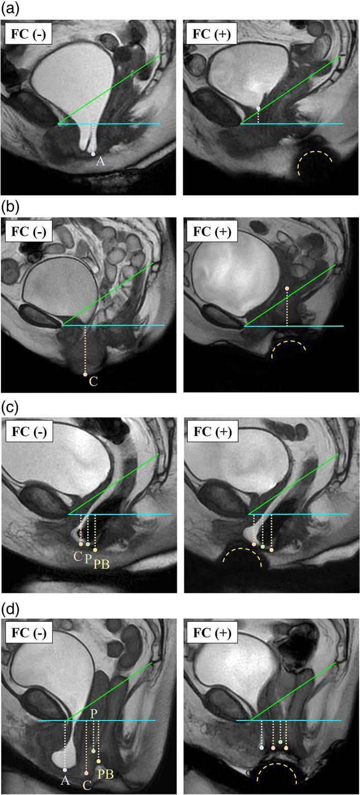
| Defecography or dynamic pelvic MRI | Complex posterior compartment symptoms, suspected enterocele, rectal prolapse, or discordance between symptoms and examination |
| Pelvic ultrasound | Suspected pelvic mass, abnormal bleeding, or other pathology, not to diagnose obvious POP |
| Renal ultrasound and renal function | Advanced prolapse with suspected upper urinary tract obstruction or hydronephrosis |
9. Management
- Symptoms and their impact
- POP compartment and stage
- Age, comorbidities, and surgical fitness
- Desire for future sexual intercourse
- Desire for uterine preservation/future fertility
- Previous treatments
- Patient preference
A. Expectant and lifestyle management
- Reassure: POP is benign and not always progressive.
- Weight reduction if overweight.
- Treat chronic cough.
- Treat constipation and avoid chronic straining.
- Avoid heavy repetitive lifting where possible.
- Smoking cessation.
- Vaginal moisturizers or topical estrogen for postmenopausal atrophy.
B. Pelvic-floor muscle training (PFMT)
- First-line for symptomatic stage I or II POP.
- Should be supervised, ideally by a pelvic-floor physiotherapist.
- NICE recommends a supervised program for at least 16 weeks for symptomatic POP-Q stage I or II prolapse, then continued exercise if beneficial. See NICE recommendations.
C. Vaginal pessary
Indications
- Symptomatic POP in a woman wishing to avoid or delay surgery
- Poor surgical candidate
- Wants future pregnancy
- Awaiting surgery
- Temporary symptom relief
- Recurrent prolapse
Common types
- Ring pessary with or without support: common first option, relatively easy to insert/remove.
- Gellhorn pessary: useful for more advanced prolapse but often harder for the patient to manage.
- Cube and shelf pessaries are other options.
Advantages
- Non-surgical
- Often gives rapid bulge symptom relief
- Can be used with PFMT
- Can be removed if poorly tolerated
Complications
- Vaginal discharge
- Odor
- Bleeding
- Vaginal erosion/ulceration
- Infection
- Rarely fistula or incarceration if neglected
D. Surgical management
Surgical principles
- Identify all compartment defects.
- Restore apical support when needed.
- Choose reconstructive versus obliterative surgery.
- Discuss recurrence, dyspareunia, urinary symptoms, bowel symptoms, mesh-related risks, and possible future surgery.
- If hysterectomy is performed for prolapse, the vaginal apex must be suspended to reduce post-hysterectomy vault prolapse risk.
Reconstructive surgery
| Defect | Common surgical options |
|---|---|
| Anterior vaginal wall prolapse | Native tissue anterior colporrhaphy; selected paravaginal repair |
| Posterior vaginal wall prolapse/rectocele | Posterior colporrhaphy, site-specific repair |
| Uterine/apical prolapse | Vaginal hysterectomy plus apical suspension, uterosacral ligament suspension, sacrospinous fixation, sacrohysteropexy |
| Post-hysterectomy vaginal vault prolapse | Sacrospinous fixation, uterosacral suspension, abdominal/laparoscopic sacrocolpopexy |
| Enterocele | Vaginal or abdominal enterocele repair/culdoplasty, usually with apical support restoration |
Uterus-preserving surgery
- Sacrohysteropexy
- Vaginal sacrospinous hysteropexy
- Other hysteropexy procedures, depending on local expertise
Sacrocolpopexy
- Usually abdominal or laparoscopic.
- Mesh is attached to the vaginal apex and anchored to the sacrum.
- Often considered durable for apical/vault prolapse.
- Risks include mesh exposure/erosion, bowel injury, vascular injury, ileus, and rare sacral osteomyelitis.
Obliterative surgery
- Suitable for frail patients with severe prolapse who no longer desire penetrative vaginal intercourse.
- Effective and relatively lower operative burden.
- Irreversible loss of vaginal intercourse must be explicitly discussed.
Mesh: an exam-safe answer
10. Complications of POP
Complications of untreated advanced prolapse
- Vaginal/cervical ulceration and bleeding
- Infection and discharge
- Voiding difficulty and urinary retention
- Recurrent UTI
- Hydroureter/hydronephrosis and, rarely, renal impairment
- Obstructed defecation
- Sexual dysfunction and impaired quality of life
Complications of pessary treatment
- Discharge, odor
- Erosion and bleeding
- Infection
- Rare fistula or impaction with neglected pessary
Surgical complications
- Hemorrhage
- Bladder, ureteric, bowel, or vascular injury
- UTI, wound infection, venous thromboembolism
- Urinary retention
- De novo stress urinary incontinence
- Dyspareunia
- Recurrence or prolapse in another compartment
- Mesh exposure/erosion where mesh is used
11. Differential diagnosis of a vaginal mass/bulge
- Prolapsed submucosal fibroid
- Cervical polyp
- Vaginal wall cyst
- Urethral diverticulum
- Vaginal or cervical malignancy
- Bartholin cyst, usually posterolateral and at the introitus rather than a true vaginal-wall prolapse
- Rectal prolapse
12. High-yield exam points
- Most specific symptom: a vaginal bulge or the feeling that “something is coming down.”
- POP involves anterior, apical, and posterior compartments.
- Most important risk factor: pregnancy and vaginal childbirth.
- Posterior vaginal wall prolapse = rectocele.
- Complete uterovaginal prolapse = procidentia.
- Post-hysterectomy apical prolapse = vaginal vault prolapse.
- POP-Q is the standard objective staging system.
- Routine imaging is not required if physical examination clearly demonstrates POP.
- Consider post-void residual in voiding difficulty or advanced prolapse.
- First-line for symptomatic stage I-II: supervised PFMT for at least 16 weeks.
- Pessary is a good non-surgical option and can be combined with PFMT.
- Surgical planning should be multicompartmental. Correct apical support when it is deficient.
- In a woman undergoing hysterectomy for prolapse, perform apical suspension to reduce later vault prolapse.
- Colpocleisis is for women who do not want future penetrative vaginal intercourse.
- Do not present transvaginal mesh as routine first-line treatment.
13. Mnemonics
Risk factors: “PUSH DOWN”
- P - Parity and pregnancy
- U - Uterine/pelvic surgery, especially hysterectomy
- S - Straining from constipation
- H - Heavy lifting
- D - Delivery trauma, especially instrumental delivery
- O - Obesity
- W - Weak connective tissue/levator ani
- N - New age: aging and menopause
Compartments: “A-A-P”
- Anterior: cystocele
- Apical: uterine or vault prolapse
- Posterior: rectocele/enterocele
POP-Q stages: “Above, At, Beyond, Out”
- Stage I: above hymen
- Stage II: at hymen
- Stage III: beyond hymen
- Stage IV: out, near-total/total eversion