
Symptoms of insulin resistance
insulin resistance symptoms acanthosis nigricans metabolic syndrome
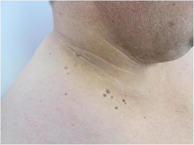
This clinical photograph displays a lateral view of a patient's neck and upper shoulder, demonstrating classic dermatological manifestations of insulin resistance. Centered on the lateral and posterior neck folds are poorly defined, symmetrical, velvety hyperpigmented plaques characteristic of acanthosis nigricans. The affected skin shows a brownish-tan discoloration with a prominent thickened, verrucous (wart-like) texture following the natural skin lines. Distributed within and adjacent to these hyperpigmented areas are multiple fibroma pendula (skin tags or acrochordons). These are small, skin-colored to slightly hyperpigmented, pedunculated papules. The educational focus of this image is the visual identification of metabolic-related skin changes commonly associated with obesity, hyperinsulinemia, and type 2 diabetes mellitus. The co-occurrence of acanthosis nigricans and multiple acrochordons in intertriginous areas is a significant clinical indicator for underlying metabolic syndrome.

Clinical photograph of the axillary skin (upper inner arm) documenting acanthosis nigricans in the setting of obesity. This close-up dermatologic image demonstrates hyperpigmented, velvety plaques within flexural skin folds, with epidermal thickening and subtle papillomatosis along the basilar fold lines. The coloration ranges from dark brown to near-black, and the texture appears markedly roughened compared with adjacent noninvolved skin. The appearance is symmetric in typical obesity‑associated acanthosis nigricans, though unilateral presentation can occur. In context, the findings reflect insulin resistance driving keratinocyte proliferation and increased dermal matrix, producing the characteristic velvety hyperpigmented plaques of the axilla. Clinically, these signs signal metabolic syndrome risk and warrant screening for impaired glucose tolerance, type 2 diabetes, dyslipidemia, and obesity‑related comorbidities. The image is not a diagnostic modality by itself but serves as documentation for dermatologic assessment, obesity management, and endocrine correlation. Differential diagnoses include frictional dermatitis, postinflammatory hyperpigmentation, and rare malignant acanthosis nigricans in atypical presentations or older patients. This dataset label supports education, clinical correlation with BMI, and research on insulin‑resistance phenotypes; useful for medical trainees, dermatology and endocrinology teaching collections, and AI model training for skin disease recognition. Annotation aids clinical teaching, biopsy decision considerations, and automated annotation pipelines for medical imaging.
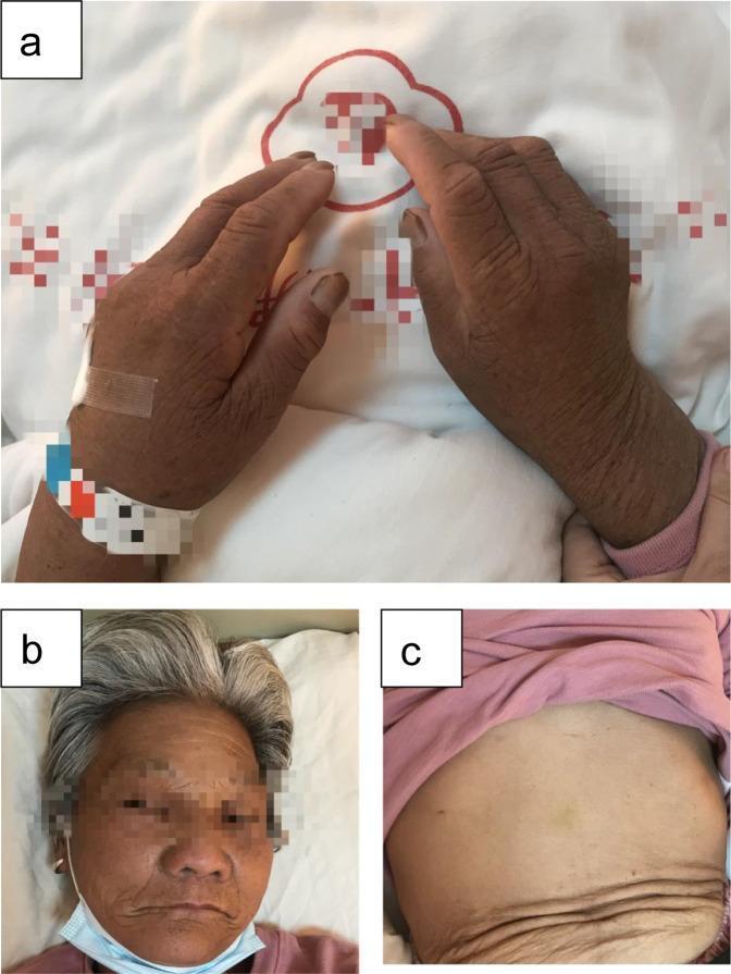
This composite of clinical photographs illustrates the dermatological manifestations of acanthosis nigricans in a patient diagnosed with Type B insulin resistance syndrome. Image (a) displays the dorsal aspect of the hands and wrists, showing significant diffuse hyperpigmentation, skin thickening, and coarsening of the skin texture (lichenification) with prominent skin folds. Image (b) shows similar hyperpigmented, velvety, and thickened skin affecting the facial region, particularly the forehead and periorbital areas. In contrast, image (c) provides a visual comparison of the abdominal skin, which lacks the intense hyperpigmentation and thickening seen on the extremities and face, though some lower abdominal skin folds are visible. These findings are characteristic of severe insulin resistance, where high circulating insulin levels stimulate keratinocytes and fibroblasts, leading to epidermal hyperplasia. The images serve as an educational example of how systemic metabolic and autoimmune disorders, such as those involving insulin receptor autoantibodies, can present with extensive cutaneous markers.
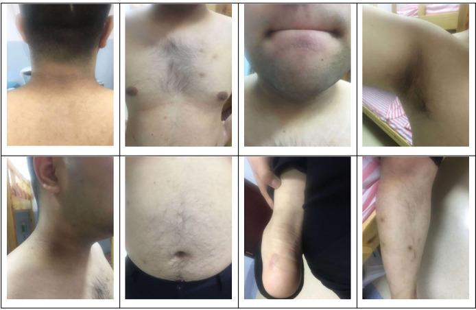
This composite of clinical photographs illustrates physical manifestations of severe insulin resistance in a 22-year-old male. The images display classic dermatological signs across several anatomical regions. Prominent findings include acanthosis nigricans, characterized by hyperpigmented, velvety, and thickened (hyperplastic) plaques localized on the posterior and lateral neck, the axilla (underarm), and the inguinal folds. Additionally, the photographs show significant hypertrichosis, with coarse, dense terminal hair growth across the chest and abdomen. Other visible clinical features include general obesity and darkened skin textures in flexural areas. These cutaneous markers are highly suggestive of systemic metabolic dysfunction, such as Type A Insulin Resistance Syndrome (TAIRS). The visual data serves as an educational tool for identifying skin-based complications of hyperinsulinemia and hormonal imbalances often encountered in endocrinology and dermatology.
"insulin resistance" symptoms signs clinical features
Symptoms of Insulin Resistance
1. Cutaneous Signs (Most Visible)
Acanthosis Nigricans
Skin Tags (Acrochordons / Fibroma Pendula)
2. Metabolic / Laboratory Symptoms
| Finding | Significance |
|---|---|
| Fasting hyperglycemia (glucose ≥100 mg/dL) | Liver fails to suppress gluconeogenesis |
| High postprandial glucose | Impaired skeletal muscle glucose uptake |
| Fasting hyperinsulinemia | Compensatory beta-cell hypersecretion |
| High triglycerides (>150 mg/dL) | Excess FFA flux from adipose to liver; hypertriglyceridemia is "an excellent marker of the insulin-resistant condition" |
| Low HDL cholesterol (<39 mg/dL men; <51 mg/dL women) | Altered HDL composition and accelerated clearance |
| Small dense LDL particles | More atherogenic LDL phenotype |
| Elevated hsCRP | Adipose-derived cytokines (IL-6, TNF-α) drive hepatic CRP production |
- Harrison's Principles of Internal Medicine 22E, block 46
- Tietz Textbook of Laboratory Medicine 7th Ed., block 19
3. Weight and Body Composition Symptoms
- Abdominal (central) obesity - visceral adipose accumulation is both a cause and effect; waist >40 inches in men, >35 inches in women is a diagnostic threshold
- Difficulty losing weight despite diet/exercise
- Weight gain concentrated around the abdomen ("apple shape")
4. Blood Pressure
- Preserved renal sodium reabsorption effect of insulin (despite resistance to its vascular/glucose effects) → increased blood volume
- Increased sympathetic nervous system activity (also preserved in insulin resistance)
- Impaired nitric oxide-mediated vasodilation in the endothelium
- Elevated angiotensin II from adipose angiotensinogen
- Blood pressure ≥130/85 mmHg is a diagnostic criterion for metabolic syndrome
5. Symptoms from Compensatory Hyperinsulinemia
- Increased hunger / reactive hypoglycemia - postprandial insulin surges can cause a glucose "crash" 2-4 hours after eating, driving cravings
- Fatigue and brain fog - unstable glucose delivery to the brain
- Polyphagia (increased appetite, especially for carbohydrates)
6. Hormonal Symptoms (in Women)
-
Irregular periods / anovulation
-
Hirsutism, acne (androgen excess from ovarian stimulation by insulin)
-
This is the pathophysiologic basis of PCOS in many patients
-
Tietz Textbook of Laboratory Medicine 7th Ed., block 19
7. Cardiovascular Symptoms
- Elevated VLDL and small dense LDL
- Endothelial dysfunction (reduced NO, increased endothelin-1)
- Pro-thrombotic state (elevated PAI-1, fibrinogen)
- Hypertension
Diagnostic Criteria: Metabolic Syndrome (≥3 of 5)
- Increased waist circumference (>88 cm/35 in women; >102 cm/40 in men)
- Triglycerides >150 mg/dL
- HDL-C <1.3 mmol/L women or <1.0 mmol/L men
- Blood pressure ≥130/85 mmHg
- Fasting plasma glucose ≥100 mg/dL
- Tietz Textbook of Laboratory Medicine 7th Ed.