
Myocardial infarction
Reading File
Searching sources...
Myocardial infarction (MI) is necrosis of cardiac muscle due to prolonged ischemia, most commonly from occlusion of a coronary artery by an atherosclerotic plaque rupture with superimposed thrombosis. Let me pull comprehensive textbook content on this.
Finding Sources
Searching PubMed
"myocardial infarction"[MeSH Terms] AND "management"[MeSH Terms]
Now let me read the key textbook sections in parallel - Braunwald's, Robbins Pathology, Ganong's, and Goldman-Cecil:
Reading File
Reading File
Reading File
Reading File
The Goldman-Cecil result was off-target. Let me read the correct MI sections - Robbins pathology continuation, and search for morphology, clinical features, and management:
Reading File
Finding Sources
Finding Sources
Excellent content. Let me read more from Robbins (morphology and complications) and fetch the Frameworks for Internal Medicine ACS section:
Reading File
Reading File
Finding Sources
Let me now get the Harrison's treatment section and the Washington Manual treatment, plus the Robbins morphology / complications section:
Reading File
Reading File
Reading File
I now have comprehensive content from multiple authoritative sources. Let me fetch one key image and get the complications section:
Reading File
Loading Image
I now have rich, comprehensive content from multiple authoritative textbooks. Let me compile the full answer.
Myocardial Infarction
Definition
Myocardial infarction (MI) is necrosis of cardiac muscle resulting from ischemia. The 2018 joint task force of U.S. and European Cardiology groups defines it as "the presence of acute myocardial injury detected by abnormal cardiac biomarkers in the setting of evidence of acute myocardial ischemia." - Robbins & Kumar Basic Pathology, p. 354
Epidemiology
- Frequency rises progressively with age and atherosclerotic risk factors
- ~10% of MIs occur before age 40; ~45% before age 65
- Men are at greater risk than women, but the gap narrows with age
- Women have relative protection during reproductive years; menopause accelerates risk
- Overall in-hospital death rate is ~7-8%; STEMI mortality ~10%, NSTEMI ~6%
- Out-of-hospital: one-third of STEMI patients die (usually from arrhythmia within 1 hour)
- Robbins & Kumar Basic Pathology, p. 354
Classification (ACS Spectrum)
| Type | Mechanism | ECG | Occlusion |
|---|---|---|---|
| STEMI | Complete coronary occlusion | ST elevation | Transmural |
| NSTEMI | Incomplete occlusion | ST depression / T changes | Subendocardial |
| Unstable Angina | Disrupted plaque + thrombus | No ST elevation | No biomarker rise |
- Frameworks for Internal Medicine, p. 33
Pathogenesis
The vast majority of MIs are caused by acute thrombosis within coronary arteries, initiated by disruption or erosion of an atheromatous plaque. The sequence of events:
- An atheromatous plaque is eroded or disrupted by endothelial injury, intraplaque hemorrhage, or mechanical forces, exposing subendothelial collagen and necrotic plaque contents to blood
- Platelets adhere, aggregate, and are activated - releasing thromboxane A2, ADP, and serotonin, causing further platelet aggregation and vasospasm
- Coagulation activation via tissue factor exposure adds to the growing thrombus
- Within minutes, the enlarging thrombus may completely occlude the coronary lumen
Angiography within 4 hours of MI demonstrates coronary thrombosis in nearly 90% of cases. In ~10% of transmural MIs, no occlusive atherosclerosis is present - causes include vasospasm, embolization from mural thrombi (e.g., atrial fibrillation), or valve vegetations. - Robbins & Kumar Basic Pathology, p. 354-355
Myocardial Response to Ischemia
| Time | Event |
|---|---|
| Seconds | Aerobic metabolism ceases; ATP drops; lactic acid accumulates |
| Minutes | Loss of contractility (reversible) |
| 20-40 min | Irreversible damage and coagulative necrosis |
| 1-3 days | Acute inflammation, neutrophil infiltration |
| 5-10 days | Macrophage infiltration, removal of necrotic myocytes |
| 1-2 weeks | Granulation tissue replacement |
| 6 weeks | Dense collagenous scar (well advanced) |
- Robbins & Kumar Basic Pathology, p. 355
Patterns of Infarction
Irreversible injury begins in the subendocardial zone (last to receive blood, exposed to highest intramural pressures). Without intervention, it expands as a wavefront of necrosis to become transmural in 3-6 hours.
| Artery Occluded | Territory Infarcted | % of MIs |
|---|---|---|
| LAD (proximal) | Anterior LV wall, anterior 2/3 of septum, apex | 40-50% |
| RCA (proximal) | Much of right ventricle, inferior/posterior LV | 30-40% |
| LCX (proximal) | Lateral left ventricle | 15-20% |
- Robbins & Kumar Basic Pathology, p. 355
Histological Features of MI Repair
The progression of changes on histology is a key diagnostic feature:
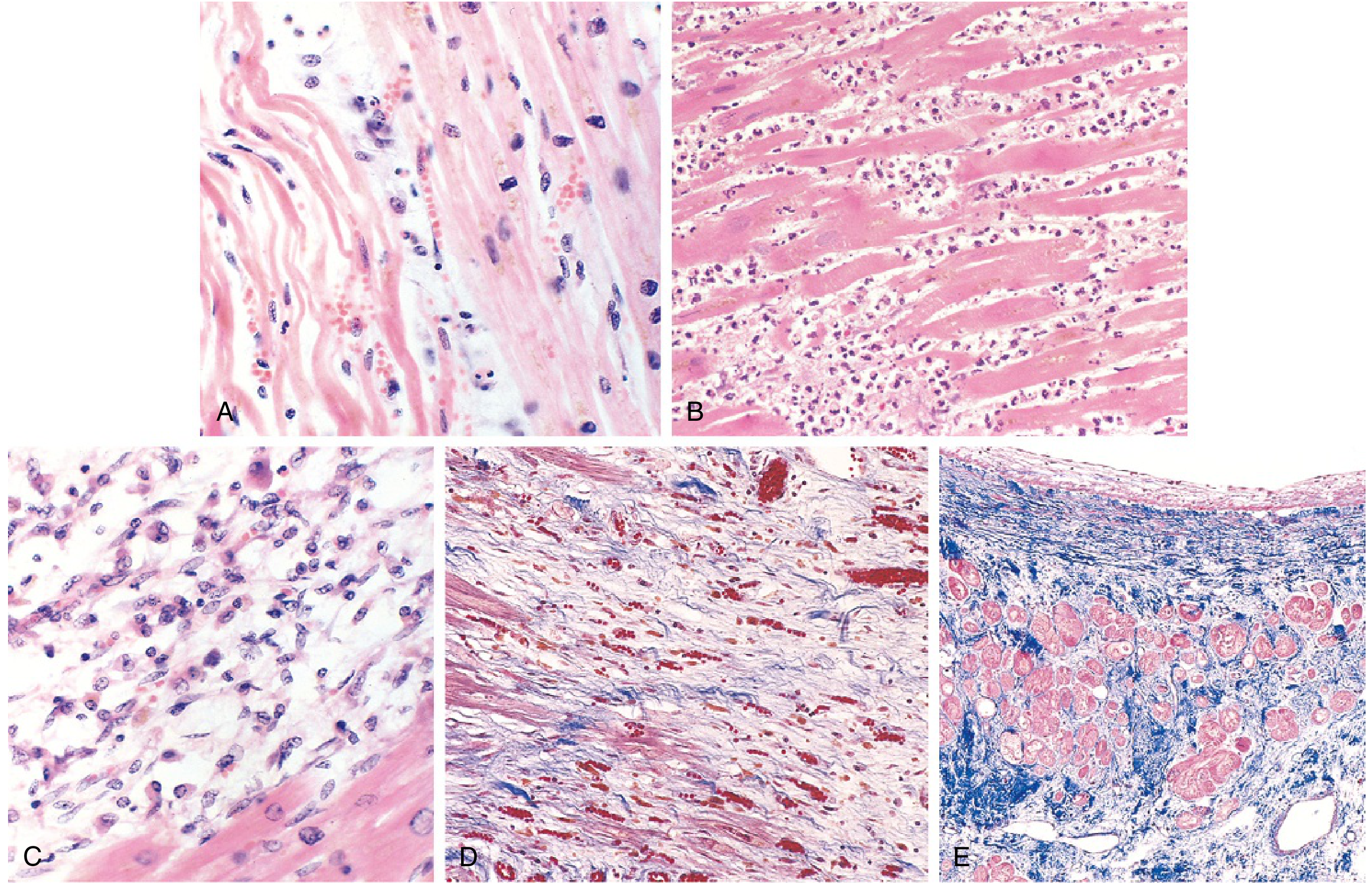
FIG: Microscopic progression of MI: (A) Day 1 - coagulative necrosis, wavy fibers; (B) Day 2-3 - dense neutrophil infiltrate; (C) Day 7-10 - macrophage removal of debris; (D) Week 1-2 - granulation tissue with new capillaries; (E) Healed - dense blue collagen scar (Masson trichrome). - Robbins & Kumar Basic Pathology
ECG Changes
The three major membrane abnormalities causing ECG changes in acute MI (Ganong's, p. 534):
| Defect in Infarcted Cells | Current Flow | ECG Change |
|---|---|---|
| Rapid repolarization | Out of infarct | ST elevation |
| Decreased resting membrane potential (K+ loss) | Into infarct | TQ depression (manifested as ST elevation) |
| Delayed depolarization | Out of infarct | ST elevation |
- Acute phase: ST segment elevation in leads over the infarct; reciprocal ST depression in opposite leads
- Hyperacute T waves: broad-based, tall, symmetrical T-waves - earliest STEMI finding
- Days to weeks later: Q waves appear (electrically silent scar fails to contribute positivity)
- STEMI = ST elevation in ≥2 contiguous leads (criteria: ≥2 mm in V1-V3; ≥1 mm in other leads)
- Ganong's Review of Medical Physiology, p. 534; Frameworks for Internal Medicine, p. 33
Cardiac Biomarkers
| Marker | Rise | Peak | Returns to Normal |
|---|---|---|---|
| Troponin I / T | 2-4 hrs | 48 hrs | 7-10 days |
| CK-MB | 2-4 hrs | 24-48 hrs | ~72 hrs |
| Myoglobin | 1-2 hrs | 6-8 hrs | 24 hrs |
- Troponins I and T are not normally in circulation; they are the most sensitive and specific markers
- They remain elevated for 7-10 days, allowing late diagnosis when CK-MB has normalized
- With reperfusion, both peak earlier due to rapid washout
- CK-MB was long the standard but is now largely replaced by troponins
- Robbins & Kumar Basic Pathology, p. 358-359
Reperfusion Injury
Restoring flow before irreversible injury is the goal ("time is myocardium"), but reperfusion carries its own risks. Contributors to reperfusion injury:
- Mitochondrial dysfunction - altered membrane permeability leads to outer membrane rupture, releasing pro-apoptotic contents
- Myocyte hypercontracture - intracellular Ca²⁺ overload causes uncontrolled myofibril contraction
- Free radicals (O₂⁻, H₂O₂, •OH) damage membrane proteins and phospholipids
- Leukocyte aggregation in reperfused vessels causes "no-reflow" phenomenon
Morphologically, reperfused infarcts show contraction bands (hypereosinophilic cross-striations) and may be hemorrhagic.
- Robbins & Kumar Basic Pathology, p. 357-358
Clinical Features
- Chest pain: severe, crushing, pressure-like, substernal - often radiates to left arm, jaw, or epigastrium; typically lasts >20 minutes
- Associated: diaphoresis, nausea, dyspnea, syncope
- "Angina equivalents" in certain populations: women, diabetics, and post-op patients may present atypically (dyspnea or fatigue alone)
- Silent MI: can occur in elderly and diabetics without pain
- Frameworks for Internal Medicine, p. 33
Treatment
Immediate Management (STEMI)
- ECG within 10 minutes of arrival
- Goal: PCI within 90 minutes (door-to-balloon) or within 120 minutes of first medical contact
- Supplemental O₂ only if SpO₂ <90%
Upstream Medical Therapy
| Medication | Dose/Notes |
|---|---|
| Aspirin | 160-325 mg chewed (buccal absorption for rapid COX-1 inhibition), then 75-162 mg daily |
| Second antiplatelet (P2Y12 inhibitor) | Clopidogrel, ticagrelor, or prasugrel |
| Anticoagulant | Heparin (UFH or LMWH) or bivalirudin |
| Nitroglycerin SL | 0.4 mg q5 min x3 for ongoing ischemia (avoid if SBP <90 mmHg, RV infarction, or PDE5 inhibitor use in prior 24h) |
| Morphine | 2-4 mg IV q5 min (effective analgesia; caution - vagotonic, reduces cardiac output) |
| IV Beta-blocker | Metoprolol 5 mg q2-5 min x3 doses (if HR >60, SBP >100 mmHg, PR interval normal) |
| Statins | High-intensity (atorvastatin 40-80 mg) |
| ACE inhibitor / ARB | Start within 24 hours (especially reduced EF, anterior MI, diabetes, hypertension) |
Reperfusion Strategies
- Primary PCI: preferred if door-to-balloon time ≤90 min and PCI-capable facility available; stent placement
- Fibrinolysis (thrombolytics): tPA, reteplase, or tenecteplase - used when PCI not available within 120 minutes; followed by transfer to PCI center
- NSTEMI: generally managed conservatively initially; early invasive strategy for high-risk patients
- Harrison's Principles of Internal Medicine 22E; Washington Manual of Medical Therapeutics; Robbins & Kumar Basic Pathology
Complications
| Complication | Timing | Notes |
|---|---|---|
| Arrhythmias | Immediate/hours | Most common cause of early death (VF in 80-90% of ischemic deaths) |
| Cardiogenic shock | Hours-days | Large infarcts (>40% LV), mortality >50% |
| Acute heart failure / pulmonary edema | Hours-days | From reduced contractility |
| Free wall rupture | 3-7 days | Hemopericardium and tamponade; peak at day 4-5 |
| Ventricular septal rupture | 3-7 days | New harsh systolic murmur |
| Papillary muscle rupture | 2-7 days | Acute mitral regurgitation |
| Ventricular aneurysm | Weeks-months | From scar thinning; predisposes to thrombus, arrhythmia |
| Mural thrombus | Days-weeks | Especially in large anterior MI; risk of embolism |
| Dressler's syndrome | Weeks | Pericarditis from autoimmune response to necrotic myocardium |
- Robbins & Kumar Basic Pathology, p. 358-360; Textbook of Clinical Echocardiography
Key Points Summary
- MI = coagulative necrosis due to sustained ischemia (>20-40 min), most often from plaque rupture + thrombosis
- LAD is the most commonly involved artery (40-50% of MIs)
- Troponin I/T is the biomarker of choice - rises at 2-4 hrs, peaks at 48 hrs, persists 7-10 days
- Hallmark ECG change = ST elevation in leads overlying infarct
- Treatment priority is rapid reperfusion: primary PCI (goal ≤90 min door-to-balloon)
- Aspirin + dual antiplatelet + anticoagulation are the pillars of antithrombotic therapy
- Complications span from early arrhythmia/shock to late aneurysm/Dressler's syndrome
Pathology oriented
Searching sources...
Finding Sources
Finding Sources
Reading File
Reading File
Excellent - rich pathology content. Now let me get the morphology section and more complications, plus fetch key pathology images:
Reading File
Reading File
Loading Image
Now let me get the detailed morphology table and reperfusion injury section from Robbins Cotran:
Reading File
Loading Image
I have the morphology table content in that truncated section. Let me read the exact morphology table:
Reading File
Loading Image
I now have everything needed for a comprehensive pathology-focused answer. Let me also get the reperfusion morphology and TTC stain image:
Reading File
Finding Sources
Let me fetch two more key pathology images - the TTC stain and the reperfusion infarct:
Loading Image
Reading File
I now have the complete, detailed pathology-oriented content from both Robbins editions plus the key images. Let me compile the full answer.
Myocardial Infarction - Pathology-Oriented Review
1. Definition & Cell Death Mechanism
MI is death of cardiac muscle due to prolonged ischemia, proceeding by ischemic coagulative necrosis as the predominant mechanism (apoptosis can also occur). Myocardial necrosis proceeds invariably to scar formation - cardiac myocytes cannot regenerate.
The critical threshold: reversible injury transitions to irreversible necrosis after 20-40 minutes of sustained ischemia.
- Robbins, Cotran & Kumar Pathologic Basis of Disease, p. 511
2. Pathogenesis of Coronary Occlusion
The sequence underlying most MIs:
- Plaque disruption - atheromatous plaque is eroded or suddenly ruptured by endothelial injury, intraplaque hemorrhage, or mechanical forces, exposing subendothelial collagen and necrotic lipid core to blood
- Platelet activation - platelets adhere, aggregate, and release thromboxane A2, ADP, and serotonin, causing further aggregation and vasospasm
- Coagulation cascade - tissue factor exposure activates coagulation, adding to the thrombus
- Occlusion - within minutes, the thrombus fully occludes the lumen
Angiography within 4 hours of MI demonstrates coronary thrombosis in nearly 90% of cases; by 12-24 hours (without intervention), only 60% - some thrombi lyse spontaneously.
Non-atherosclerotic causes (~10% of MIs)
- Vasospasm (cocaine, ephedrine, catecholamines)
- Embolism (from left atrial thrombus in AF, infective endocarditis vegetations, prosthetic valves, patent foramen ovale)
- Vasculitis, amyloid deposition, sickle cell disease (intramural arterioles)
- Microfocal infarction from microembolization
- Robbins, Cotran & Kumar, p. 511
3. Patterns of Infarction
Distribution by Vessel
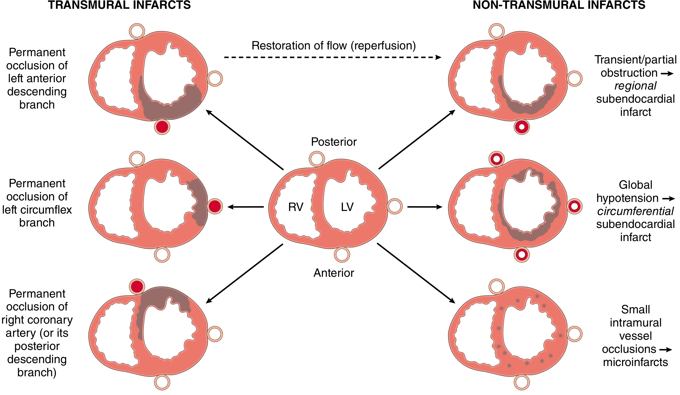
| Artery | Territory | Frequency |
|---|---|---|
| LAD | Anterior LV wall (near apex), anterior 2/3 of interventricular septum, apex circumferentially | 40-50% |
| RCA | Inferior/posterior LV wall, posterior 1/3 of septum, inferior/posterior RV free wall (15-30%) | 30-40% |
| LCX | Lateral LV wall (excluding apex) | 15-20% |
Types of Infarction
| Type | Mechanism | Pattern |
|---|---|---|
| Transmural | Complete epicardial vessel occlusion (without reperfusion) | Full-wall thickness, in territory of occluded artery |
| Subendocardial (regional) | Transient/partial obstruction, or reperfusion before full-wall necrosis | Subendocardial zone, regional |
| Subendocardial (circumferential) | Global hypotension superimposed on chronic stenoses | Circumferential, not limited to one coronary territory |
| Multifocal microinfarction | Small intramural vessel occlusion (vasculitis, microemboli, cocaine spasm) | Scattered small foci |
Why is the subendocardium most vulnerable? It is the last zone to receive blood from epicardial vessels and is exposed to the highest intramural pressures - both factors reduce perfusion. A narrow rim (~0.1 mm) immediately under the endocardium is spared by diffusion of O2 from ventricular lumen.
- Robbins, Cotran & Kumar, p. 512-513
4. Gross and Microscopic Morphology - Evolution Over Time
Gross Visualization
Early infarcts (<12 hours) are not grossly apparent on standard examination. To identify:
- Triphenyl tetrazolium chloride (TTC) stain: colors intact myocardium brick-red (dehydrogenases intact); infarcted zone appears pale/unstained because LDH leaks out through damaged membranes. Works on infarcts >3 hours old.
- Old scars appear white and glistening
Complete Morphological Timeline (Table 9.2 / Table 12.5)
| Time | Gross Features | Light Microscopy | Electron Microscopy |
|---|---|---|---|
| 0-0.5 hr (reversible) | None | None | Relaxation of myofibrils; glycogen loss; mitochondrial swelling |
| 0.5-4 hr | None | Usually none; variable waviness of fibers at border | Sarcolemmal disruption; mitochondrial amorphous densities |
| 4-12 hr | Occasional dark mottling | Onset coagulative necrosis; edema; hemorrhage | - |
| 12-24 hr | Dark mottling | Ongoing coagulative necrosis; pyknosis of nuclei; hypereosinophilic myocytes; marginal contraction band necrosis; early neutrophilic infiltrate | - |
| 1-3 days | Mottling with yellow-tan infarct center | Coagulative necrosis with loss of nuclei and striations; brisk neutrophilic infiltrate | - |
| 3-7 days | Hyperemic border; central yellow-tan softening | Disintegration of dead myofibers; dying neutrophils; early macrophage phagocytosis at border; hyperemic border = early granulation tissue | - |
| 7-10 days | Maximally yellow-tan and soft, depressed red-tan margins | Well-developed phagocytosis by macrophages; granulation tissue at margins | - |
| 10-14 days | Red-gray depressed infarct borders | Well-established granulation tissue with new vessels and collagen deposition | - |
| 2-8 weeks | Gray-white scar, progressive from border | Dense collagen scar, progressive from outside in | - |
| >8 weeks | Completed scarring | Dense fibrous scar | - |
- Robbins & Kumar Basic Pathology, Table 9.2; Robbins, Cotran & Kumar, Table 12.5
Microscopic Histology Images
From left to right: (A) Day 1: coagulative necrosis + wavy fibers; (B) Day 2-3: neutrophilic infiltrate; (C) Day 7-10: macrophage phagocytosis; (D) Week 1-2: granulation tissue; (E) Completed scar - dense blue collagen on Masson trichrome
Key histological features to recognize:
- Wavy fibers (0.5-4 hr): viable myocardium stretches and buckles non-contractile dead fibers
- Coagulative necrosis with hypereosinophilic cytoplasm and pyknotic nuclei (4-24 hr)
- Neutrophil infiltrate peaking day 1-3, then dying
- Macrophage phagocytosis of debris, days 5-10 (most pronounced)
- Granulation tissue with capillary ingrowth, weeks 1-2
- Dense fibrous scar by 6-8 weeks (indistinguishable in age regardless of whether 8 weeks or 10 years old)
5. Reperfusion - Morphological Changes
Restoring blood flow modifies the morphology of irreversibly injured cells:
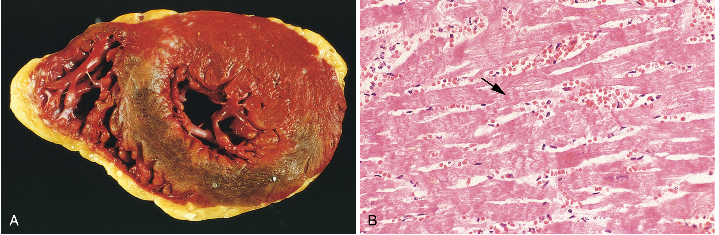
Hallmark of reperfused infarct:
- Contraction band necrosis (CBN): intense eosinophilic transverse bands of hypercontracted sarcomeres across myofibers - caused by calcium influx through damaged sarcolemma, triggering uncontrolled actin-myosin interactions. Without ATP, sarcomeres lock in this agonal tetanic state
- Infarct is typically hemorrhagic (vascular injury and leakiness)
- Corresponds to grossly visible brick-red/dark hemorrhagic zone
Stunned vs. Hibernating Myocardium
| State | Mechanism | Duration | Outcome |
|---|---|---|---|
| Stunned myocardium | Short-term ischemia + reperfusion; persistent biochemical abnormalities | Days to weeks | Spontaneous recovery of contractile function |
| Hibernating myocardium | Chronic sublethal ischemia; low metabolism/function | Months | Restored by revascularization (CABG, PCI) |
- Robbins, Cotran & Kumar, p. 515-516
6. Consequences and Complications
Nearly three-quarters of patients experience one or more complications after acute MI.
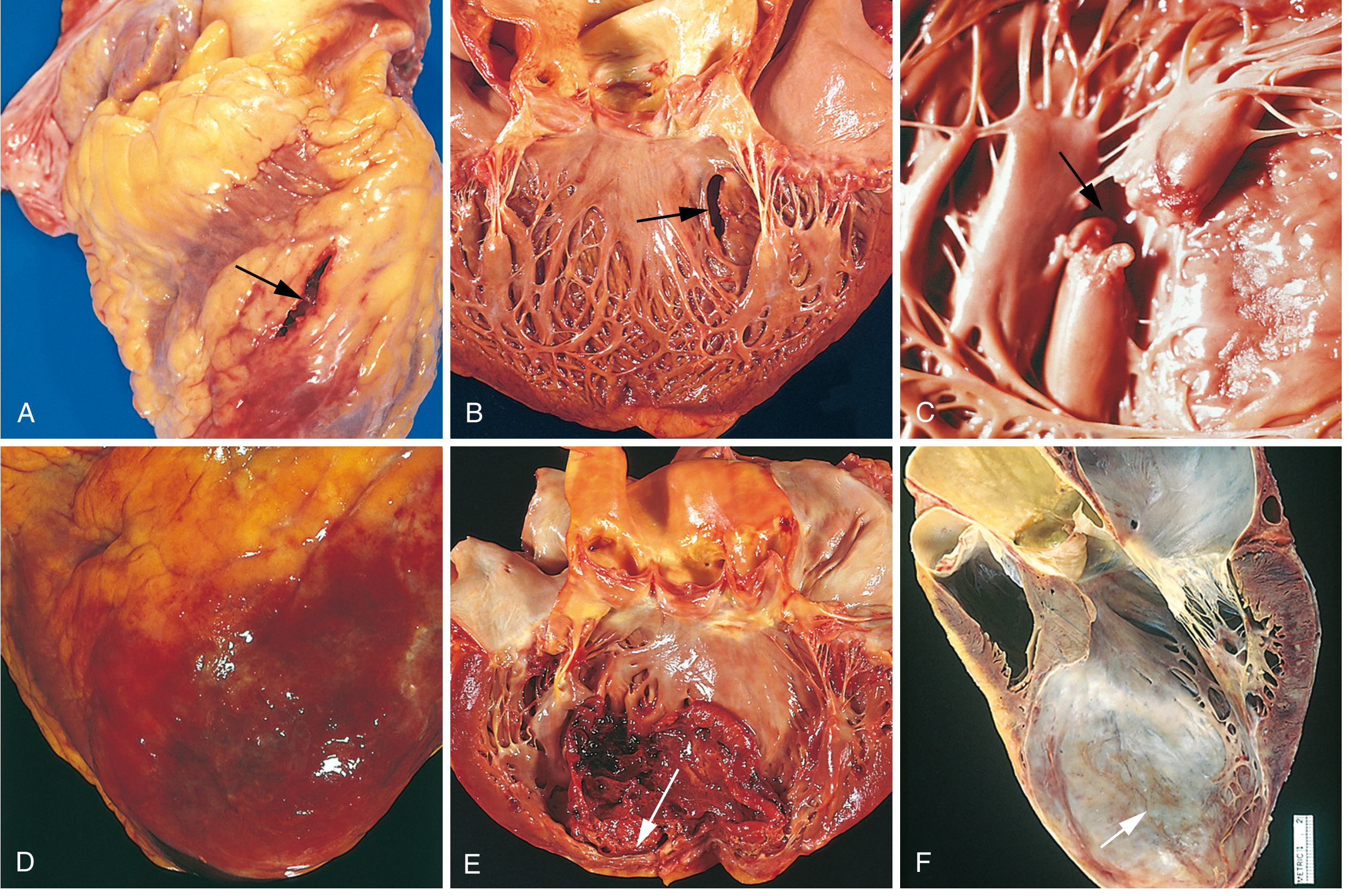
Complications - Organized by Timing
| Complication | Timing | Pathological Basis | Notes |
|---|---|---|---|
| Arrhythmias | Immediate - 24 hr | Electrical instability/irritability of ischemic myocardium | ~90% of patients; VF risk highest in first hour; most common cause of sudden cardiac death |
| Contractile dysfunction / cardiogenic shock | Hours - days | LV pump failure proportional to volume of damage | Cardiogenic shock in ~10% of transmural MIs; associated with damage to ≥40% of LV |
| Papillary muscle dysfunction | 2-7 days | Ischemia → poor contractility → mitral regurgitation | Rupture uncommon but catastrophic |
| Free wall rupture | 3-7 days (peak day 4-5) | Necrotic wall thinning | 1-3% of MIs; leads to hemopericardium and cardiac tamponade |
| Ventricular septal rupture | 3-7 days | Necrosis of septum | Creates VSD with left-to-right shunt; new harsh holosystolic murmur |
| Pericarditis | 2-3 days | Epicardial manifestation of transmural myocardial inflammation | Fibrinohemorrhagic pericarditis; friction rub; resolves within days |
| Mural thrombus | Days - weeks | Stasis (reduced contractility) + endocardial damage (thrombogenic surface) + chamber dilation | Risk of systemic embolism; especially anteroapical MI |
| Ventricular aneurysm | Weeks - months | Large transmural anteroseptal infarct heals as thinned scar wall | Does NOT rupture (scar is fibrous); causes mural thrombus, arrhythmia, heart failure |
| Dressler syndrome | Weeks after MI | Autoimmune - antibodies against damaged pericardium/myocardium | Intense pericarditis, fever; rare |
Risk Stratification by Infarct Location
| Infarct Type | Characteristic Complications |
|---|---|
| Anterior transmural | Free wall rupture, chamber dilation, mural thrombus, aneurysm |
| Posterior transmural | Conduction blocks, RV involvement; posterior VSD harder to manage |
| Large transmural (any) | Cardiogenic shock, arrhythmias, late CHF |
| Subendocardial | Pericarditis, rupture, and aneurysm rarely occur |
- Robbins, Cotran & Kumar, p. 517-519
7. Ventricular Remodeling
After MI, the non-infarcted segments undergo compensatory hypertrophy and dilation - termed ventricular remodeling. Initially hemodynamically beneficial, but:
- Ventricular dilation + increased O2 demand can worsen ischemia
- Shape changes and scar stiffening further reduce cardiac output
- ACE inhibitors reduce these deleterious effects
- Robbins, Cotran & Kumar, p. 519
Summary: The Pathologist's Approach to MI
| Question | Key Answer |
|---|---|
| What is the cell death type? | Coagulative necrosis (predominantly) |
| What is the earliest detectable microscopic change? | Wavy fibers at border (0.5-4 hr) |
| How do you confirm an infarct <12 hours old? | TTC stain - infarcted zone = pale/unstained |
| What marks reperfusion? | Contraction band necrosis + hemorrhagic infarct |
| What is the most vulnerable zone? | Subendocardium |
| What is the hallmark of a healed infarct? | Dense collagenous scar (cannot age it once complete) |
| Most common coronary artery involved? | LAD (40-50%) |
| Why can't myocardium regenerate? | Cardiac myocytes are terminally differentiated |
This is a shared conversation. Sign in to Orris to start your own chat.